

New method to visualize neurons with DAT in slices of rat VTA using fluorescent substrate for DAT, ASP+
- PMID: 24052926
- PMCID: PMC3773718
- DOI: 10.1166/jnsne.2013.1040
New method to visualize neurons with DAT in slices of rat VTA using fluorescent substrate for DAT, ASP+
Abstract
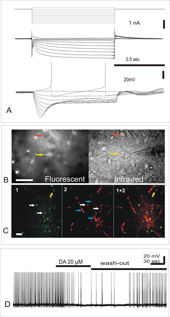
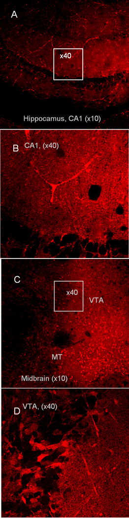
The ventral tegmental area (VTA), and in particular dopamine (DA) neurons in this region of midbrain, has been shown to play an important role in motivation (goal-directed behavior), reward, and drug addiction. Most evidence that implicates VTA DA neurons in these functions are based on widely accepted but indirect electrophysiological characterization, including the hyperpolarization activated non-specific cation current (Ih), spike frequency, and inhibition by D2 receptor agonists. In this study, we used a known neuronal dopamine transporter (DAT) fluorescent substrate [4-(4- (dimethylamino) styryl)-N-methylpyridinium iodide] (ASP+) to visualize DAT-containing cell bodies of DA neurons in VTA region in rat brain slices. Uptake of 100 nM of ASP+ in brain slices of rat VTA region marked 38% of visible neurons, while other neurons from this region and 100% neurons from hippocampus slices were not fluorescent. Using patch-clamp techniques, we have found that pronounced Ih current was present in all fluorescent neurons from VTA area, also spike frequency was similar to the widely accepted values for DA neurons. Furthermore, additional study has shown that there are 84% coincidence of ASP+ fluorescence in neuronal cell bodies and Falck-Hillarp labeling of DA cells. Electrophysiological recordings during ASP+ application have confirmed that low concentrations (100 nM) of ASP+ have no visible effect on neuronal activity during 1-2 hours after staining. Thus, uptake of fluorescent monoamine analog ASP+ by DAT can be an additional criterion for identification of DAT-containing neurons in slices.
Keywords: DAT fluorescent substrate; dopamine neurons; ventral tegmental area.
Figures
References
-
- Grace AA, Floresco SB, Goto Y, Lodge DJ. Regulation of firing of dopaminergic neurons and control of goal-directed behaviors. Trends Neurosci. 2007;30:220–227. - PubMed
-
- Aghajanian G, Bunney BC. Central dopaminergic neurons: Neurophysiologic identification and responses to drugs. In: Usdin E, Snyder SH, editors. Frontiers in catecholamine research; proceedings. Elmsford, N.Y.: Pergamon Press; 1973. pp. 643–648.
-
- Grace AA, Bunney BS. Intracellular and extracellular electrophysiology of nigral dopaminergic neurons--1. Identification and characterization. Neuroscience. 1983;10:301–315. - PubMed
-
- Cameron DL, Wessendorf MW, Williams JT. A subset of ventral tegmental area neurons is inhibited by dopamine, 5-hydroxytryptamine and opioids. Neuroscience. 1997;77:155–166. - PubMed
Grants and funding
LinkOut - more resources
Full Text Sources
Other Literature Sources