. 2013 Jun 4;1(1):19-27.
doi: 10.1016/j.stemcr.2013.04.001. eCollection 2013.

The interfollicular epidermis of adult mouse tail comprises two distinct cell lineages that are differentially regulated by Wnt, Edaradd, and Lrig1

Affiliations

The interfollicular epidermis of adult mouse tail comprises two distinct cell lineages that are differentially regulated by Wnt, Edaradd, and Lrig1

Céline Gomez et al. Stem Cell Reports. .

Abstract

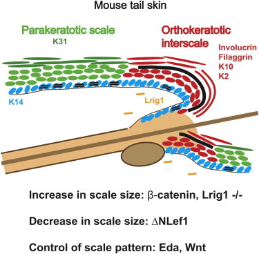
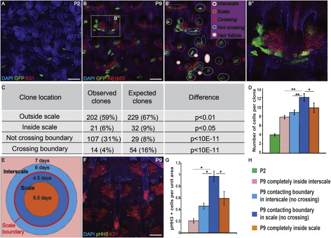
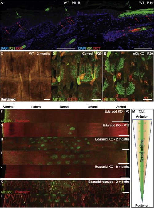
Current models of how mouse tail interfollicular epidermis (ife) is maintained overlook the coexistence of two distinct terminal differentiation programs: parakeratotic (scale) and orthokeratotic (interscale). lineage tracing and clonal analysis revealed that scale and interscale are maintained by unipotent cells in the underlying basal layer, with scale progenitors dividing more rapidly than interscale progenitors. Although scales are pigmented and precisely aligned with hair follicles, melanocytes and follicles were not necessary for scale differentiation. Epidermal Wnt signaling was required for scale enlargement during development and for postnatal maintenance of scale-interscale boundaries. Loss of Edaradd inhibited ventral scale formation, whereas loss of Lrig1 led to scale enlargement and fusion. In wild-type skin, Lrig1 was not expressed in IFE but was selectively upregulated in dermal fibroblasts underlying the interscale. We conclude that the different IFE differentiation compartments are maintained by distinct stem cell populations and are regulated by epidermal and dermal signals.


Figures

None
Graphical abstract
Figure 1
Figure 1
Differentiation of Scale and Interscale IFE (A–I) Sagittal sections of WT tail skin. (A–C) Hematoxylin and eosin (H&E) staining. (D–F) Immunostaining for FLG (red) and K31 (green). (G–I) Immunostaining for K10 (red) and AB1653 (green). Insets are single-stained, enlarged views of the boxed areas. Interscale (arrows); scale (arrowheads); DAPI counterstain (blue); dermo-epidermal junction (white dashed lines). (J–Q) Whole mounts of WT tail epidermis. (J–L) Immunostaining for K2 (red) and AB1653 (green). (M–Q) AB1653 immunostaining (green) with phalloidin (red) counterstain. Images were obtained from the anterior part of the tail. (R) Scales are induced along the dorsal midline at P3 and spread laterally to reach the ventral midline by P14. (S and T) Schematics showing sagittal (S) and whole-mount (T) views of scale and interscale IFE in 2-month-old mice. In all panels, scales are green and interscales are red. Scale bars, 100 μm (A–L) and 500 μm (M–Q). See also Figure S1.
Figure 2
Figure 2
Clonal Analysis of Scale and Interscale Epidermis (A and B) Tail epidermal whole mounts of K14CreER × CagcatGFP mice immunostained for GFP and K31 at the stages indicated. (B′) Analysis of GFP-positive clones in (B). GFP clones: interscale (pink), scale (orange), encompassing scale and interscale (red), and bordering scale without crossing (blue) or HF (solid pink). (B″) Enlargement of the boxed region in (B). (C and D) Quantitation of observed and expected clone types (C) and number of cells per clone (D). t test, *p < 0.05, **p < 0.001. Error bars represent SEM. (E) Rates of cell division in different areas. (F) Immunostaining for K31 (red) and pHH3 (green) of WT P9 tail epidermis. (G) Spatial distribution of pHH3 positive cells. z test, *p < 0.05. Error bars represent SD. (H) Legends and color code for (D), (E), and (G). Scale bars, 100 μm. See also Figure S2.
Figure 3
Figure 3
Scale Formation Occurs in the Absence of Melanocytes and HF (A and B) Immunostaining of WT tail skin sagittal sections with DCT and K31 antibodies. Melanocytes (red arrows); scale differentiation (green arrows); DAPI counterstain (blue). (C) Bright-field view of WT 2-month-old tail epidermal whole mount showing scale pigmentation. (D and E) Immunostaining for AB1653 (green) and SOX10 (red) in P20 epidermal whole mount of ckit knockout (E) and WT littermate (D) mice. (F–J) Edaradd knockout tail epidermis labeled with AB1653 (green) and phalloidin (red). (F) n = 6 mice, (G) n = 4, (H and I) n = 10, (J) n = 3. (K and L) Two-month-old Edaradd knockout rescued with K14::Edaradd transgene, labeled with AB1653 (green) and phalloidin (red) (n = 5). (M) Diagram of scale induction in Edaradd knockout. Scale bars, 100 μm (A–E) and 500 μm (F–L). See also Figure S3.
Figure 4
Figure 4
Wnt and Lrig1 Signaling Controls Scale Borders (A) WT tail skin labeled with LEF1 and K31 antibodies and DAPI counterstain. (B and C) Differences in scale size (B) and distance between rows of HF (C) in K14ΔNLef1 and control littermates. Error bars represent SEM. (D–O) Immunostaining with AB1653 (green) and phalloidin counterstain (red) of tail epidermal whole mounts: (D) 2-month-old control and (E–F) K14ΔNLef1 littermates (dorsal [E] and ventral [F] views), (G) control and (H–I) 4OHT-treated K14ΔNβ-cateninER transgenics, Lrig1 knockouts (M–O), and WT littermates (J–L). (P) LRIG1 immunostaining of P1 WT tail dermis. (Q) AB1653 and LRIG1 immunostaining of WT P5 tail skin. Epidermal basal layer (dotted lines); HF (solid lines); dermal LRIG1 expression (yellow arrows). Scale bars, 100 μm. See also Figure S4.

References

    1. Arwert E.N., Hoste E., Watt F.M. Epithelial stem cells, wound healing and cancer. Nat. Rev. Cancer. 2012;12:170–180. - PubMed
    1. Braun K.M., Niemann C., Jensen U.B., Sundberg J.P., Silva-Vargas V., Watt F.M. Manipulation of stem cell proliferation and lineage commitment: visualisation of label-retaining cells in wholemounts of mouse epidermis. Development. 2003;130:5241–5255. - PubMed
    1. Brown S.J., McLean W.H. One remarkable molecule: filaggrin. J. Invest. Dermatol. 2012;132:751–762. - PMC - PubMed
    1. Cable J., Jackson I.J., Steel K.P. Mutations at the W locus affect survival of neural crest-derived melanocytes in the mouse. Mech. Dev. 1995;50:139–150. - PubMed
    1. Carroll J.M., Romero M.R., Watt F.M. Suprabasal integrin expression in the epidermis of transgenic mice results in developmental defects and a phenotype resembling psoriasis. Cell. 1995;83:957–968. - PubMed

Publication types

MeSH terms

LinkOut - more resources