. 2013 Jul 25;1(2):105-13.
doi: 10.1016/j.stemcr.2013.06.007. eCollection 2013.

Functional vascular endothelium derived from human induced pluripotent stem cells

Affiliations

Functional vascular endothelium derived from human induced pluripotent stem cells

William J Adams et al. Stem Cell Reports. .

Abstract

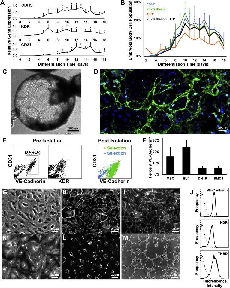
Vascular endothelium is a dynamic cellular interface that displays a unique phenotypic plasticity. This plasticity is critical for vascular function and when dysregulated is pathogenic in several diseases. Human genotype-phenotype studies of endothelium are limited by the unavailability of patient-specific endothelial cells. To establish a cellular platform for studying endothelial biology, we have generated vascular endothelium from human induced pluripotent stem cells (iPSCs) exhibiting the rich functional phenotypic plasticity of mature primary vascular endothelium. These endothelial cells respond to diverse proinflammatory stimuli, adopting an activated phenotype including leukocyte adhesion molecule expression, cytokine production, and support for leukocyte transmigration. They maintain dynamic barrier properties responsive to multiple vascular permeability factors. Importantly, biomechanical or pharmacological stimuli can induce pathophysiologically relevant atheroprotective or atheroprone phenotypes. Our results demonstrate that iPSC-derived endothelium possesses a repertoire of functional phenotypic plasticity and is amenable to cell-based assays probing endothelial contributions to inflammatory and cardiovascular diseases.


Figures

None
Graphical abstract
Figure 1
Figure 1
Derivation of iPSC-ECs (A) Gene expression time profiles of CDH5 (VE-cadherin), KDR, and CD31 measured by quantitative real-time PCR (n = 3) with the asterisks () denoting values different than day 0 (p < 0.05). (B) CD31, VE-cadherin, and KDR expression in EB cells measured by flow cytometry (n = 3). (C) Phase-contrast image of an EB after 10 days of differentiation. (D) Reconstruction of two-photon confocal microscopy images of VE-cadherin (green) and nuclei (blue) within an EB after 10 days of differentiation. (E) The isolation of VE-cadherin+ cells from the EB with magnetic bead sorting. (F–M) Fraction of EB cells expressing VE-cadherin from different iPSC lines (F; n = 4). Isolated iPSC-ECs characterized by phase contrast (G), immunofluorescence for VE-cadherin (H) and CD31 (I), flow cytometry (J) for VE-cadherin, KDR, and thrombomodulin (sample in solid line; isotype control in dotted), immunofluorescence for eNOS (K) and endocytosed acetylated-LDL (L), and a phase-contrast image of the network formation on Matrigel (M). All pooled data are represented as mean ± SD. See also Figure S1.
Figure 2
Figure 2
Assessment of Endothelial Activation in iPSC-ECs (A) Surface expression of E-selectin, ICAM-1, and VCAM-1 after 6 or 24 hr of 10 U/ml IL-1β, 10 ng/ml TNF-α, or 1 μg/ml LPS treatment measured by flow cytometry. (B) Culture supernatant concentration of chemokines after 24 hr treatment of TNF-α, IL-1β, IFNγ, LPS, or TNF-α+IFNγ with the asterisks () indicating difference to vehicle (veh) (p < 0.05; n = 3–4). (C) Time profile of transmigrated neutrophils and T cells after 4 hr TNF-α treatment. (D) Fraction of neutrophils or T cells transmigrated across TNF-α- stimulated iPSC-ECs and HUVECs (n = 3; n.s., not significant). (E–H) Transmigration of neutrophils after pretreatment of neutrophils with anti-CD18 antibody (E) or pretreatment of iPSC-ECs with anti- ICAM-1 antibody (F). Transmigration of T cells after pretreatment of T cells with anti-CD18 antibody (G) or pretreatment of iPSC-ECs with anti- ICAM-1 antibody (H). A binding nonblocking anti-MHC class I antibody was used as a control (n = 2). All pooled data are represented as mean ± SD. See also Figure S2 and Movies S1 and S2.
Figure 3
Figure 3
Barrier Properties of iPSC-ECs (A) Changes in transendothelial electrical resistance after treatment with 10 μM histamine, 100 ng/ml VEGF, 200 ng/ml prostaglandin E2, 0.5 μg/ml sphingosine-1-phosphate, or 100 μM O-Me. Data are represented as mean ± SD (n = 3) with the asterisks () denoting different from control (p < 0.05). Arrows indicate time of addition of compound. (B) VE-cadherin and actin after 24 hr treatment with 100 μM O-Me seen by immunofluorescence microscopy. See also Figure S3.
Figure 4
Figure 4
Acquisition of Atheroprotective and Atheroprone Endothelial Phenotypes (A–C) A single period of atheroprotective and atheroprone shear stress waveforms (A). iPSC-ECs imaged by phase contrast after 72 hr of atheroprone (B) or atheroprotective flow (C; arrow indicates direction of flow). (D) Relative (Rel.) gene expression after 72 hr of atheroprotective or atheroprone shear stress. (E) Relative gene expression after 24 hr treatment with 10 μM simvastatin. The asterisks () indicate statistical significance with p < 0.05 (n = 4). All pooled data are represented as mean ± SD. See also Figure S4.

References

    1. Adams W.J., García-Cardeña G. Novel stem cell-based drug discovery platforms for cardiovascular disease. J. Biomol. Screen. 2012;17:1117–1127. - PubMed
    1. Bos J.L. Epac proteins: multi-purpose cAMP targets. Trends Biochem. Sci. 2006;31:680–686. - PubMed
    1. Chen X.L., Nam J.O., Jean C., Lawson C., Walsh C.T., Goka E., Lim S.T., Tomar A., Tancioni I., Uryu S. VEGF-induced vascular permeability is mediated by FAK. Dev. Cell. 2012;22:146–157. - PMC - PubMed
    1. Chiu J.J., Chien S. Effects of disturbed flow on vascular endothelium: pathophysiological basis and clinical perspectives. Physiol. Rev. 2011;91:327–387. - PMC - PubMed
    1. Choi K.D., Yu J., Smuga-Otto K., Salvagiotto G., Rehrauer W., Vodyanik M., Thomson J., Slukvin I. Hematopoietic and endothelial differentiation of human induced pluripotent stem cells. Stem Cells. 2009;27:559–567. - PMC - PubMed

Publication types

LinkOut - more resources