. 2013 Jul 24;1(2):198-208.
doi: 10.1016/j.stemcr.2013.07.001. eCollection 2013.

Do pluripotent stem cells exist in adult mice as very small embryonic stem cells?

Affiliations

Do pluripotent stem cells exist in adult mice as very small embryonic stem cells?

Masanori Miyanishi et al. Stem Cell Reports. .

Abstract

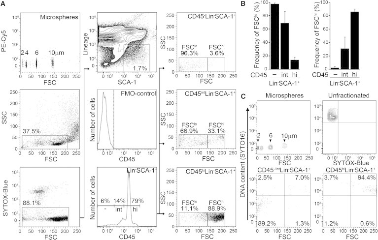
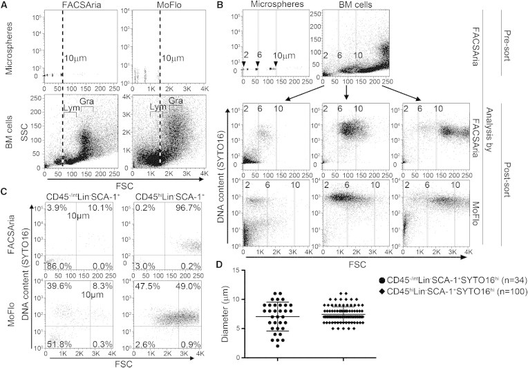
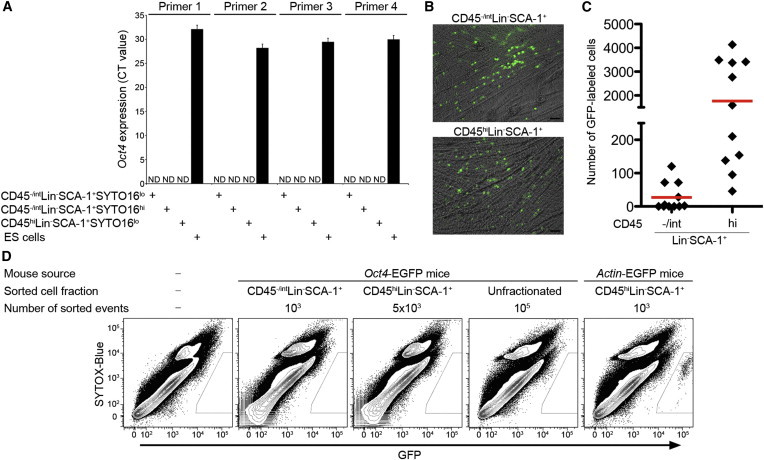
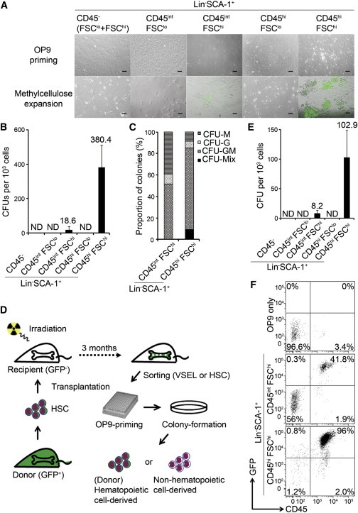
Very small embryonic-like stem cells (VSELs) isolated from bone marrow (BM) have been reported to be pluripotent. Given their nonembryonic source, they could replace blastocyst-derived embryonic stem cells in research and medicine. However, their multiple-germ-layer potential has been incompletely studied. Here, we show that we cannot find VSELs in mouse BM with any of the reported stem cell potentials, specifically for hematopoiesis. We found that: (1) most events within the "VSEL" flow-cytometry gate had little DNA and the cells corresponding to these events (2) could not form spheres, (3) did not express Oct4, and (4) could not differentiate into blood cells. These results provide a failure to confirm the existence of pluripotent VSELs.


Figures

Figure 1
Figure 1
Adult Mouse Bone Marrow CD45LinSCA-1+ Cells Enriched in the FSClo Region Contain Little DNA (A) FACS plots of BM cells from wild-type mice. The initial gate (left middle) was based on defined-size microspheres (left upper) and expected to include both HSCs and VSELs. After excluding dead cells (left bottom), we focused on the LinSCA-1+ fraction (center upper), subdividing these into FMO-defined CD45 (dashed line in center middle), CD45int, and CD45hi fractions (center bottom). The frequency of FSClo (<10 μm microspheres) and FSChi (>10 μm microspheres) cells in each subfraction is indicated (right panels). (B) Frequency of FSClo (left) or FSChi (right) cells among the CD45, CD45int, and CD45hi subfractions of LinSCA-1+ events. The mean ± SD of data from 22 independent experiments are shown. (C) Analysis of DNA content by SYTO16 staining. The threshold of high SYTO16 positivity was determined to include 99% of unfractionated BM cells (upper right). The vertical line in the lower plots indicates the position of 10 μm microspheres. See also Tables S1 and S2.
Figure 2
Figure 2
Evaluation of Cell Size by Different Cell Sorters (A) Defined-size microspheres (upper) and whole BM cells (lower) were analyzed by FACSAria (left) and MoFlo (right). Dashed lines indicate the FSC of 10 μm microspheres. The positions of lymphocytes (Lym) and granulocytes (Gra) are indicated in the lower panels. (B) On a FACSAria, BM cells were sorted by expected size (2–6, 6–10, and >10 μm) based on their FSC relative to microspheres (upper) and then reanalyzed on the same FACSAria (middle) and on a MoFlo (lower). Vertical lines indicate the positions of 2, 6, and 10 μm microspheres on each machine. (C) FACS plots of diploid (SYTO16hi) CD45−/intLinSCA-1+ cells (candidate VSELs; left) and CD45hiLinSCA-1+ cells (HSCs; right) analyzed by FACSAria (upper) or MoFlo (lower). Vertical lines indicate the position of 10 μm microspheres in each sorter, and horizontal lines indicate the threshold of SYTO16 positivity. (D) Comparison of the sizes of candidate VSELs (n = 34) and HSCs (n = 100) determined with an image flow cytometer. Each dot represents a cell, and each line with error bars represents the mean ± SD. See also Figure S1.
Figure 3
Figure 3
No Evidence for the Pluripotency of CD45−/intLinSCA-1+ Cells (A) Real-time RT-PCR analysis of Oct4 expression in subfractions of LinSCA-1+ cells and in ESCs (BM cells were prepared as a pool of four mice and then sorted in quadruplicate and subjected to RT-PCR; the data are shown as mean ± SD). ND, not detected. (B) Representative images of 8 day progeny of FACS-purified LinSCA-1+ populations from Actin-EGFP mice. Cells were cocultured with C2C12 cells in DMEM supplemented with 2% FCS. Scale bar, 50 μm. (C) The total number of GFP+ cells detected by FACS analysis at day 8 coculture of either 1,000 CD45−/intLinSCA-1+ events or 1,000 CD45hiLinSCA-1+ events (n = 11 for each from three independent experiments); the red line indicates the mean. (D) FACS analysis of Oct4-derived EGFP expression on culture day 8. On day 0, the following were plated on C2C12 cells: no cells (far left column) or BM cells from Oct4-EGFP or Actin-EGFP mice, sorted by the indicated phenotypes. Data shown are representative of three independent experiments. See also Figure S2 and Table S3.
Figure 4
Figure 4
HSCs Are the Only Contributor to Postnatal Mouse Hematopoiesis (A) Representative images on culture days 5 and 10. Each FACS-purified BM LinSCA-1+ fraction from Actin-EGFP mice was cocultured with OP9 cells for the first 5 days (i.e., OP9 priming) and transferred to methylcellulose for an additional 5 days (i.e., methylcellulose expansion). The threshold between FSClo and FSChi was defined by 10 μm microspheres. Scale bar, 100 μm. (B) The number of colony-forming units (CFUs) from 103 cells of each LinSCA-1+ fraction. Data shown are mean ± SD of four independent experiments. ND, not detected. (C) Proportion of colonies. From five independent colony assays, 199 colonies derived from CD45intLinSCA-1+FSChi cells and 250 colonies derived from CD45hiLinSCA-1+FSChi cells were picked up, cytospun, and stained with the May-Giemsa method to determine the cell types included. CFU-M, CFU-macrophage; CFU-G, CFU-granulocyte; CFU-GM, CFU-granulocyte/macrophage; CFU-Mix, CFU-erythroid and myeloid cells. (D) Schematic of in vivo experiments. (E) The number of CFUs from 103 cells of each LinSCA-1+ fraction. Data shown here are mean ± SD. (F) Ten day progeny of CD45intLinSCA-1+FSChi or CD45hiLinSCA-1+FSChi cells were harvested with OP9 stromal cells and analyzed by FACS. Live cells were gated and tested for the expression of CD45 and GFP. A total of 5 × 104 events were recorded. Data were similar in three independent experiments. See also Figures S3 and S4.

References

    1. Begley C.G., Ellis L.M. Drug development: Raise standards for preclinical cancer research. Nature. 2012;483:531–533. - PubMed
    1. Beltrami A.P., Cesselli D., Bergamin N., Marcon P., Rigo S., Puppato E., D’Aurizio F., Verardo R., Piazza S., Pignatelli A. Multipotent cells can be generated in vitro from several adult human organs (heart, liver, and bone marrow) Blood. 2007;110:3438–3446. - PubMed
    1. Bhartiya D., Shaikh A., Nagvenkar P., Kasiviswanathan S., Pethe P., Pawani H., Mohanty S., Rao S.G., Zaveri K., Hinduja I. Very small embryonic-like stem cells with maximum regenerative potential get discarded during cord blood banking and bone marrow processing for autologous stem cell therapy. Stem Cells Dev. 2012;21:1–6. - PubMed
    1. Check E. Stem cells: the hard copy. Nature. 2007;446:485–486. - PubMed
    1. Danova-Alt R., Heider A., Egger D., Cross M., Alt R. Very small embryonic-like stem cells purified from umbilical cord blood lack stem cell characteristics. PLoS ONE. 2012;7:e34899. - PMC - PubMed

Publication types

Substances