. 2014 Feb;50(2):389-97.
doi: 10.1165/rcmb.2012-0529OC.

Increased concentration of iodide in airway secretions is associated with reduced respiratory syncytial virus disease severity

Affiliations

Increased concentration of iodide in airway secretions is associated with reduced respiratory syncytial virus disease severity

Rachel J Derscheid et al. Am J Respir Cell Mol Biol. 2014 Feb.

Abstract

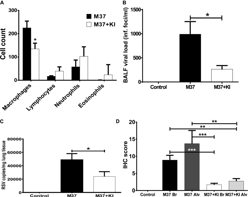
Recent studies have revealed that the human and nonrodent mammalian airway mucosa contains an oxidative host defense system. This three-component system consists of the hydrogen peroxide (H2O2)-producing enzymes dual oxidase (Duox)1 and Duox2, thiocyanate (SCN(-)), and secreted lactoperoxidase (LPO). The LPO-catalyzed reaction between H2O2 and SCN(-) yields the bactericidal hypothiocyanite (OSCN(-)) in airway surface liquid (ASL). Although SCN(-) is the physiological substrate of LPO, the Duox/LPO/halide system can generate hypoiodous acid when the iodide (I(-)) concentration is elevated in ASL. Because hypoiodous acid, but not OSCN(-), inactivates respiratory syncytial virus (RSV) in cell culture, we used a lamb model of RSV to test whether potassium iodide (KI) could enhance this system in vivo. Newborn lambs received KI by intragastric gavage or were left untreated before intratracheal inoculation of RSV. KI treatment led to a 10-fold increase in ASL I(-) concentration, and this I(-) concentration was approximately 30-fold higher than that measured in the serum. Also, expiratory effort, gross lung lesions, and pulmonary expression of an RSV antigen and IL-8 were reduced in the KI-treated lambs as compared with nontreated control lambs. Inhibition of LPO activity significantly increased lesions, RSV mRNA, and antigen. Similar experiments in 3-week-old lambs demonstrated that KI administration was associated with reduced gross lesions, decreased RSV titers in bronchoalveolar lavage fluid, and reduced RSV antigen expression. Overall, these data indicate that high-dose KI supplementation can be used in vivo to lessen the severity of RSV infections, potentially through the augmentation of mucosal oxidative defenses.


Figures

Figure 1.
Figure 1.
Iodide (I) concentrations in the airway secretions and serum of lambs after systemic administration of I. (A) I concentration in nasal secretions of lambs at the indicated time points after intravenous injection of NaI (closed circles; n = 5) or PBS (open circles; n = 5) at 0 hours. (B) I concentrations in the serum of lambs at the indicated time points after intravenous (i.v.) administration of NaI (closed triangles; n = 5) or PBS (open triangles; n = 5) at 0 hours. (C) I concentration in the nasal and tracheobronchial secretions of lambs (closed circles and closed squares, respectively) at the indicated time points after intragastric potassium iodide (KI) administration at 0 hours. The numbers of animals used for the collection of nasal secretions were 5 (0 and 4 h), 4 (12 h), and 3 (36 h). The numbers of animals used for the collection of tracheal secretions were 1 (4 h), 1 (12 h), and 3 (36 h). (D) I concentration in the serum of lambs (n ≥ 3) at the indicated time points after intragastric delivery of KI. Error bars = SEM.
Figure 2.
Figure 2.
Effect of oral KI supplementation on human respiratory syncytial virus (hRSV) strain A2 mRNA levels and airway surface liquid (ASL) [I]. Respiratory syncytial virus (RSV) replication (A) and [I] in tracheal ASL (B) were evaluated 6 days after inoculating lambs with hRSV A2; the indicated groups of animals also received daily doses of KI (1.8 mg/kg BW). (A) RSV mRNA signal normalized to total RNA loaded per RT-qPCR: control, n = 5; RSV+KI, n = 5; RSV, n = 5. (B) No KI, n = 5; KI, n = 5. Error bars = SEM. *P < 0.05.
Figure 3.
Figure 3.
The effect of KI on RSV M37 infection newborn (2- to 3-d-old) lambs. (A) Enhanced expiratory effort (forced expiration). Lambs inoculated with RSV M37 and receiving KI had significantly reduced expiratory efforts at Days 3, 4, and 5 after inoculation compared with RSV-inoculated lambs lacking KI and control lambs. Control group, n = 6; M37 group, n = 11; M37 + KI group, n = 10. Error bars = SEM. *P < 0.05; **P < 0.01; ***P < 0.001. (B) Lambs receiving KI had significantly reduced gross lesions compared with RSV-inoculated lambs lacking KI and control lambs. RSV M37–infected lambs receiving KI and dapsone had significantly increased gross lesions compared with control and RSV-infected (no KI). Control group, n = 14; M37 group, n = 11; M37 + KI group, n = 10; M37 + KI + dapsone group, n = 5. Error bars = SEM. *P < 0.05; ***P < 0.001. The minor control group lesions were found (by immunohistochemistry) to not be RSV. (C) Lambs receiving KI had a trend of reduced RSV M37 mRNA compared with lambs lacking KI, whereas lambs receiving RSV M37, KI, and dapsone had significantly increased RSV M37 mRNA levels compared with lambs treated with M37 alone and M37 + KI. Error bars = SEM. *P < 0.05; ***P < 0.001. (D) Newborn lambs inoculated with RSV M37 and receiving KI had significantly reduced levels of viral antigen in alveolar regions compared with RSV-inoculated lambs lacking KI. In bronchioles, RSV M37 antigen was not significantly altered; control lambs lacked RSV antigen. RSV M37-infected lambs receiving KI and dapsone had significantly increased RSV antigen compared with control and RSV-infected (no KI). Control group, n = 14; M37 group, n = 11; M37 + KI group, n = 10; M37 + KI + dapsone group, n = 5. Error bars = SEM. *P < 0.05; ***P < 0.001. (E, F). Lung from newborn lambs infected with RSV M37 and stained for RSV antigen. (E) Lung of lamb lacking KI administration in which there is abundant RSV antigen (brown) in most airway epithelial cells. (F) Lung of lamb that received KI; a few cells contain RSV antigen.
Figure 4.
Figure 4.
The effect of KI on RSV M37 infection in 3-week-old lambs. (A) Effect of KI treatment on the cellular composition of bronchoalveolar lavage fluid (BALF) in RSV-infected 3-week-old lambs. Cytospin preparations of BALF were stained with modified Wright’s, and 300 cell differentials were performed to assess relative differences in populations of inflammatory cells. Values are expressed as mean ± SD. *P < 0.01 based on ANOVA followed by Tukey-Kramer multiple comparisons test. (B) (BALF) titers of RSV in 3-week-old lambs. Lambs receiving KI had significantly less viable RSV than lambs lacking KI. Control lambs lacked RSV titers. Control group, n = 4; M37 group, n = 5; M37 + KI group, n = 5. Error bars = SEM. *P < 0.05. (C) RSV M37 mRNA levels in lungs of lambs receiving KI trended toward significant reductions compared with RSV-inoculated lambs lacking KI; controls lack RSV mRNA. Control group, n = 8; M37 group, n = 10; M37 + KI group, n = 10. Error bars = SEM. *P < 0.05. (D) Three-week-old lambs receiving KI had significantly reduced levels of RSV antigen in bronchiolar and alveolar regions compared with RSV-inoculated lambs lacking KI; controls lacked RSV antigen. Control bronchioles and bronchi (Br), n = 8; control alveoli (Alv), n = 8; M37 Br, n = 30; M37 Alv, n = 30; M37 + KI Br, n = 30; M37 + KI Alv, n = 30. Error bars = SEM. **P < 0.01; ***P < 0.001. IHC, immunohistochemistry.
Figure 5.
Figure 5.
Ontogeny of lung expression of lactoperoxidase (LPO) and dual-functioning oxidases (Duox)1 and Duox2 in lambs as assessed by RT-qPCR. (A) Expression of LPO mRNA at various stages of lamb development: 115 days of gestation group, n = 5; 130 days of gestation group, n = 4; term lamb group, n = 4; adult lamb group, n = 4. Error bars = SEM. *P < 0.05. (B) Expression of Duox1 mRNA at various stages of lamb development. (C) Expression of Duox2 mRNA at various stages of lamb development: 115 days of gestation group, n = 3; 130 days of gestation group, n = 3; term lamb group, n = 4; adult lamb group, n = 4. Error bars = SEM. *P < 0.05.

References

    1. Collins PL, Melero JA. Progress in understanding and controlling respiratory syncytial virus: still crazy after all these years. Virus Res. 2011;162:80–99. - PMC - PubMed
    1. Welliver RC, Sr, Checchia PA, Bauman JH, Fernandes AW, Mahadevia PJ, Hall CB. Fatality rates in published reports of RSV hospitalizations among high-risk and otherwise healthy children. Curr Med Res Opin. 2010;26:2175–2181. - PubMed
    1. Sommer C, Resch B, Simões EA. Risk factors for severe respiratory syncytial virus lower respiratory tract infection. Open Microbiol J. 2011;5:144–154. - PMC - PubMed
    1. Abed Y, Boivin G. Treatment of respiratory virus infections. Antiviral Res. 2006;70:1–16. - PMC - PubMed
    1. Empey KM, Peebles RS, Jr, Kolls JK. Pharmacologic advances in the treatment and prevention of respiratory syncytial virus. Clin Infect Dis. 2010;50:1258–1267. - PMC - PubMed

Publication types

MeSH terms