. 2014 Apr 10;20(11):1693-708.
doi: 10.1089/ars.2013.5219. Epub 2013 Dec 18.

Nrf2 regulates angiogenesis: effect on endothelial cells, bone marrow-derived proangiogenic cells and hind limb ischemia

Affiliations

Nrf2 regulates angiogenesis: effect on endothelial cells, bone marrow-derived proangiogenic cells and hind limb ischemia

Urszula Florczyk et al. Antioxid Redox Signal. .

Abstract

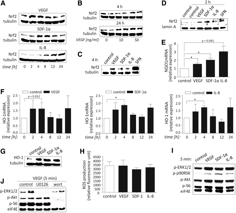
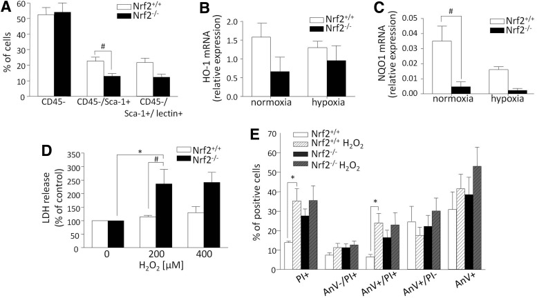
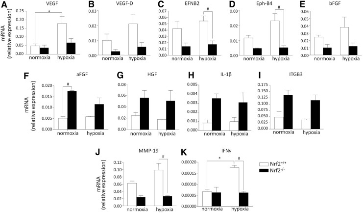
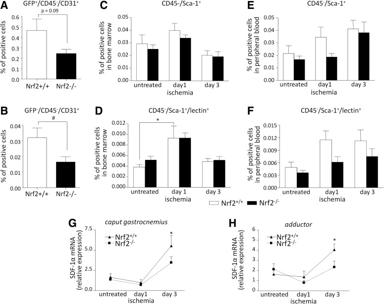
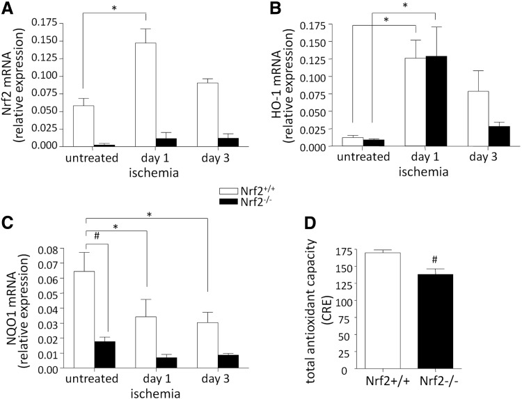
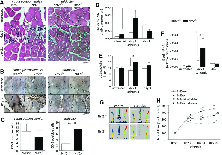
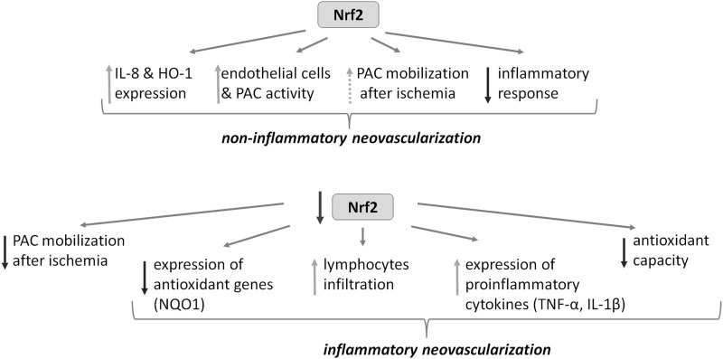
Aims: Nuclear factor E2-related factor 2 (Nrf2), a key cytoprotective transcription factor, regulates also proangiogenic mediators, interleukin-8 and heme oxygenase-1 (HO-1). However, hitherto its role in blood vessel formation was modestly examined. Particularly, although Nrf2 was shown to affect hematopoietic stem cells, it was not tested in bone marrow-derived proangiogenic cells (PACs). Here we investigated angiogenic properties of Nrf2 in PACs, endothelial cells, and inflammation-related revascularization.

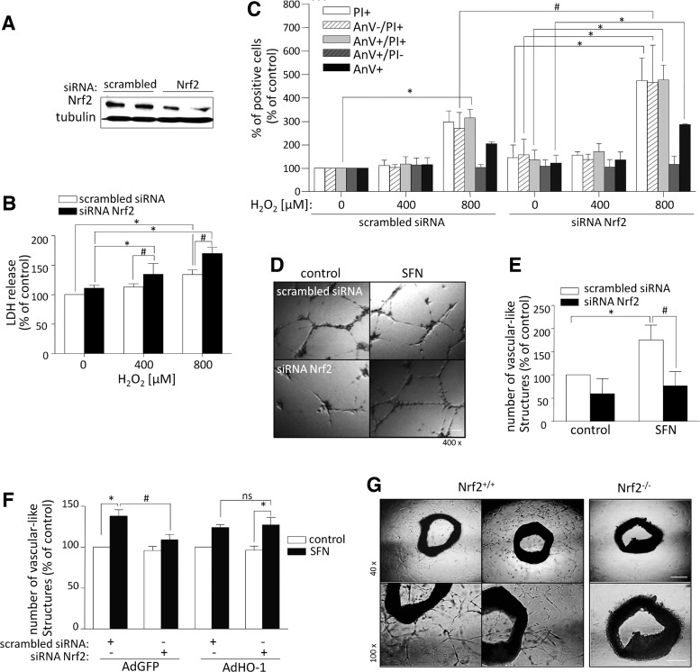
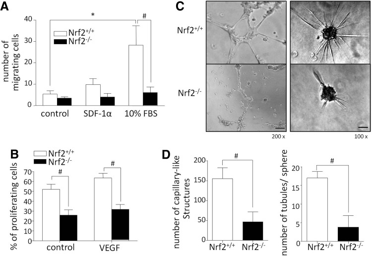
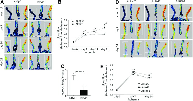
Results: Treatment of endothelial cells with angiogenic cytokines increased nuclear localization of Nrf2 and induced expression of HO-1. Nrf2 activation stimulated a tube network formation, while its inhibition decreased angiogenic response of human endothelial cells, the latter effect reversed by overexpression of HO-1. Moreover, lack of Nrf2 attenuated survival, proliferation, migration, and angiogenic potential of murine PACs and affected angiogenic transcriptome in vitro. Additionally, angiogenic capacity of PAC Nrf2(-/-) in in vivo Matrigel assay and PAC mobilization in response to hind limb ischemia of Nrf2(-/-) mice were impaired. Despite that, restoration of blood flow in Nrf2-deficient ischemic muscles was better and accompanied by increased oxidative stress and inflammatory response. Accordingly, the anti-inflammatory agent etodolac tended to diminish blood flow in the Nrf2(-/-) mice.

Innovation: Identification of a novel role of Nrf2 in angiogenic signaling of endothelial cells and PACs.

Conclusion: Nrf2 contributes to angiogenic potential of both endothelial cells and PACs; however, its deficiency increases muscle blood flow under tissue ischemia. This might suggest a proangiogenic role of inflammation in the absence of Nrf2 in vivo, concomitantly undermining the role of PACs in such conditions.


Figures

<b>FIG. 1.</b>
FIG. 1.
Nrf2 is activated in response to proangiogenic stimulation of HMEC-1. HMEC-1 were stimulated with growth factors VEGF (50 ng/ml), SDF-1α (100 ng/ml), and IL-8 (200 ng/ml) for indicated time (2, 4, 8, 12, or 24 h). (A) The level of Nrf2 protein is increased after stimulation with growth factors. (B) The effect of VEGF on Nrf2 protein tends to be dose dependent after 24 h of treatment. HMEC-1 were treated with 10 or 50 ng/ml VEGF for 4 or 24 h. (C) The level of Nrf2 protein is increased after starvation of cells in 2% FBS-containing medium for 24 h and further growth factor stimulation. (A–C) Western blot. Tubulin: control constitutive protein. SFN (2.5 μM): positive control. (D) Growth factors induce Nrf2 localization in the nucleus. Western blot. SFN: positive control. Lamin A: control constitutive protein of nuclear fraction. (E–G) The expression of Nrf2 target genes, NQO1 and HO-1, is increased in response to stimulation with growth factors. (E) 24 h, (F) different time points. RT-PCR; (G) 4 h. Western blot. Tubulin: control constitutive protein. (H) Incubation with growth factors for 2 h does not change the ROS production. DCF fluorescence measurement. (I) Akt and ERK1/2 are activated and their downstream targets S6 and p90RSK tend to be phosphorylated after 5 min of stimulation with growth factors. (J) pAkt/pS6 and pERK1/2 are decreased after incubation of VEGF-stimulated cells with wortmannin and U0126, respectively. Western blot. eIF4E: control constitutive protein; Western blot: representative experiments (nJ=experiment in duplicate); each bar represents the mean±SEM of three to four independent experiments (nH=2) performed in duplicates. *p<0.05, control versus growth factor stimulation. ERK-1/2, extracellular signal-regulated kinase-1/2; HMEC-1, human microvascular endothelial cells; HO-1, heme oxygenase-1; IL-8, interleukin-8; NQO1, NAD(P)H:quinone oxidoreductase 1; Nrf2, nuclear factor E2-related factor 2; ROS, reactive oxygen species; SDF-1, stromal cell-derived factor-1; SFN, sulforaphane; VEGF, vascular endothelial growth factor.
<b>FIG. 2.</b>
FIG. 2.
Silencing of Nrf2 decreases angiogenic potential of HMEC-1 and diminishes angiogenic response of murine aortic endothelium. (A) Nrf2 protein level is decreased after transfection of HMEC-1 for 64 h with siRNA against human Nrf2 mRNA (50 nM). Western blot. Tubulin: control constitutive protein. Representative experiment. (B–E) HMEC-1 were transfected for 48 h with siRNA against human Nrf2 mRNA (50 nM) or scrambled siRNA (50 nM). Nrf2 deficiency increases mortality and the rate of apoptosis/necrosis of HMEC-1 under oxidative stress conditions. (B) LDH release after stimulation with H2O2 for 24 h. (C) The percentage of apoptotic/necrotic cells after stimulation with H2O2 for 24 h. Staining, respectively, for annexin V (AnV) and propidium iodide (PI). Flow cytometry. (D, E) The number of vascular-like structures formed by SFN-treated Nrf2-silenced HMEC-1 is reduced. (D) Representative pictures of vascular-like structures formed by HMEC-1 with silenced Nrf2 and/or after stimulation with 2.5 μM SFN. Culture on Matrigel for 16 h in 2% FBS-containing medium. Magnification 400×(scale bar: 20 μm). (E) Quantitative analysis. (F) HO-1 overexpression reverses the inhibitory effect of siRNA Nrf2 on network formation on Matrigel. Cells were transfected with siRNA Nrf2 for 24 h and then transduced with AdHO-1 or AdGFP as a control for the next 24 h and seeded on Matrigel in the presence of SFN for 16 h. Quantitative analysis. (G) Nrf2 deficiency diminishes capillary formation from murine aortic rings. Aortic rings were isolated from Nrf2+/+ and Nrf2−/− mice and incubated on Matrigel in 2% FBS-containing medium for 12 days. Representative images of vascular structures. Magnification 40×(top panel, scale bar: 200 μm), 100×(bottom panel, scale bar: 100 μm); each bar represents the mean±SEM of three to five independent experiments performed in duplicates or triplicates. *p<0.05, control versus H2O2/SFN; #p<0.05, scrambled siRNA versus siRNA Nrf2.
<b>FIG. 3.</b>
FIG. 3.
PACs lacking Nrf2 reveal lower expression of antioxidant genes and higher mortality under oxidative stress conditions. Mononuclear cells were isolated from bone marrow of Nrf2+/+ and Nrf2−/− mice and cultured for 7–10 days in vitro. (A) Analysis of surface marker expression. Flow cytometry (nNrf2+/+=3, nNrf2−/−=3); Nrf2 deficiency in PACs affects the mRNA level of (B) HO-1 and (C) NQO1. PACs were cultured for 24 h under normoxia or hypoxia (0.5% O2). RT-PCR (nNrf2+/+=3, nNrf2−/−=3). (D, E) PAC survival under conditions of oxidative stress is impaired in the absence of Nrf2. (D) LDH release after stimulation with H2O2 for 24 h (nNrf2+/+=3–9, nNrf2−/−=2–8). (E) The percentage of apoptotic/necrotic cells after stimulation with 200 μM H2O2 for 24 h. Staining, respectively, for annexin V (AnV) and propidium iodide (PI). Flow cytometry (nNrf2+/+=6, nNrf2−/−=6); each bar represents the mean±SEM. *p<0.05, control versus H2O2; #p<0.05, Nrf2+/+ versus Nrf2−/−. PACs, proangiogenic cells.
<b>FIG. 4.</b>
FIG. 4.
PACs lacking Nrf2 reveal defective angiogenic properties. (A) PAC migration is inhibited in the absence of Nrf2. Cells were cultured for 24 h in Boyden chambers in 2% FBS-containing medium. As a chemoattractant, in the bottom of the chamber, 100 ng/ml of SDF-1α or 10% FBS was used. Quantitative analysis of migrating cells. Staining with crystal violet (nNrf2+/+=7, nNrf2−/−=6–7). (B) Proliferation of PAC Nrf2−/− is inhibited. Cells were cultured for 24 h in 2% FBS-containing medium and 50 ng/ml VEGF. Quantitative analysis of proliferating cells (PCNA+) (nNrf2+/+=6, nNrf2−/−=5). (C) The number of vascular-like structures formed by PAC Nrf2−/− is reduced. Representative pictures of networks after 12 h of culture on Matrigel (left) and capillaries after 48 h of spheroidal culture in collagen gel (right) in medium containing 2% FBS. Magnification 200×(left panel, scale bar: 50 μm), 100×(right panel, scale bar: 100 μm). (D) Quantitative analysis of vascular-like structures formed on Matrigel (left) (nNrf2+/+=10, nNrf2−/−=7) and in collagen gel (right) (nNrf2+/+=3, nNrf2−/−=3); each bar represents the mean±SEM. *p<0.05, control versus 10% FBS; #p<0.05, Nrf2+/+ versus Nrf2−/−.
<b>FIG. 5.</b>
FIG. 5.
Nrf2 level influences the expression of genes involved in neovascularization. PACs were cultured for 24 h under normoxia or hypoxia (0.5% O2). The mRNA level of several genes differentially regulated in PAC Nrf2+/+ and Nrf2−/− is shown (A–K). Transcriptome analysis, PCR array. Each bar represents the mean±SEM (nNrf2+/+=2–3; nNrf2−/−=2–3). *p<0.05, normoxia versus hypoxia; #p<0.05, Nrf2+/+ versus Nrf2−/−.
<b>FIG. 6.</b>
FIG. 6.
Proangiogenic response and mobilization of PACs are disrupted in Nrf2−/− mice. (A, B) 500,000 of PACs (Nrf2+/+ or Nrf2−/−) were mixed with Matrigel and injected subcutaneously at the ventral side of GFP transgenic mice. At day 15 the percentage of endothelial cells defined as (A) GFP+/CD45/CD31+ and (B) GFP/CD45/CD31+ was determined in the Matrigel plugs. Flow cytometry (nNrf2+/+=8; nNrf2−/−=10). (C–F) Nrf2+/+ and Nrf2−/− mice were subjected to FAL. Preoperatively (untreated) and at days 1 and 3 after surgery, the percentage of the progenitors defined as CD45/Sca-1+ and CD45/Sca-1+/lectin+ was determined in the bone marrow (C, D) (nNrf2+/+=8; nNrf2−/−=6–8) and peripheral blood (E, F) (nNrf2+/+=10–12; nNrf2−/−=10–11). Flow cytometry. (G, H) SDF-1α gradient in response to FAL is impaired in Nrf2−/− mice. The ratio of SDF-1α expression at the mRNA level (RT-PCR) in caput gastrocnemius (G) (nNrf2+/+=8–11; nNrf2−/−=9–10) and adductor (H) (nNrf2+/+=5–8; nNrf2−/−=6–8) muscles to the mRNA level of SDF-1α in the bone marrow; each bar represents the mean±SEM. *p<0.05, untreated versus ischemia (appropriate time point), #p<0.05, Nrf2+/+ versus Nrf2−/−. FAL, femoral artery ligation.
<b>FIG. 7.</b>
FIG. 7.
Nrf2 deficiency increases revascularization after FAL. Nrf2+/+ and Nrf2−/− mice were subjected to FAL. (A, B) The ratio of blood flow in the left limb (ischemic) to right limb (non-ischemic) is higher in Nrf2−/− mice. Blood flow was measured 30 min after surgery (day 0) and at days 7, 14, and 21. Laser Doppler perfusion imaging. (A) Representative pictures. (B) Quantitative analysis (nNrf2+/+=15, nNrf2−/−=11). (C) The number of necrotic toes at day 21 after FAL is slightly higher in Nrf2+/+ mice (nNrf2+/+=15, nNrf2−/−=11). (D, E) The rate of perfusion did not change significantly in Nrf2−/− mice after AdNrf2 or AdHO-1 transduction compared with control vectors (AdLacZ). Blood flow was measured 30 min after surgery and injection of adenoviral vectors (day 0) and at days 7 and 14. Laser Doppler perfusion imaging. (D) Representative pictures. (E) Quantitative analysis (n=5–6); each bar/point represents the mean±SEM. *p<0.05, day 0 versus appropriate time point; #p<0.05, Nrf2+/+ versus Nrf2−/−. To see this illustration in color, the reader is referred to the web version of this article at www.liebertpub.com/ars
<b>FIG. 8.</b>
FIG. 8.
Lack of Nrf2 changes its target gene expression and antioxidant capacity in ischemic muscles after FAL. Nrf2+/+ and Nrf2−/− mice were subjected to FAL. (A–C) Preoperatively (untreated) and at days 1 and 3 after surgery the mRNA level of, respectively, Nrf2, HO-1, and NQO1 was checked in caput gastrocnemius. RT-PCR (nNrf2+/+=10–12, nNrf2−/−=10–12). (D) Total antioxidant capacity (copper reducing equivalents [CREs]) is lower in Nrf2−/− ischemic muscles (nNrf2+/+=4, nNrf2−/−=4); each bar represents the mean±SEM. *p<0.05, untreated versus ischemia (appropriate time point); #p<0.05, Nrf2+/+ versus Nrf2−/−.
<b>FIG. 9.</b>
FIG. 9.
Lack of Nrf2 increases inflammatory response after FAL. Nrf2+/+ and Nrf2−/− mice were subjected to FAL. (A) Representative pictures of the morphology of muscles (caput gastrocnemiusleft panel, adductorright panel), untreated and ischemic (days 3 and 21 after FAL). Hematoxylin/eosin staining. Arrows indicate centrally located nuclei of muscle fibers, which suggest intensive regeneration of tissue. Magnification 1000×(scale bar: 10 μm). (B, C) The lack of Nrf2 tends to increase the degree of T cell infiltration into ischemic muscle. (B) Representative pictures of CD3 antigen staining at day 3 after FAL (caput gastrocnemiusleft panel, adductorright panel). Magnification 1000×(scale bar: 10 μm). Immunohistochemistry. (C) Quantitative analysis of CD-3-positive cells (nNrf2+/+=2–3, nNrf2−/−=2). (D–F) Preoperatively (untreated) and at days 1 and 3 after FAL the expression of TNF-α and E-sel (D, F respectively, RT-PCR) was examined in caput gastrocnemius (nNrf2+/+=10–12; nNrf2−/−=10–11), while the protein level of IL-1β was tested in the plasma (E) (Luminex) (nNrf2+/+=11; nNrf2−/−=10–12). (G, H) Nrf2+/+ and Nrf2−/− mice were subjected to FAL and etodolac treatment [10 mg/(kg bw·day−1)]. The ratio of blood flow in the left limb (ischemic) to right limb (non-ischemic) is higher in Nrf2−/− mice, but tends to be reversed after etodolac treatment. Blood flow was measured 30 min after surgery (day 0) and at days 7, 14, and 20. Laser Doppler perfusion imaging. (G) Representative pictures. (H) Quantitative analysis (nNrf2+/+=6, nNrf2−/−=5–6); each bar/point represents the mean±SEM. *p<0.05, untreated versus ischemia (appropriate time point) or day 0 versus appropriate time point; $p<0.05, control (olive oil) versus etodolac; #p<0.05, Nrf2+/+ versus Nrf2−/−; ##p<0.05, Nrf2+/+ etodolac versus Nrf2−/− etodolac. E-sel, E-selectin. To see this illustration in color, the reader is referred to the web version of this article at www.liebertpub.com/ars
<b>FIG. 10.</b>
FIG. 10.
Simplified diagram of Nrf2 participation in the processes of neovascularization and the effects of its deficiency. Nrf2 regulating proangiogenic genes, taking part in the functioning of endothelial cells and BM-derived PACs, and regulating the expression of cytoprotective genes may be an important mediator of neovascularization. However, in the absence of Nrf2 in vivo, following ischemia, angiogenesis associated with oxidative stress and inflammatory response could occur, despite an impaired function and mobilization of PACs. BM, bone marrow.

References

    1. Ades EW, Candal FJ, Swerlick RA, George VG, Summers S, Bosse DC, and Lawley TJ. HMEC-1: establishment of an immortalized human microvascular endothelial cell line. J Invest Dermatol 99: 683–690, 1992 - PubMed
    1. Afonyushkin T, Oskolkova OV, Philippova M, Resink TJ, Erne P, Binder BR, and Bochkov VN. Oxidized phospholipids regulate expression of ATF4 and VEGF in endothelial cells via NRF2-dependent mechanism: novel point of convergence between electrophilic and unfolded protein stress pathways. Arterioscler Thromb Vasc Biol 30: 1007–1013, 2010 - PubMed
    1. Aicher A, Heeschen C, Mildner-Rihm C, Urbich C, Ihling C, Technau-Ihling K, Zeiher AM, and Dimmeler S. Essential role of endothelial nitric oxide synthase for mobilization of stem and progenitor cells. Nat Med 9: 1370–1376, 2003 - PubMed
    1. Alam J, Stewart D, Touchard C, Boinapally S, Choi AM, and Cook JL. Nrf2, a Cap'n'Collar transcription factor, regulates induction of the heme oxygenase-1 gene. J Biol Chem 274: 26071–26078, 1999 - PubMed
    1. Asahara T, Masuda H, Takahashi T, Kalka C, Pastore C, Silver M, Kearne M, Magner M, and Isner JM. Bone marrow origin of endothelial progenitor cells responsible for postnatal vasculogenesis in physiological and pathological neovascularization. Circ Res 85: 221–228, 1999 - PubMed

Publication types

MeSH terms