

Radiofrequency ablation (RFA) of renal cell carcinoma (RCC): experience in 200 tumours
- PMID: 24053769
- PMCID: PMC4233988
- DOI: 10.1111/bju.12349
Radiofrequency ablation (RFA) of renal cell carcinoma (RCC): experience in 200 tumours
Abstract
Objectives: To evaluate our clinical experience with percutaneous image-guided radiofrequency ablation (RFA) of 200 renal tumours in a large tertiary referral university institution.
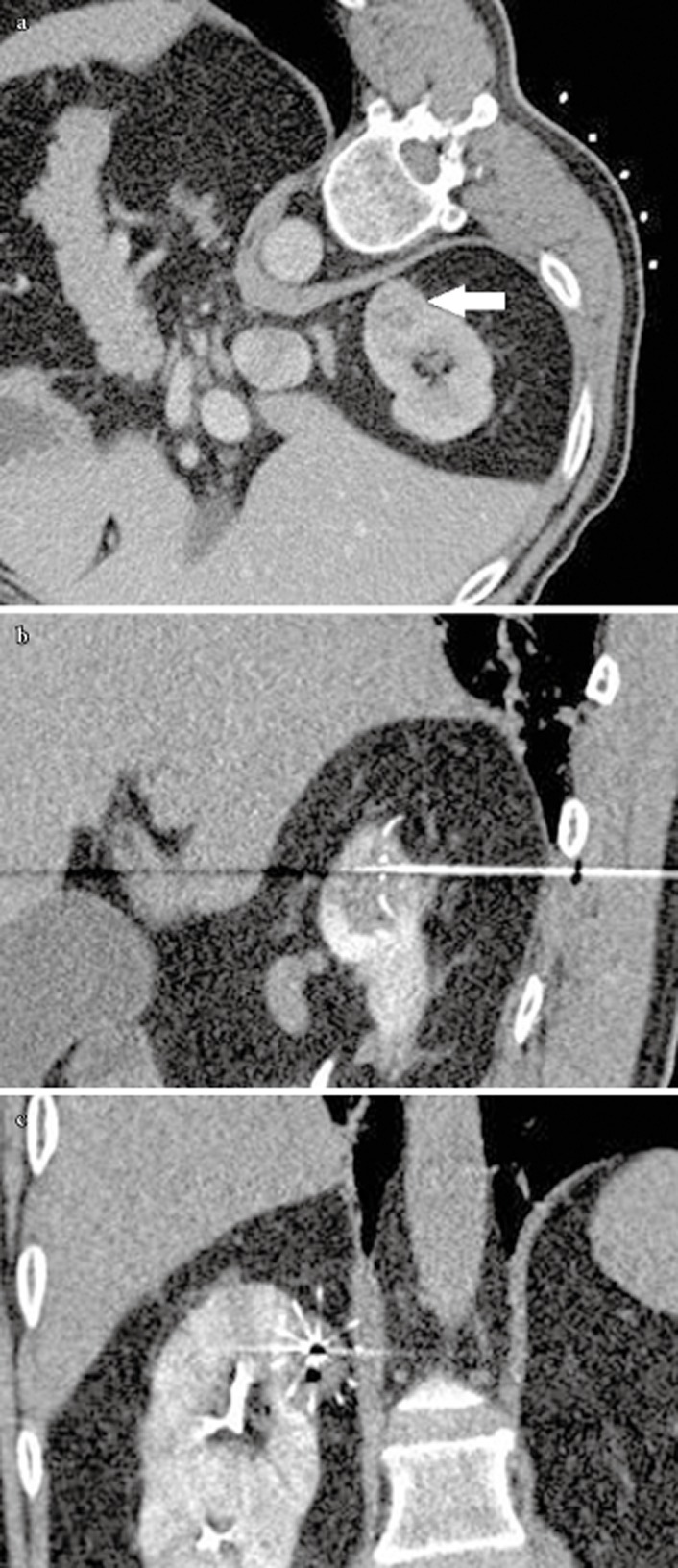
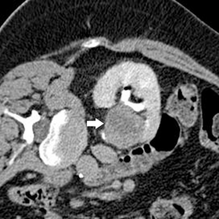
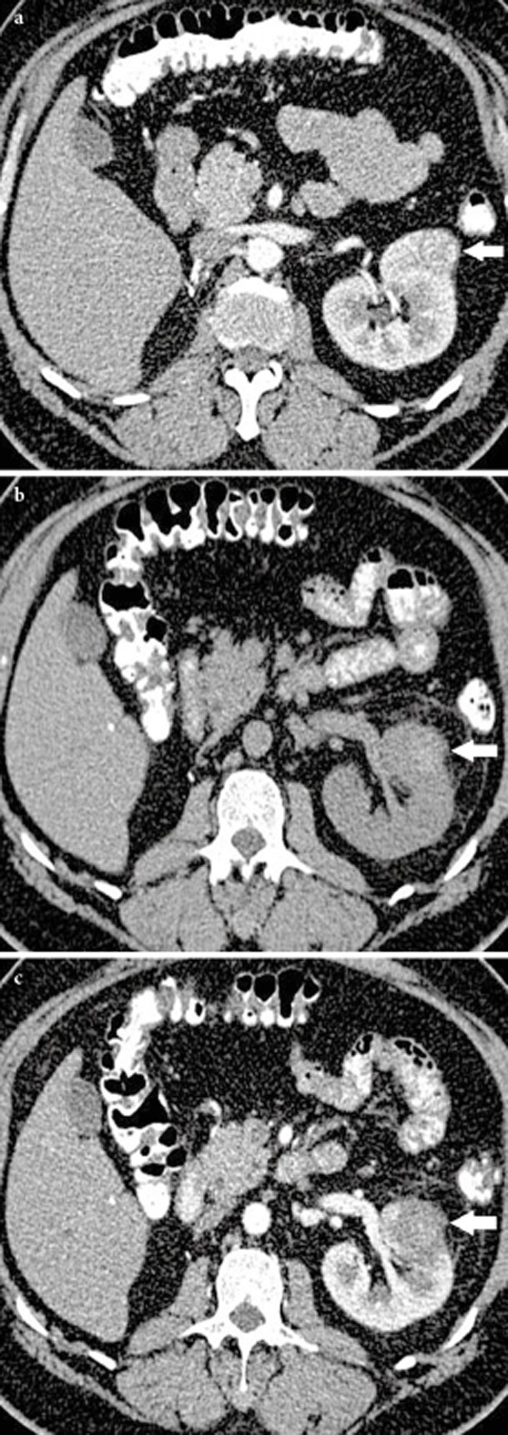
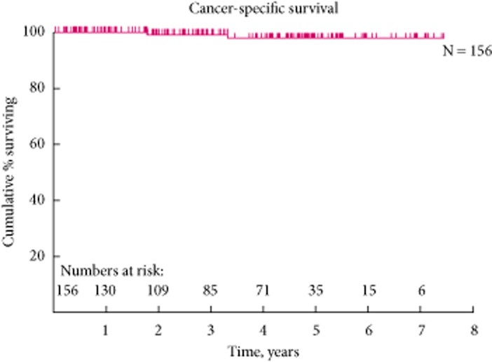
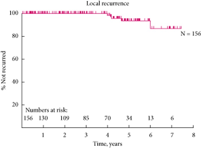
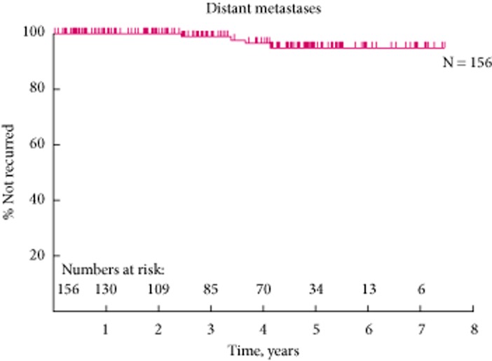
Patients and methods: Image-guided RFA (ultrasonography or computed tomography [CT]) of 200 renal tumours in 165 patients from June 2004 to 2012 was prospectively evaluated. Institutional Review Board approval was granted. The treatment response and technical success were defined by absence of contrast enhancement within the tumour on contrast enhanced CT or magnetic resonance imaging. Both major and minor complications, glomerular filtration rate (GFR) before and after RFA, the management and outcomes of the complications, as well as oncological outcome were prospectively documented. Multivariate analysis was used to determine variables associated with major complications and also the percentage GFR change after RFA. The overall (OS), 5-year cancer-specific (CSS), local recurrence-free (LRFS) and metastasis-free survival (MFS) rates are presented using the Kaplan-Meier curves.
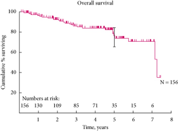
Results: In all, 200 tumours were RF ablated with a mean (range) tumour size of 2.9 (1-5.6) cm and the mean (range) patient age was 67.7 (21-88.6) years with a mean follow-up period of 46.1 months. The primary technical and overall technical success rate was 95.5% and 98.5%, respectively. Two independent predictors of successful RFA in a single sitting were tumour size (<3 cm) and exophytic location in multivariate logistic regression analysis. Major complications included ureteric injury (six patients), calyceal-cutaneous fistula (one), acute tubular necrosis (one) and abscess (two). Two independent predictors of ureteric injury were central location and lower pole position. Within this cohort of patients, only four patients developed significant renal function deterioration i.e. >25% decreased in GFR. In all, 161 (98%) patients of the 165 patients have preservation of renal function. Any change in renal function after RFA was not influenced by tumour factors or solitary kidney status. In our clinical series, this yielded a 5-year OS, CSS, LRFS and MFS rates of 75.8%, 97.9%, 93.5% and 87.7% respectively.
Conclusions: Image-guided RFA is a safe, nephron sparing and effective treatment for small renal cell carcinoma (RCC) tumours with a low rate of recurrence and has good 5-year CSS and MFS rates.
Keywords: complication; radiofrequency ablation; renal cell carcinoma; survival rates.
© 2013 The Authors. BJU International published by John Wiley & Sons Ltd on behalf of BJU International.
Figures
References
-
- Jemal A, Tiwari RC, Murray T. Cancer statistics, 2004. CA Cancer J Clin. 2004;54:8–29. - PubMed
-
- Office for National Statistics. Cancer Statistics Registrations: Registrations of Cancer Diagnosed in 2008, England. London: Office for National Statistics, National Statistics; 2010.
-
- 2008. Globocan.
-
- Zagoria RJ. Imaging of small renal masses: a medical success story. AJR Am J Roentgenol. 2000;175:945–955. - PubMed
-
- Jayson M, Sanders H. Increased incidence of serendipitously discovered renal cell carcinoma. Urology. 1998;51:203–205. - PubMed
MeSH terms
LinkOut - more resources
Full Text Sources
Other Literature Sources
Medical
