. 2013 Sep 26;4(6):1213-23.
doi: 10.1016/j.celrep.2013.08.024. Epub 2013 Sep 19.

Membrane-sculpting BAR domains generate stable lipid microdomains

Affiliations

Membrane-sculpting BAR domains generate stable lipid microdomains

Hongxia Zhao et al. Cell Rep. .

Abstract

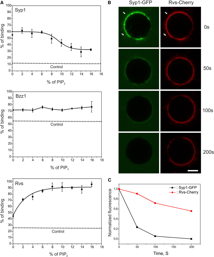
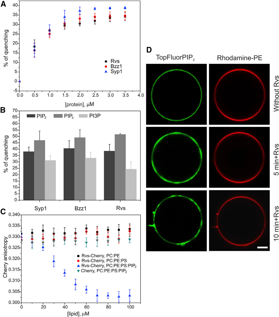
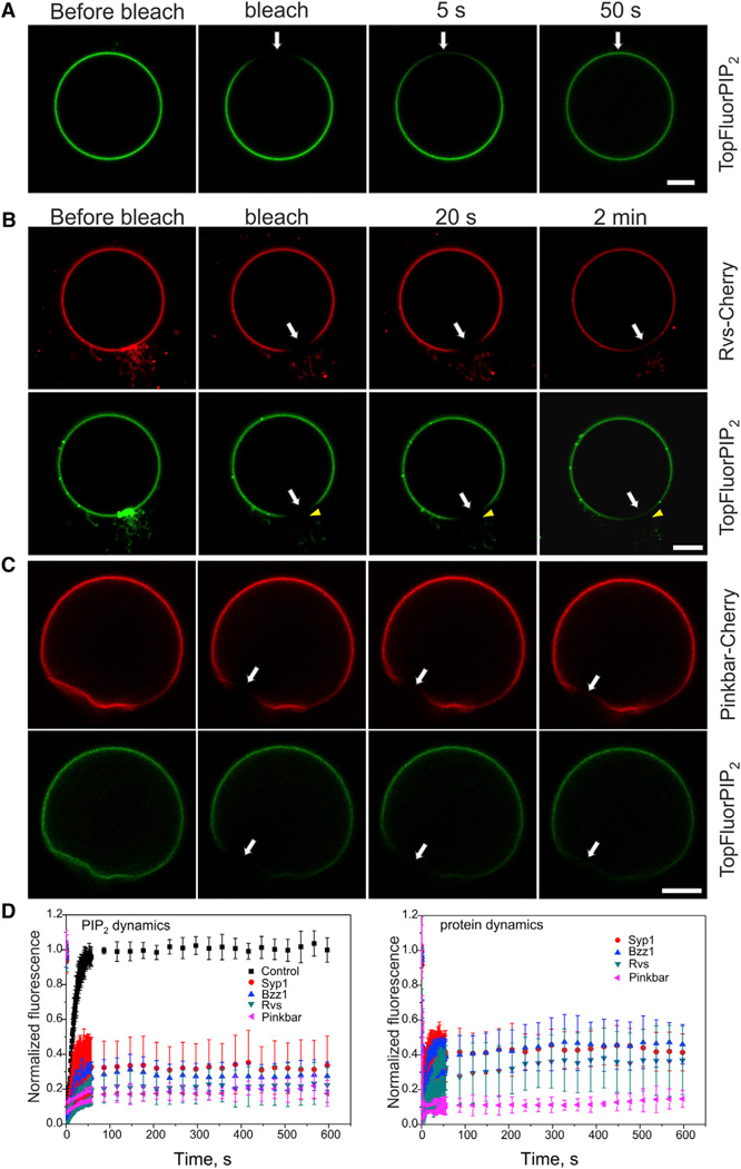
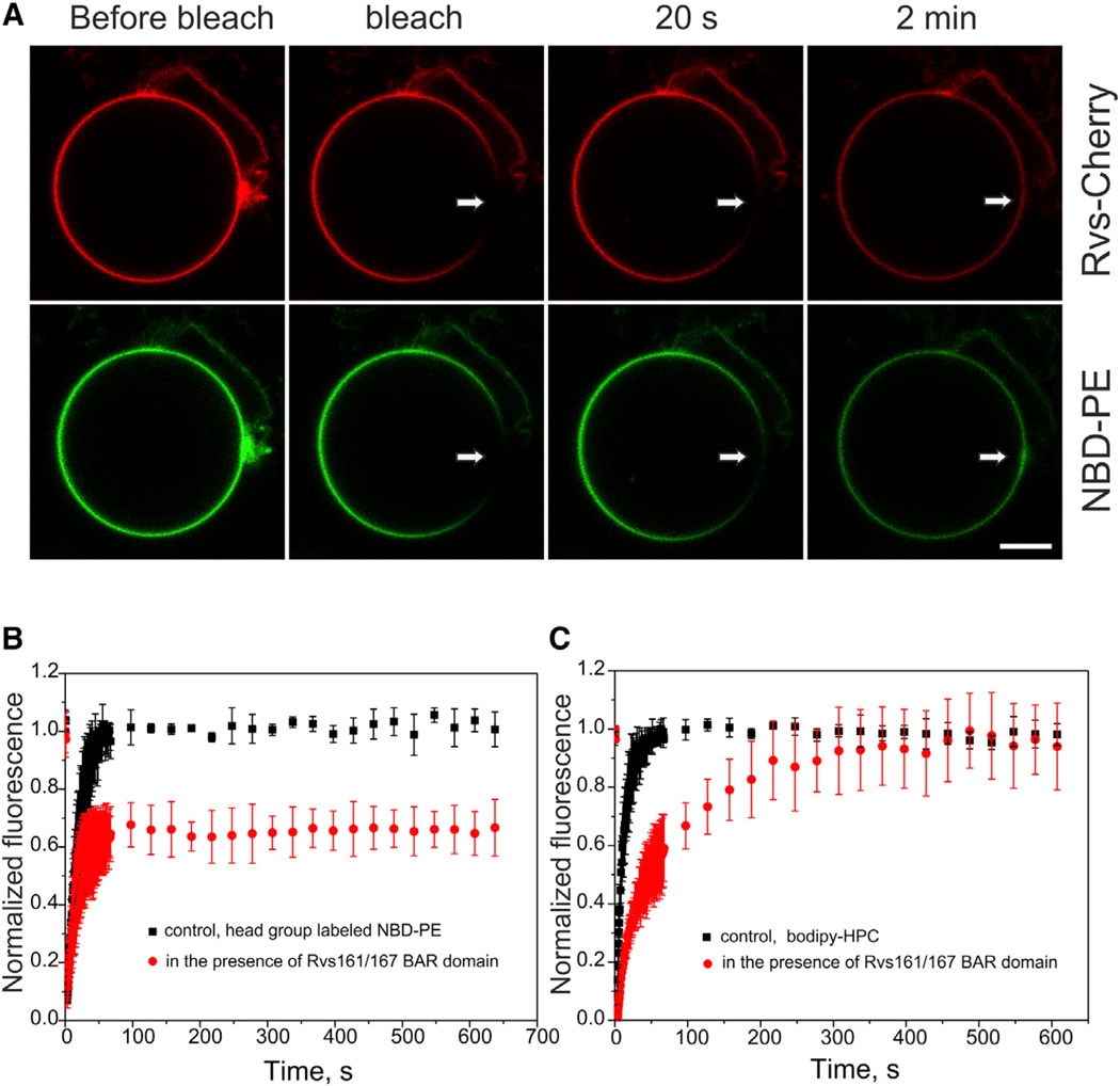
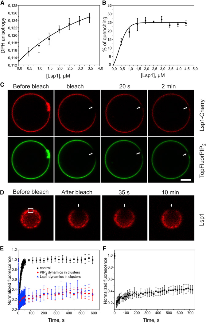
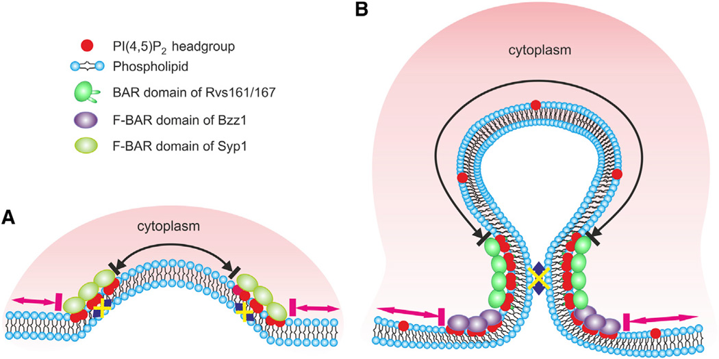
Bin-Amphiphysin-Rvs (BAR) domain proteins are central regulators of many cellular processes involving membrane dynamics. BAR domains sculpt phosphoinositide-rich membranes to generate membrane protrusions or invaginations. Here, we report that, in addition to regulating membrane geometry, BAR domains can generate extremely stable lipid microdomains by "freezing" phosphoinositide dynamics. This is a general feature of BAR domains, because the yeast endocytic BAR and Fes/CIP4 homology BAR (F-BAR) domains, the inverse BAR domain of Pinkbar, and the eisosomal BAR protein Lsp1 induced phosphoinositide clustering and halted lipid diffusion, despite differences in mechanisms of membrane interactions. Lsp1 displays comparable low diffusion rates in vitro and in vivo, suggesting that BAR domain proteins also generate stable phosphoinositide microdomains in cells. These results uncover a conserved role for BAR superfamily proteins in regulating lipid dynamics within membranes. Stable microdomains induced by BAR domain scaffolds and specific lipids can generate phase boundaries and diffusion barriers, which may have profound impacts on diverse cellular processes.


Figures

Figure 1
Figure 1. Yeast Endocytic BAR/F-BAR Domains Display Differences in Their Lipid Specificities
(A) The F-BAR/BAR domains of Syp1, Bzz1, and Rvs161/167 display different PI(4,5)P2 specificities, as measured by a vesicle cosedimentation assay. The membrane binding of Syp1 F-BAR domain was slightly inhibited at high (>8%) PI(4,5)P2 density. In contrast, interaction of the BAR domain of Rvs161/167 with vesicles was significantly enhanced by PI(4,5)P2, whereas the membrane binding of Bzz1 F-BAR domains was not augmented by high PI(4,5)P2 density in the membranes. The data are from three independent experiments, and the error bar indicates ± SD. The dotted line (control) indicates the amount of protein sedimenting in the absence of lipids. (B) When added simultaneously on vesicles, the BAR/F-BAR domains of Rvs161/167 (Rvs-Cherry) and Syp1 (Syp1-GFP) initially bind to GUVs and colocalize to PI(4,5)P2-rich tubules (indicated by arrows). However, the Syp1 F-BAR domain dissociates from the membrane within 1 to 2 min, whereas the BAR domain of Rvs 161/167 remains bound to the surface of GUVs. The scale bar represents 10 µM. (C) Quantification of the relative fluorescence intensities of the two domains on GUVs demonstrates that Rvs161/167 BAR domain replaces the Syp1 F-BAR domain from the membrane. Please note that the slight decrease in the Rvs161/167 mCherry fluorescence most likely results from photobleaching during the monitoring period. The lipid composition was POPC:POPE:POPS:PI(4,5)P2 = 50:20:20:10. The concentration of the proteins was 1 µM.
Figure 2
Figure 2. Yeast Endocytic BAR/F-BAR Domains Promote PI(4,5)P2 Clustering and Induce the Formation of Lipid Microdomains
(A) PI(4,5)P2 clustering examined at the nanometer scale by measuring the quenching of BODIPY-TMR-PI(4,5)P2. The F-BAR/BAR domains of Syp1, Bzz1, and Rvs161/167 promoted the quenching of BODIPY-conjugated PI(4,5)P2 in a concentration-dependent manner, indicating that they induce phosphoinositide clustering by bringing BODIPY-TMR-PI(4,5)P2 molecules in close proximity to each other. The lipid composition was POPC: POPE:POPS:PI(4,5)P2:bodipy-TMR-PI(4,5)P2 = 50:20:20:9.5:0.5. All error bars indicate ± SD. (B) The F-BAR/BAR domains of Syp1, Bzz1, and Rvs161/167 clustered PI(4,5)P2, PI(3,4,5)P3, and PI3P with an efficiency of PI(3,4,5)P3 > PI(4,5)P2 > PI3P. The lipid composition was POPC:POPE: POPS:BODIPY-phosphoinositide = 59:20:20:1, and the lipid concentration was 40 µM. (C) The steady-state homo-FRET fluorescence anisotropy of Rvs-Cherry decreased in the presence of PI(4,5)P2-containing vesicles, suggesting that Rvs161/167 BAR domain self-assembled into oligomers in the presence of PI(4,5)P2. The concentration of Rvs-Cherry was 0.5 µM. (D) Formation of lipid microdomains at micrometer scale revealed by light microscopy imaging of GUVs. The F-BAR/BAR domains of Syp1, Bzz1, and Rvs161/167 generated visible TopFluor-PI(4,5)P2 clusters on GUVs. However, the zwitterionic lipid phosphatidylethanolamine (PE) remained mostly uniformly distributed on the GUV membrane with some PE clustering to the membrane tubules. The lipid composition was POPC:POPE:POPS:PI(4,5)P2:TopFluor-PI(4,5)P2 :Rhodamine-PE = 50:19:20:9:1: 1. The final concentration of the Rvs161/167 BAR domain was 1 µM. All experiments were carried out at room temperature. The scale bar represents 10 µM.
Figure 3
Figure 3. BAR Domains Induce the Formation of Stable Lipid Microdomains by Significantly Diminishing the Lateral Diffusion of PI(4,5)P2
(A) In a control vesicle, rapid recovery of TopFluor-PI(4,5)P2 was observed during the 50 s period following photobleaching. (B) Both PI(4,5)P2 and associated proteins displayed very slow recovery in membrane clusters and tubular regions induced by the Rvs161/167 BAR domain. Although partial recovery could be observed in the planar region of the photobleached area 2 min after the photobleaching, no recovery was detected at the membrane tubules. Please note that the unbleached tips of the tubules remain green during the recovery period, but the green TopFluor-PI(4,5)P2 does not detectably diffuse into the BAR domain-induced membrane tubules during the recovery period (arrowhead). Thus, the PI(4,5)P2 molecules do not display detectable diffusion within the BAR domain-induced membrane tubules. (C) The BAR domain of Pinkbar and PI(4,5)P2 displayed very slow recovery on planar membranes, suggesting that the BAR domain of Pinkbar forms stable scaffolds on membrane that efficiently decrease the diffusion of phosphoinositides. (D) Quantification of the recovery of TopFluor-PI(4,5)P2 and protein fluorescence in control vesicles in BAR/F-BAR domain cluster/tubule, as well as in planar membranes induced by the BAR domain of Pinkbar. In each case, the data are mean of at least five independent experiments and the error bars indicate ± SD. Further information on the analysis of FRAP data can be found in Figure S6. The lipid composition was POPC: POPE:POPS:PI(4,5)P2:TopFluor-PI(4,5)P2 = 50: 20:20:9:1. The protein concentrations were 1 µM. The scale bar represents 10 µM.
Figure 4
Figure 4. The F-BAR/BAR Domains of Syp1, Bzz1, and Rvs161/167 Inhibit the Lateral Diffusion of Zwitterionic Lipid PE in the Membrane Clusters and Tubules
(A) A FRAP assay measuring the diffusion of NBD head group-labeled PE revealed that the lateral diffusion of this zwitterionic lipid was diminished in membrane clusters/tubules induced by the Rvs161/167 BAR domain. (B) Quantification of NBD-PE fluorescence recovery in control vesicles and in Rvs161/167 BAR domain-induced membrane tubules/clusters. It is important to note that a smaller fraction of NBD head group-labeled PE compared to PI(4,5)P2 (~40% versus 75%) displayed very slow lateral diffusion in BAR domain-induced structures. This is most likely due to less extensive enrichment of PE in BAR domain-induced membrane tubules compared to PI(4,5)P2 (see Figure 2D). (C) Dynamics of the acylchain labeled PC displayed only approximately 5-fold decrease in lateral diffusion in BAR domain clusters compared to control vesicles, and unlike head group labeled PE, it reached full fluorescence recovery during the 10 min monitoring period. In all cases, the data are mean of at least five independent experiments and the error bar indicates ± SD. The lipid composition was POPC:POPE:POPS:PI(4,5) P2:NBD-PE/BODIPY-HPC = 50:19:20:10:1. The concentration of the BAR domain of Rvs161/167 was 1 µM. The scale bar represents 10 µM.
Figure 5
Figure 5. The BAR Domain of Eisosomal Protein Lsp1 Forms Stable Membrane Microdomains, and the Protein Displays Slow Dynamics at the Plasma Membrane of Yeast Cells
(A) The BAR domain of Lsp1 decreased membrane fluidity in a concentration-dependent manner, as indicated by the increase in steady-state DPH anisotropy. This suggests that the domain not only interacts with the lipid head groups but also penetrates into the acyl-chain region of the bilayer. The lipid composition was POPC:POPE:POPS: PI(4,5)P2 = 50:20:20:10. DPH was incorporated at 1/500 ratio, and the lipid concentration was 40 µM. (B) The Lsp1 BAR domain efficiently induced the clustering of PI(4,5)P2, as measured by the self-quenching of BODIPY-TMR-PI(4,5)P2. The lipid composition was as described in Figure 2A. (C) FRAP analysis on GUVs revealed that the BAR domain of Lsp1 forms stable protein scaffolds and efficiently inhibits the lateral diffusion of PI(4,5)P2. The scale bar represents 10 µM. The lipid composition and protein concentration were as described in Figure 3. (D) In agreement with the biochemical data, full-length Lsp1 displayed similar slow dynamics at the plasma membrane of yeast cells. (E) Quantification of the fluorescence recovery of TopFluor-PI(4,5)P2 and the BAR domain of Lsp1 in membrane clusters/tubules. Furthermore, quantification of the fluorescence recovery of TopFluor-PI(4,5)P2 in control vesicles in the absence of the BAR domain is shown. The values in the graph are mean of at least five independent experiments, and the error bar represents ± SD. (F) Quantification of the fluorescence recovery of full-length Lsp1 at the plasma membrane of yeast cells. The values in the graph are mean of ten independent FRAP experiments, and the error bars represent ± SD.
Figure 6
Figure 6. A Schematic Model of the Effects of F-BAR/BAR Domains on the Distribution and Dynamics of PI(4,5)P2 in Endocytic Invaginations
(A) Syp1 is the first BAR superfamily protein to arrive at the sites of endocytosis in budding yeast. Although the F-BAR domain of Syp1 does not display specificity for PI(4,5)P2, it efficiently inhibits the lateral diffusion of phosphoinositides in the clusters (blue arrow). Thus, the Syp1 oligomer may form a lipid diffusion barrier between the tip of the invagination (black arrow) and the surrounding regions of the plasma membrane (pink arrows). (B) During the subsequent phase of endocytosis, Bzz1 binds at the invagination base through its F-BAR domain to stabilize the endocytic site with Rvs161/167, which localizes along the membrane tubule through its BAR domain. The Bzz1-Rvs161/167 scaffold efficiently inhibits the lateral diffusion of PI(4,5)P2 in this region (blue arrow) and may thus be involved in vesicle scission. The phosphoinositides and membrane proteins outside this region can diffuse freely (pink arrows) but cannot enter to the neck region, due to a lipid diffusion barrier formed by Bzz1 and Rvs161/167. Similarly, phosphoinositides and membrane proteins can diffuse within the tip of the endocytic bud (black arrow) but cannot exit the site, due to the lipid diffusion barrier.

References

    1. Antonny B. Mechanisms of membrane curvature sensing. Annu. Rev. Biochem. 2011;80:101–123. - PubMed
    1. Bhatia VK, Madsen KL, Bolinger PY, Kunding A, Hedegaård P, Gether U, Stamou D. Amphipathic motifs in BAR domains are essential for membrane curvature sensing. EMBO J. 2009;28:3303–3314. - PMC - PubMed
    1. Boucrot E, Pick A, Çamdere G, Liska N, Evergren E, McMahon HT, Kozlov MM. Membrane fission is promoted by insertion of amphipathic helices and is restricted by crescent BAR domains. Cell. 2012;149:124–136. - PMC - PubMed
    1. Brach T, Specht T, Kaksonen M. Reassessment of the role of plasma membrane domains in the regulation of vesicular traffic in yeast. J. Cell Sci. 2011;124:328–337. - PubMed
    1. Cho H, Kim YA, Yoon JY, Lee D, Kim JH, Lee SH, Ho WK. Low mobility of phosphatidylinositol 4,5-bisphosphate underlies receptor specificity of Gq-mediated ion channel regulation in atrial myocytes. Proc. Natl. Acad. Sci. USA. 2005;102:15241–15246. - PMC - PubMed

Publication types

MeSH terms