

Design and application of combined 8-channel transmit and 10-channel receive arrays and radiofrequency shimming for 7-T shoulder magnetic resonance imaging
- PMID: 24056112
- PMCID: PMC4036121
- DOI: 10.1097/RLI.0b013e3182a5662d
Design and application of combined 8-channel transmit and 10-channel receive arrays and radiofrequency shimming for 7-T shoulder magnetic resonance imaging
Abstract
Objective: The objective of the study was to investigate the feasibility of 7-T shoulder magnetic resonance imaging by developing transmit and receive radiofrequency (RF) coil arrays and exploring RF shim methods.
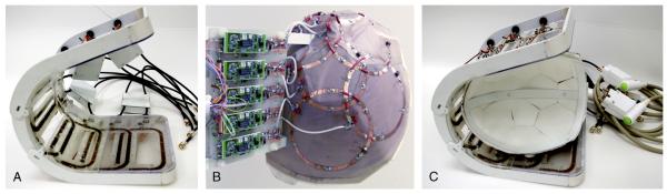
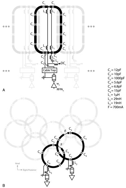
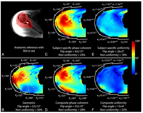
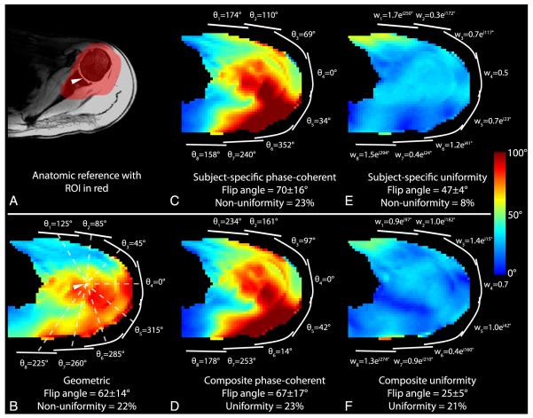
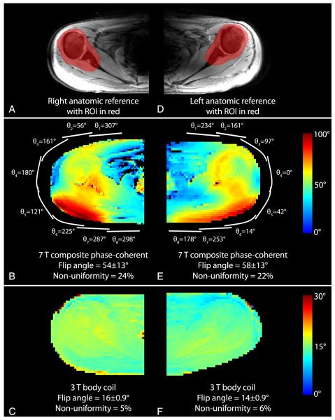
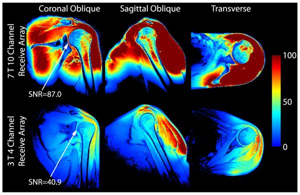
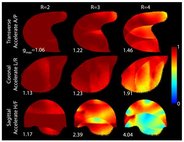
Materials and methods: A mechanically flexible 8-channel transmit array and an anatomically conformable 10-channel receive array were designed and implemented. The transmit performance of various RF shim methods was assessed through local flip angle measurements in the right and left shoulders of 6 subjects. The receive performance was assessed through signal-to-noise ratio measurements using the developed 7-T coil and a baseline commercial 3-T coil.
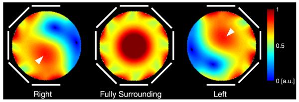
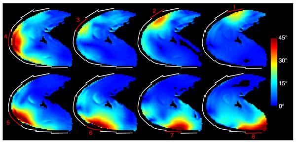
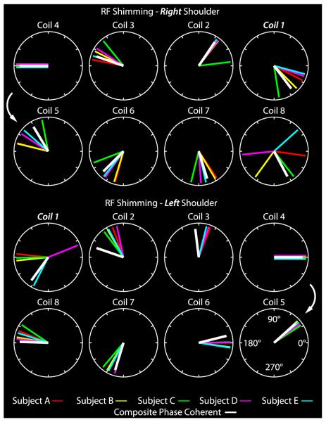
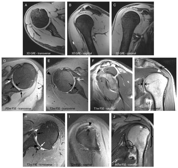
Results: The 7-T transmit array driven with phase-coherent RF shim weights provided adequate B₁⁺ efficiency and uniformity for turbo spin echo shoulder imaging. B₁⁺ twisting that is characteristic of high-field loop coils necessitates distinct RF shim weights in the right and left shoulders. The 7-T receive array provided a 2-fold signal-to-noise ratio improvement over the 3-T array in the deep articular shoulder cartilage.
Conclusions: Shoulder imaging at 7-T is feasible with a custom transmit/receive array either in a single-channel transmit mode with a fixed RF shim or in a parallel transmit mode with a subject-specific RF shim.
Figures
Similar articles
-
A 32-channel combined RF and B0 shim array for 3T brain imaging.Magn Reson Med. 2016 Jan;75(1):441-51. doi: 10.1002/mrm.25587. Epub 2015 Feb 17. Magn Reson Med. 2016. PMID: 25689977 Free PMC article.
-
A 16-channel dual-row transmit array in combination with a 31-element receive array for human brain imaging at 9.4 T.Magn Reson Med. 2014 Feb;71(2):870-9. doi: 10.1002/mrm.24726. Magn Reson Med. 2014. PMID: 23483645
-
Development of a 7 T RF coil system for breast imaging.NMR Biomed. 2017 Jan;30(1):10.1002/nbm.3664. doi: 10.1002/nbm.3664. Epub 2016 Nov 11. NMR Biomed. 2017. PMID: 27859861 Free PMC article.
-
An illustrated comparison of processing methods for MR phase imaging and QSM: combining array coil signals and phase unwrapping.NMR Biomed. 2017 Apr;30(4):e3601. doi: 10.1002/nbm.3601. Epub 2016 Sep 13. NMR Biomed. 2017. PMID: 27619999 Free PMC article. Review.
-
RF coils: A practical guide for nonphysicists.J Magn Reson Imaging. 2018 Jun 13;48(3):590-604. doi: 10.1002/jmri.26187. Online ahead of print. J Magn Reson Imaging. 2018. PMID: 29897651 Free PMC article. Review.
Cited by
-
Sparse parallel transmission on randomly perturbed spiral k-space trajectory.Quant Imaging Med Surg. 2014 Apr;4(2):106-11. doi: 10.3978/j.issn.2223-4292.2014.04.12. Quant Imaging Med Surg. 2014. PMID: 24834422 Free PMC article.
-
Dependence of B1+ and B1- Field Patterns of Surface Coils on the Electrical Properties of the Sample and the MR Operating Frequency.Concepts Magn Reson Part B Magn Reson Eng. 2016 Feb;46(1):25-40. doi: 10.1002/cmr.b.21319. Epub 2016 Feb 4. Concepts Magn Reson Part B Magn Reson Eng. 2016. PMID: 27795697 Free PMC article.
-
Magnetic Resonance Imaging of Phosphocreatine and Determination of BOLD Kinetics in Lower Extremity Muscles using a Dual-Frequency Coil Array.Sci Rep. 2016 Jul 28;6:30568. doi: 10.1038/srep30568. Sci Rep. 2016. PMID: 27465636 Free PMC article.
-
7-T clinical MRI of the shoulder in patients with suspected lesions of the rotator cuff.Eur Radiol Exp. 2020 Feb 7;4(1):10. doi: 10.1186/s41747-019-0142-1. Eur Radiol Exp. 2020. PMID: 32030499 Free PMC article.
-
Characterization of a dielectric phantom for high-field magnetic resonance imaging applications.Med Phys. 2014 Oct;41(10):102303. doi: 10.1118/1.4895823. Med Phys. 2014. PMID: 25281973 Free PMC article.
References
-
- Magee TH, Williams D. Sensitivity and specificity in detection of labral tears with 3.0-T MRI of the shoulder. AJR Am J Roentgenol. 2006;187:1448–1452. - PubMed
-
- Magee T, Williams D. 3.0-T MRI of the supraspinatus tendon. AJR Am J Roentgenol. 2006;187:881–886. - PubMed
-
- La Rocca Vieira R, Rybak LD, Recht M. Technical update on magnetic resonance imaging of the shoulder. Magn Reson Imaging Clin N Am. 2012;20:149–161. - PubMed
-
- Juras V, Welsch G, Bar P, et al. Comparison of 3T and 7T MRI clinical sequences for ankle imaging. Eur J Radiol. 2012;81:1846–1850. - PubMed
-
- Regatte RR, Schweitzer ME. Ultra-high-field MRI of the musculoskeletal system at 7.0T. J Magn Reson Imaging. 2007;25:262–269. - PubMed
Publication types
MeSH terms
Grants and funding
LinkOut - more resources
Full Text Sources
Other Literature Sources
Medical
