

Differential immunotoxicities of poly(ethylene glycol)- vs. poly(carboxybetaine)-coated nanoparticles
- PMID: 24056145
- PMCID: PMC3858532
- DOI: 10.1016/j.jconrel.2013.09.010
Differential immunotoxicities of poly(ethylene glycol)- vs. poly(carboxybetaine)-coated nanoparticles
Abstract
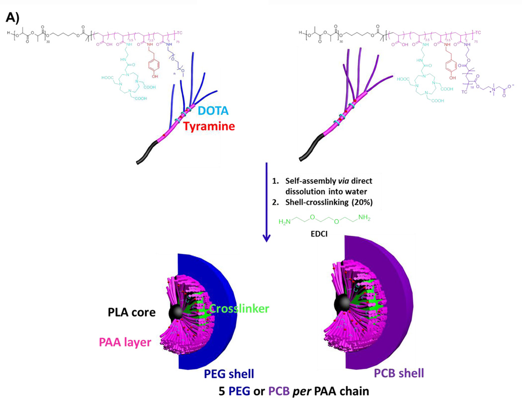
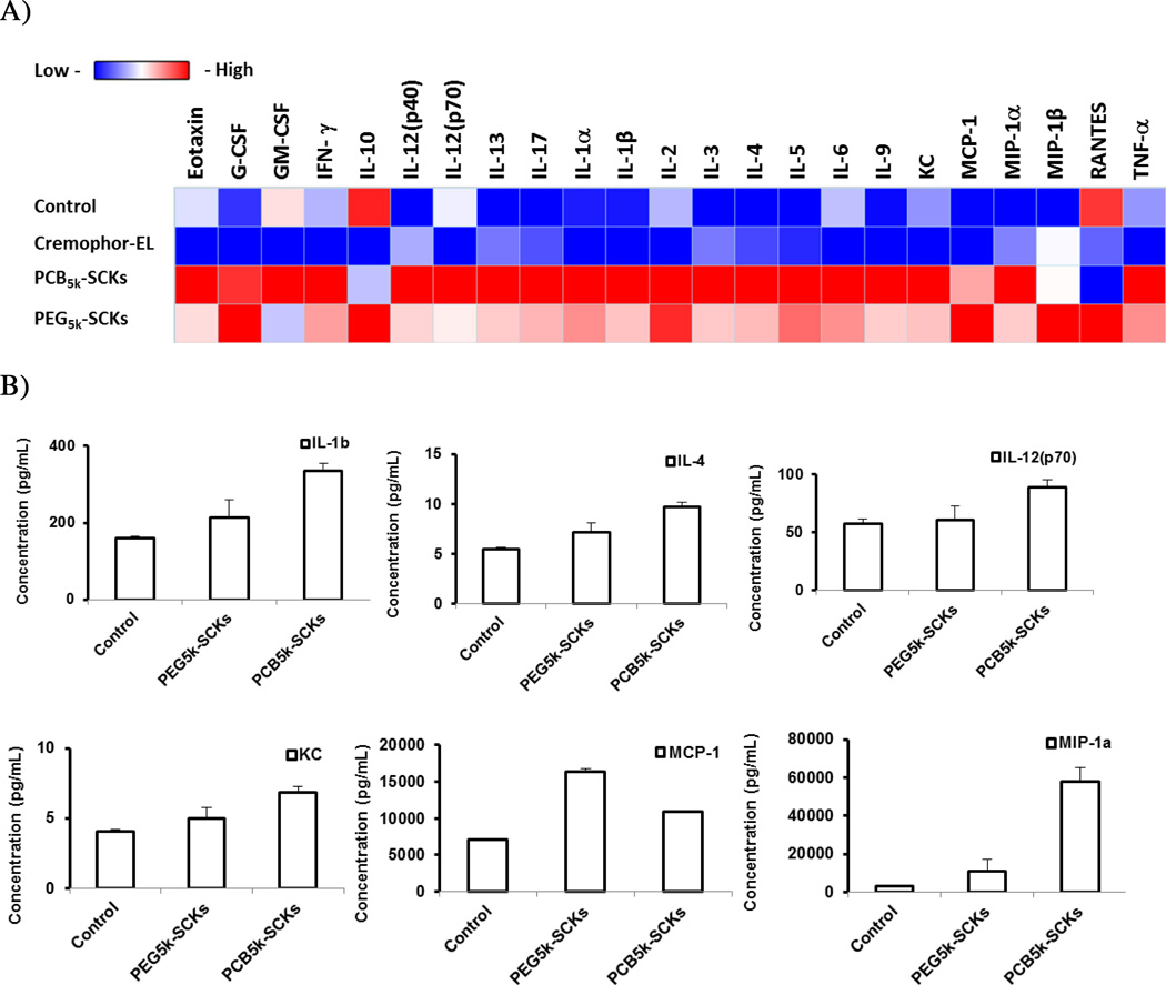
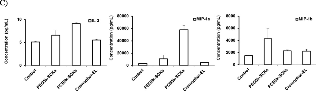
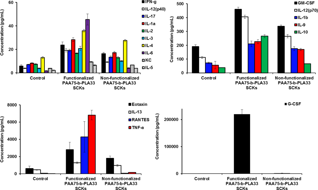
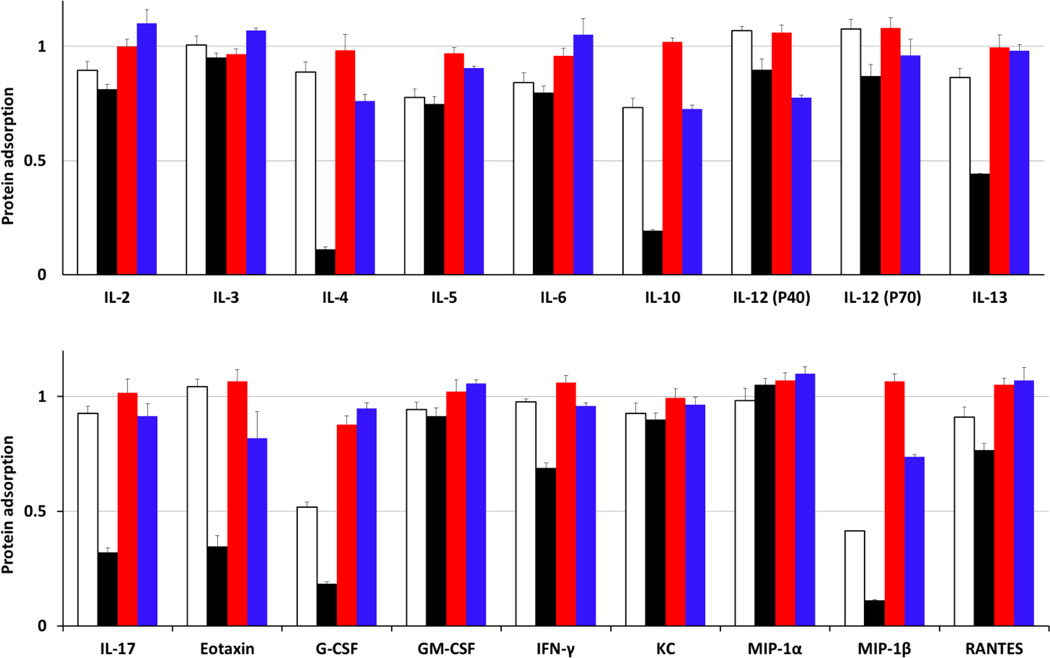
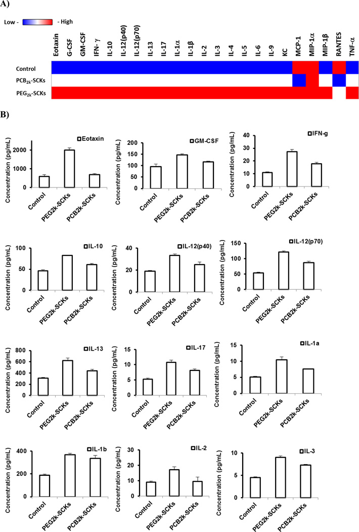
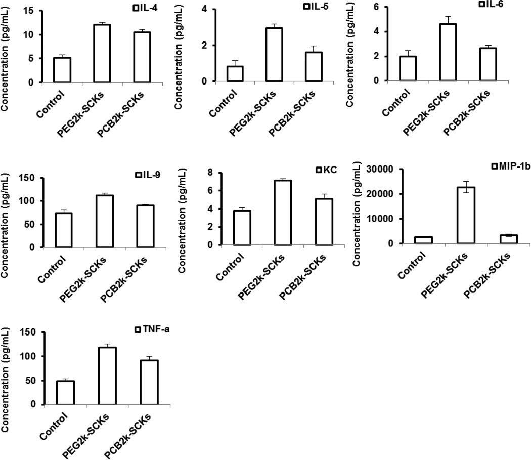
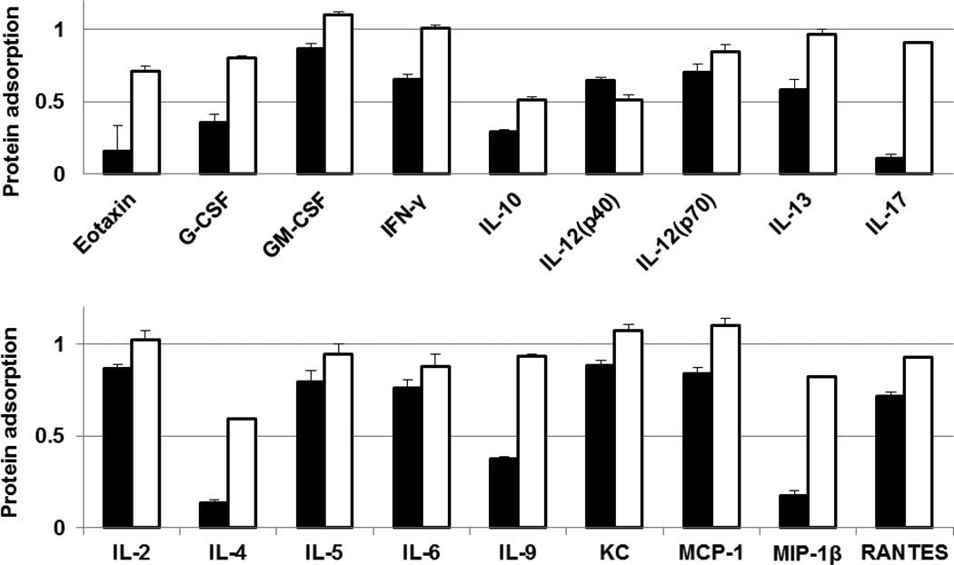
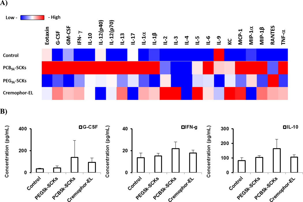
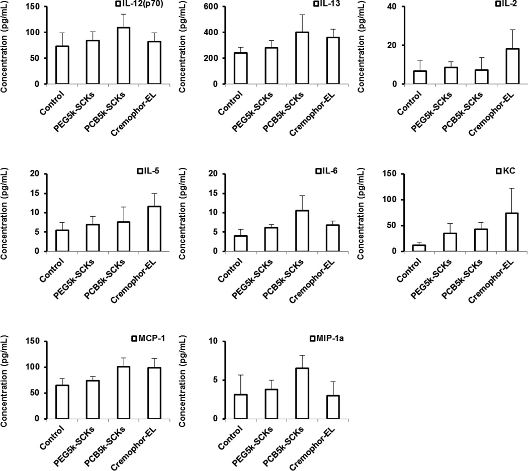
Although the careful selection of shell-forming polymers for the construction of nanoparticles is an obvious parameter to consider for shielding of core materials and their payloads, providing for prolonged circulation in vivo by limiting uptake by the immune organs, and thus, allowing accumulation at the target sites, the immunotoxicities that such shielding layers elicit is often overlooked. For instance, we have previously performed rigorous in vitro and in vivo comparisons between two sets of nanoparticles coated with either non-ionic poly(ethylene glycol) (PEG) or zwitterionic poly(carboxybetaine) (PCB), but only now report the immunotoxicity and anti-biofouling properties of both polymers, as homopolymers or nanoparticle-decorating shell, in comparison to the uncoated nanoparticles, and Cremophor-EL, a well-known low molecular weight surfactant used for formulation of several drugs. It was found that both PEG and PCB polymers could induce the expression of cytokines in vitro and in vivo, with PCB being more immunotoxic than PEG, which corroborates the in vivo pharmacokinetics and biodistribution profiles of the two sets of nanoparticles. This is the first study to report on the ability of PEG, the most commonly utilized polymer to coat nanomaterials, and PCB, an emerging zwitterionic anti-biofouling polymer, to induce the secretion of cytokines and be of potential immunotoxicity. Furthermore, we report here on the possible use of immunotoxicity assays to partially predict in vivo pharmacokinetics and biodistribution of nanomaterials.
Keywords: Cremophor-EL; Immunotoxicity; Nanoparticles; Poly(carboxybetaine); Poly(ethylene glycol); Protein adsorption.
© 2013 Elsevier B.V. All rights reserved.
Figures
Similar articles
-
Synthesis and in vivo pharmacokinetic evaluation of degradable shell cross-linked polymer nanoparticles with poly(carboxybetaine) versus poly(ethylene glycol) surface-grafted coatings.ACS Nano. 2012 Oct 23;6(10):8970-82. doi: 10.1021/nn303030t. Epub 2012 Oct 8. ACS Nano. 2012. PMID: 23043240 Free PMC article.
-
The effects of poly(zwitterions)s versus poly(ethylene glycol) surface coatings on the biodistribution of protein nanoparticles.Biomater Sci. 2016 Aug 16;4(9):1351-60. doi: 10.1039/c6bm00201c. Biomater Sci. 2016. PMID: 27426309
-
Mesoscopic Structures of Poly(carboxybetaine) Block Copolymer and Poly(ethylene glycol) Block Copolymer in Solutions.Langmuir. 2017 Aug 1;33(30):7575-7582. doi: 10.1021/acs.langmuir.7b01610. Epub 2017 Jul 17. Langmuir. 2017. PMID: 28689413
-
Preferential tumor accumulation and desirable interstitial penetration of poly(lactic-co-glycolic acid) nanoparticles with dual coating of chitosan oligosaccharide and polyethylene glycol-poly(D,L-lactic acid).Acta Biomater. 2016 Jan;29:248-260. doi: 10.1016/j.actbio.2015.10.017. Epub 2015 Oct 22. Acta Biomater. 2016. PMID: 26476340
-
Lipid-polymer hybrid nanoparticles as a new generation therapeutic delivery platform: a review.Eur J Pharm Biopharm. 2013 Nov;85(3 Pt A):427-43. doi: 10.1016/j.ejpb.2013.07.002. Epub 2013 Jul 17. Eur J Pharm Biopharm. 2013. PMID: 23872180 Review.
Cited by
-
To PEGylate or not to PEGylate: Immunological properties of nanomedicine's most popular component, polyethylene glycol and its alternatives.Adv Drug Deliv Rev. 2022 Jan;180:114079. doi: 10.1016/j.addr.2021.114079. Epub 2021 Dec 10. Adv Drug Deliv Rev. 2022. PMID: 34902516 Free PMC article. Review.
-
Mechanisms Underlying Cytotoxicity Induced by Engineered Nanomaterials: A Review of In Vitro Studies.Nanomaterials (Basel). 2014 Jun 12;4(2):454-484. doi: 10.3390/nano4020454. Nanomaterials (Basel). 2014. PMID: 28344232 Free PMC article. Review.
-
Polycarboxybetaine in advanced drug delivery systems: From structure-function relationship to therapeutic applications.Int J Pharm X. 2025 Mar 29;9:100329. doi: 10.1016/j.ijpx.2025.100329. eCollection 2025 Jun. Int J Pharm X. 2025. PMID: 40236609 Free PMC article. Review.
-
Some Nanocarrier's Properties and Chemical Interaction Mechanisms with Flavones.Molecules. 2023 Mar 22;28(6):2864. doi: 10.3390/molecules28062864. Molecules. 2023. PMID: 36985836 Free PMC article. Review.
-
Data Mining as a Guide for the Construction of Cross-Linked Nanoparticles with Low Immunotoxicity via Control of Polymer Chemistry and Supramolecular Assembly.Acc Chem Res. 2015 Jun 16;48(6):1620-30. doi: 10.1021/acs.accounts.5b00066. Epub 2015 May 26. Acc Chem Res. 2015. PMID: 26011318 Free PMC article.
References
-
- Le Garrec D, Gori S, Luo L, Lessard D, Smith DC, Yessine MA, Ranger M, Leroux JC. Poly(N-vinylpyrrolidone)-block-poly(D,L-lactide) as a new polymeric solubilizer for hydrophobic anticancer drugs: in vitro and in vivo evaluation. J Control Release. 2004;99:83–101. - PubMed
Publication types
MeSH terms
Substances
Grants and funding
LinkOut - more resources
Full Text Sources
Other Literature Sources
