Review
. 2013 Dec 13;425(24):4872-92.
doi: 10.1016/j.jmb.2013.09.015. Epub 2013 Sep 20.

Paramyxovirus activation and inhibition of innate immune responses

Affiliations
Review

Paramyxovirus activation and inhibition of innate immune responses

Griffith D Parks et al. J Mol Biol. .

Abstract

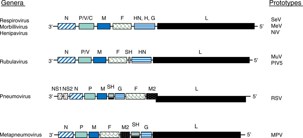
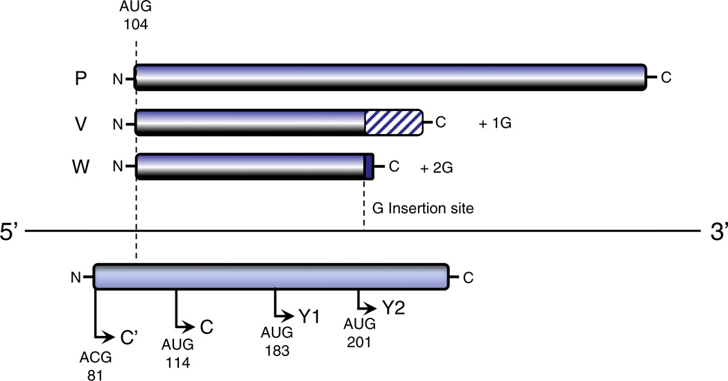
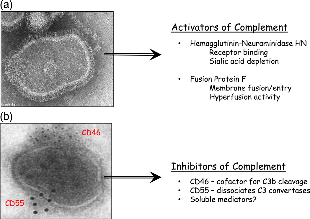
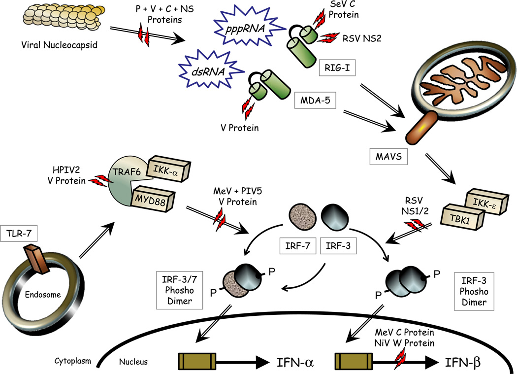
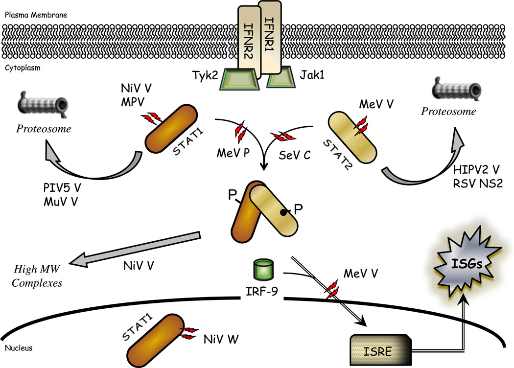
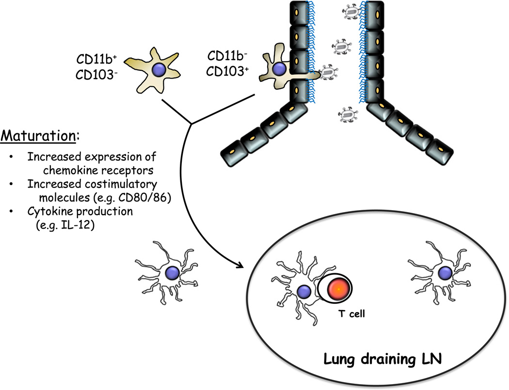
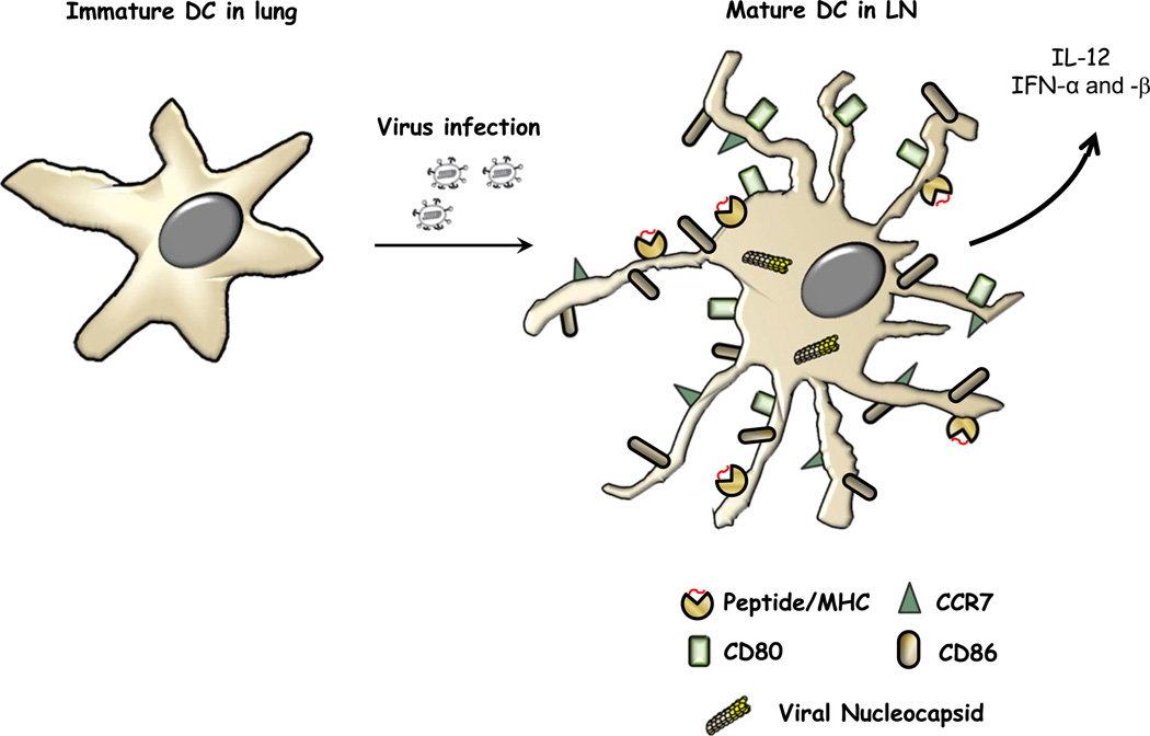
Paramyxoviruses represent a remarkably diverse family of enveloped nonsegmented negative-strand RNA viruses, some of which are the most ubiquitous disease-causing viruses of humans and animals. This review focuses on paramyxovirus activation of innate immune pathways, the mechanisms by which these RNA viruses counteract these pathways, and the innate response to paramyxovirus infection of dendritic cells (DC). Paramyxoviruses are potent activators of extracellular complement pathways, a first line of defense that viruses must face during natural infections. We discuss mechanisms by which these viruses activate and combat complement to delay neutralization. Once cells are infected, virus replication drives type I interferon (IFN) synthesis that has the potential to induce a large number of antiviral genes. Here we describe four approaches by which paramyxoviruses limit IFN induction: by limiting synthesis of IFN-inducing aberrant viral RNAs, through targeted inhibition of RNA sensors, by providing viral decoy substrates for cellular kinase complexes, and through direct blocking of the IFN promoter. In addition, paramyxoviruses have evolved diverse mechanisms to disrupt IFN signaling pathways. We describe three general mechanisms, including targeted proteolysis of signaling factors, sequestering cellular factors, and upregulation of cellular inhibitors. DC are exceptional cells with the capacity to generate adaptive immunity through the coupling of innate immune signals and T cell activation. We discuss the importance of innate responses in DC following paramyxovirus infection and their consequences for the ability to mount and maintain antiviral T cells.

Keywords: DC; ISG; ORF; WT; dendritic cells; innate immunity; interferon; interferon-stimulated gene; moDC; monocyte-derived DC; open reading frame; pDC; paramyxovirus; plasmacytoid DC; wild type.


Figures

Fig. 1
Fig. 1
Schematic diagram of representative paramyxovirus genomes. Four paramyxovirus genome structures are categorized by the differential coding strategies for viral antagonists of innate immune responses, including V protein, C proteins, SH protein, and NS proteins. Genomes are shown schematically in a 3′-to-5′ orientation with boxes indicating the approximate size of ORFs for viral proteins and thin lines indicating intergenic regions. Prototype members of the paramyxovirus genera are listed on the right.
Fig. 2
Fig. 2
Schematic diagram of coding strategy in the SeV P/V/C gene. The position of the common initiating AUG codon for the P, V, and W ORFs at base 104 is shown above a line indicating the viral mRNA. Hatched and black boxes indicate the V protein Cys-rich C-terminal domain that is fused to the shared P N-terminal domain by addition of a G residue during viral transcription and the short W domain that is accessed by insertion of two G residues, respectively. The four in-frame initiation codons for the COOH-terminal nested set of C′, C, Y1, and Y2 ORFs are shown as rightward arrows below the P gene mRNA.
Fig. 3
Fig. 3
Activation and inhibition of complement by paramyxovirus envelope proteins. Electron micrographs of purified PIV5 are shown (~55,000×) to illustrate the spike glycoproteins HN and F that radiate from the virion surface and are potent activators of C′ pathways (a). An electron micrograph of purified PIV5 that has been immunogold labeled with anti-CD46 and anti-CD55 antibodies is shown (b) to illustrate the apparent polarized distribution of C′ regulators in the virion envelope.
Fig. 4
Fig. 4
Activation and inhibition of IFN-β and IFN-α synthesis pathways during paramyxovirus replication. The viral nucleocapsid template is shown on the upper left with RNA products such as dsRNA and pppRNA that can activate latent factors MDA-5 and RIG-I. This in turn signals through MAVS to activate a cytoplasmic kinase complex composed in part of TBK-1 and IKK-ε. Alternatively, in some cell types, viral single-stranded RNA can activate endosomal TLR-7, leading to signaling to a complex composed in part by TRAF6/MYD88/IKK-α. Latent transcription factors IRF-3 and IRF-7 are phosphorylated and assembled into homo-dimers (IRF-3) or hetero-dimers (IRF-3 and IRF-7) that function with other transcription factors to activate the IFN-β or IFN-α promoters, respectively. Steps in the signaling pathways that are blocked by select paramyxovirus proteins are indicated by dual red lightning bolts.
Fig. 5
Fig. 5
Paramyxovirus inhibition of IFN signaling pathways. IFN binding to the extracellular domain of the plasma-membrane-localized IFN receptor complex results in phosphorylation of intracellular STAT1 and STAT2 by the Tyk2/Jak1 kinase and hetero-dimerization. Following association with IRF-9, the complex is translocated to the nucleus to activate transcription of ISGs containing an interferon-stimulated response element (ISRE). Steps and targets in the signaling pathway that are blocked by select paramyxovirus proteins are indicated by dual red lightning bolts.
Fig. 6
Fig. 6
DC survey the lung airway and parenchyma for the presence of viral pathogens. DC in the lung are a heterogenous population that includes two major DC types important for innate sensing, antigen acquisition, and subsequent T cell activation. These two subtypes can be differentiated by the distinct anatomic niches in which they reside, the airway versus the parenchyma, and by the cell surface molecules they express. Airway DC are marked by the expression of CD103, while parenchymal DC express CD11b. Following innate immune signals that occur as a result of paramyxovirus infection or encounter with virus components, DC undergo maturation, leave the lung, and migrate to the draining lymph node. There they engage naïve T cells to induce activation and differentiation into effector cells.
Fig. 7
Fig. 7
DC maturation results in upregulation of costimulatory markers and chemokine receptors together with the production of cytokines. DC (left side) reside in the lung in an immature state. As a result of virus infection or exposure to viral constituents, DC undergo an innate immune response termed maturation. This involves upregulated expression of the chemokine receptor CCR7, which promotes efficient trafficking to the lymph node, as well as costimulatory molecules (e.g., CD80 and CD86) that facilitate T cell interactions, expansion, and survival. In addition, mature DC produce cytokines that promote the acquisition of effector function in activated T cells (IL-12 and IFN-α/β). In the context of these innate responses, mature DC present antigen (red lines) in the context of surface complexes (yellow circles).

References

    1. Lamb RA, Parks GD. Paramyxoviridae: the viruses and their replication. In: Knipe DM, Howley PM, editors. Fields Virology. 6th ed. Philadelphia, PA: Wolters Kluwer and Lippincott Williams and Wilkins; 2013. pp. 957–995.
    1. Rubin SA, Sauder CJ, Carbone KM. Mumps virus. In: Knipe DM, Howley PM, editors. Fields Virology. 6th ed. Philadelphia, PA: Wolters Kluwer and Lippincott Williams and Wilkins; 2013. pp. 1024–1041.
    1. Griffin DE. Measles virus. In: Knipe DM, Howley PM, editors. Fields Virology. 6th ed. Philadelphia, PA: Wolters Kluwer and Lippincott Williams and Wilkins; 2013. pp. 1042–1069.
    1. Parks GD, Manuse MJ, Johnson JJ. The parainfluenza virus simian virus 5. In: Samal S, editor. The Biology of Paramyxoviruses. Norfolk, UK: Caister Academic Press; 2011. pp. 37–68.
    1. Collins PL. Human respiratory syncytial virus. In: Samal S, editor. The Biology of Paramyxoviruses. Norfolk, UK: Caister Academic Press; 2011. pp. 341–410.

Publication types

MeSH terms

LinkOut - more resources