. 2013 Nov;92(11):970-5.
doi: 10.1177/0022034513505772. Epub 2013 Sep 20.

Dental pulp tissue engineering in full-length human root canals

Affiliations

Dental pulp tissue engineering in full-length human root canals

V Rosa et al. J Dent Res. 2013 Nov.

Abstract

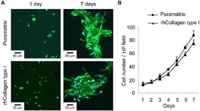
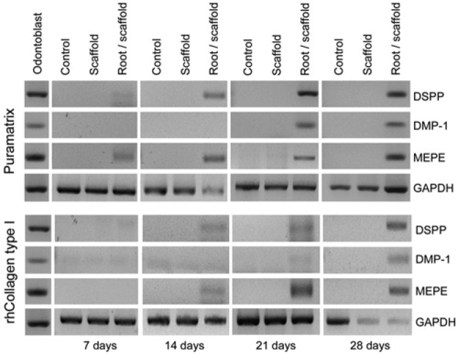
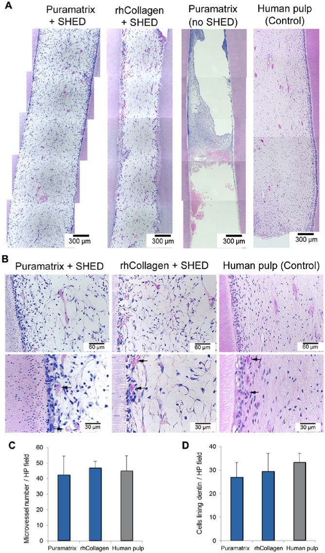
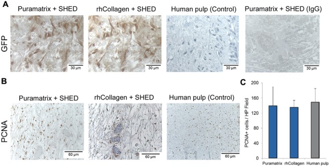
The clinical translation of stem-cell-based dental pulp regeneration will require the use of injectable scaffolds. Here, we tested the hypothesis that stem cells from exfoliated deciduous teeth (SHED) can generate a functional dental pulp when injected into full-length root canals. SHED survived and began to express putative markers of odontoblastic differentiation after 7 days when mixed with Puramatrix™ (peptide hydrogel), or after 14 days when mixed with recombinant human Collagen (rhCollagen) type I, and injected into the root canals of human premolars in vitro. Roots of human premolars injected with scaffolds (Puramatrix™ or rhCollagen) containing SHED were implanted subcutaneously into immunodeficient mice (CB-17 SCID). We observed pulp-like tissues with odontoblasts capable of generating new tubular dentin throughout the root canals. Notably, the pulp tissue engineered with SHED injected with either Puramatrix™ or rhCollagen type I presented similar cellularity and vascularization when compared with control human dental pulps. Analysis of these data, collectively, demonstrates that SHED injected into full-length human root canals differentiate into functional odontoblasts, and suggests that such a strategy might facilitate the completion of root formation in necrotic immature permanent teeth.

Keywords: biocompatible materials; dentinogenesis; neovascularization; odontoblasts; stem cells; tissue scaffolds.


Conflict of interest statement

The authors declare no potential conflicts of interest with respect to the authorship and/or publication of this article.

Figures

Figure 1.
Figure 1.
SHED morphology and proliferation in injectable scaffolds in vitro. (A) SHED are dispersed and present a round morphology when cultured in Puramatrix™ or rhCollagen type I for 1 day. Cells proliferate, elongate, and form clusters after 7 days. Green depicts Actin-F staining, and blue depicts DAPI staining (nuclei). (B) Graph depicting proliferation of SHED over time when mixed with Puramatrix™ or rhCollagen type I. Cell number throughout the experiment was not statistically different when cells were cultured in Puramatrix™ or rhCollagen type I, as determined by t test (p > .05).
Figure 2.
Figure 2.
Expression of putative markers of odontoblastic differentiation by SHED in vitro. RT-PCR analysis of markers of odontoblastic differentiation (DMP-1, DSPP, MEPE) in SHED mixed with scaffolds (Puramatrix™, rhCollagen type I) and injected into human root canals. As controls, we evaluated SHED cultured in standard tissue culture plates (Control) or SHED cultured in the injectable scaffolds (Puramatrix™ or rhCollagen type I) but in the absence of tooth structure (Scaffold) for up to 28 days.
Figure 3.
Figure 3.
Dental pulp tissue engineering with SHED injected into human root canals and transplanted into immunodeficient mice. (A) Low-magnification and (B) high-magnification images of tissues formed when SHED mixed with scaffolds (Puramatrix™, rhCollagen type I groups) were injected into full-length root canals of human premolars. A vascularized connective tissue occupied the full extension of the root canal. Cell densification and many blood vessels were observed along dentin walls. Scaffolds (Puramatrix™) injected into the root canals without cells were used as controls for SHED. Freshly extracted human premolars were used as tissue controls. Black arrows point to blood vessels close to the odontoblastic layer. (C) Graph depicting microvessel density and (D) cellular density of dental pulp tissues engineered with SHED injected into full human root canals. Microvessel density and cellular density were similar in both experimental conditions and the control group (human pulp), as determined by one-way ANOVA.
Figure 4.
Figure 4.
Characterization of the tissues engineered with SHED. SHED stably tranduced with green fluorescence protein (GFP) were mixed with scaffolds (Puramatrix™ or rhCollagen type I) and injected into full-length root canals of human premolars. (A) Immunohistochemistry for GFP (brown) confirmed that tissues were formed primarily with SHED transplanted into the root canal. Images were randomly captured from the center of engineered dental pulps or control human pulps. Immunohistochemistry with GFP of human dental pulps, and non-specific isotype-matched IgG, were used as controls for this experiment. (B) Immunohistochemistry with PCNA (brown) to evaluate cell proliferation in pulps engineered with Puramatrix™ or rhCollagen type I. Immunohistochemistry with PCNA of human dental pulps was used as control for this experiment. (C) Graph depicting the number of PCNA-positive cells in tissues formed with SHED injected into full-length human root canals. The number of PCNA-positive cells was similar in both experimental conditions and the control group (human pulp), as determined by one-way ANOVA.

References

    1. Bento LW, Zhang Z, Imai A, Nör F, Dong Z, Shi S, et al. (2013). Endothelial differentiation of SHED requires MEK1/ERK signaling. J Dent Res 92:51-57. - PMC - PubMed
    1. Casagrande L, Demarco FF, Zhang Z, Araujo FB, Shi S, Nör JE. (2010). Dentin-derived BMP-2 and odontoblastic differentiation of SHED. J Dent Res 89:603-608. - PubMed
    1. Cavalcanti BN, Zeitlin BD, Nör JE. (2013). A hydrogel scaffold that maintains viability and supports differentiation of dental pulp stem cells. Dent Mater 29:97-102. - PMC - PubMed
    1. Chadipiralla K, Yochim JM, Bahuleyan B, Huang CY, Garcia-Godoy F, Murray PE, et al. (2010). Osteogenic differentiation of stem cells derived from human periodontal ligaments and pulp of human exfoliated deciduous teeth. Cell Tissue Res 340:323-333. - PubMed
    1. Cordeiro MM, Dong Z, Kaneko T, Zhang Z, Miyazawa M, Shi S, et al. (2008). Dental pulp tissue engineering with stem cells from exfoliated deciduous teeth. J Endod 34:962-969. - PubMed

Publication types

MeSH terms