

Cavernous sinus syndrome secondary to intracranial lymphoma in a cat
- PMID: 24056274
- PMCID: PMC11112182
- DOI: 10.1177/1098612X13505580
Cavernous sinus syndrome secondary to intracranial lymphoma in a cat
Abstract
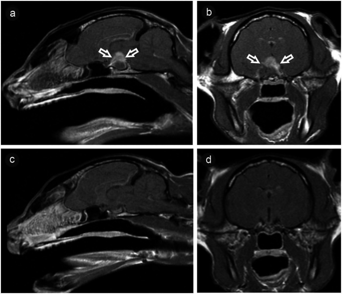
Cavernous sinus syndrome is characterised by internal and external ophthalmoplegia and sensory deficits over the head due to combined deficits of the three cranial nerves (CNs) responsible for the eye movements and pupil function (CN III, IV, VI) and at least one branch of the trigeminal nerve (CN V). It has rarely been described in cats and may occur secondarily to inflammatory, infectious or neoplastic lesions within the region of the cavernous sinus on the ventral aspect of the calvarium. This report describes the clinical and magnetic resonance imaging findings in a 14-year-old domestic shorthair cat with neurological deficits compatible with cavernous sinus syndrome caused by presumptive extranodal lymphoma. Treatment with chemotherapy resulted in clinical and imaging remission. Identification of the neurological deficits in cavernous sinus syndrome allows accurate neuroanatomical localisation in order to target diagnostic imaging studies.
© ISFM and AAFP 2013.
Conflict of interest statement
The authors do not have any potential conflicts of interest to declare.
Figures

Similar articles
-
Peripheral cranial neuropathies consistent with cavernous sinus syndrome caused by extracranial nasopharyngeal lymphoma in a cat.Can Vet J. 2019 Nov;60(11):1156-1160. Can Vet J. 2019. PMID: 31692620 Free PMC article.
-
A retrospective study of cavernous sinus syndrome in 4 dogs and 8 cats.J Vet Intern Med. 1996 Mar-Apr;10(2):65-71. doi: 10.1111/j.1939-1676.1996.tb02029.x. J Vet Intern Med. 1996. PMID: 8683482
-
Clinical and magnetic resonance imaging features of nasopharyngeal lymphoma in two cats with concurrent intracranial mass.J Small Anim Pract. 2006 Nov;47(11):678-81. doi: 10.1111/j.1748-5827.2006.00151.x. J Small Anim Pract. 2006. PMID: 17076793
-
[Cavernous sinus syndrome].Ned Tijdschr Geneeskd. 2000 Jan 22;144(4):156-60. Ned Tijdschr Geneeskd. 2000. PMID: 10668540 Review. Dutch.
-
Parasellar syndromes.Curr Neurol Neurosci Rep. 2002 Sep;2(5):423-31. doi: 10.1007/s11910-002-0069-3. Curr Neurol Neurosci Rep. 2002. PMID: 12169223 Review.
Cited by
-
Peripheral cranial neuropathies consistent with cavernous sinus syndrome caused by extracranial nasopharyngeal lymphoma in a cat.Can Vet J. 2019 Nov;60(11):1156-1160. Can Vet J. 2019. PMID: 31692620 Free PMC article.
-
Cavernous sinus syndrome in dogs and cats: case series (2002-2015).Open Vet J. 2018;8(2):186-192. doi: 10.4314/ovj.v8i2.12. Epub 2018 May 26. Open Vet J. 2018. PMID: 29911023 Free PMC article.
-
Clinical and magnetic resonance imaging features of lymphoma involving the nervous system in cats.J Vet Intern Med. 2022 Mar;36(2):679-693. doi: 10.1111/jvim.16350. Epub 2022 Jan 20. J Vet Intern Med. 2022. PMID: 35048412 Free PMC article.
References
-
- Theisen SK, Podell M, Schneider T, et al.. A retrospective study of cavernous sinus syndrome in 4 dogs and 8 cats. J Vet Intern Med 1996; 10: 65–71. - PubMed
-
- Dennis R, Kirberger RM, Barr F, et al.. Urogenital tract. In: Dennis R, Kirberger RM, Barr F, Wrigley RH. (eds). Handbook of small animal radiology and ultrasound: techniques and differential diagnosis. 2nd ed. London: Elsevier, 2010, pp 297–330.
-
- Troxel MT, Vite CH, Massicotte C, et al.. Magnetic resonance imaging features of feline intracranial neoplasia: retrospective analysis of 46 cats. J Vet Intern Med 2004; 18: 176–189. - PubMed
-
- Ropper AH, Brown RH. Disorders of ocular movement and pupillary function. In: Ropper AH, Brown RH. (eds). Adams and Victor’s principles of neurology. 8th ed. London: McGraw-Hill, 2005, pp 222–245.
-
- Penderis J. Disorders of the eyes and vision. In: Platt SR, Olby NJ. (eds). BSAVA manual of canine and feline neurology. 4th ed. Gloucester: BSAVA, 2013, pp 167–194.
Publication types
MeSH terms
LinkOut - more resources
Full Text Sources
Other Literature Sources
Medical
Miscellaneous
