. 2013 Nov;9(11):715-20.
doi: 10.1038/nchembio.1340. Epub 2013 Sep 22.

A propofol binding site on mammalian GABAA receptors identified by photolabeling

Affiliations

A propofol binding site on mammalian GABAA receptors identified by photolabeling

Grace M S Yip et al. Nat Chem Biol. 2013 Nov.

Abstract

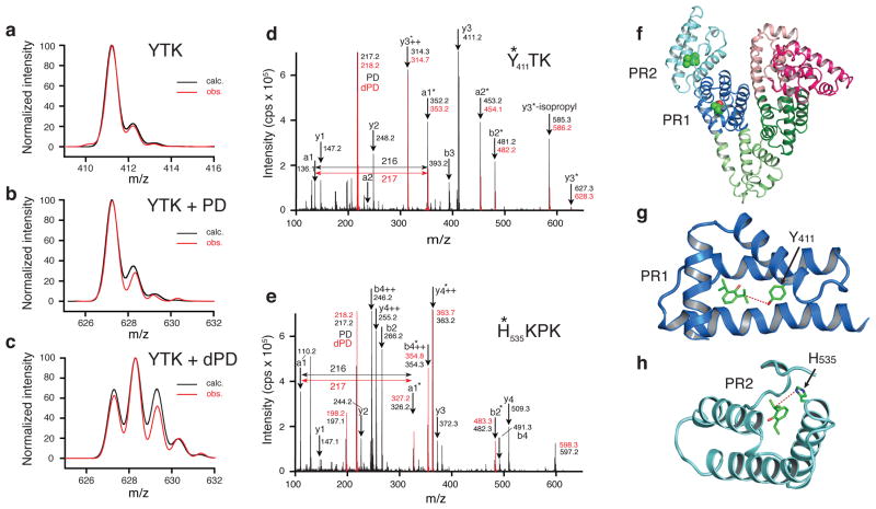
Propofol is the most important intravenous general anesthetic in current clinical use. It acts by potentiating GABAA (γ-aminobutyric acid type A) receptors, but where it binds to this receptor is not known and has been a matter of some debate. We synthesized a new propofol analog photolabeling reagent whose biological activity is very similar to that of propofol. We confirmed that this reagent labeled known propofol binding sites in human serum albumin that have been identified using X-ray crystallography. Using a combination of protiated and deuterated versions of the reagent to label mammalian receptors in intact membranes, we identified a new binding site for propofol in GABAA receptors consisting of both β3 homopentamers and α1β3 heteropentamers. The binding site is located within the β subunit at the interface between the transmembrane domains and the extracellular domain and lies close to known determinants of anesthetic sensitivity in the transmembrane segments TM1 and TM2.


Conflict of interest statement

Competing financial interests The authors declare no competing financial interests.

Figures

Figure 1
Figure 1. Characterization of ortho-propofol diazirine
(a) The chemical structures of propofol (left) and ortho-propofol diazirine (1). (b) The percentage potentiation of GABA-evoked currents by propofol (3 μM; n=12), ortho-propofol diazirine (5 μM: n=12), para-methylene-propofol diazirine (4; 50 μM; n=7) and meta-propofol diazirine (2; 200 μM; n=3). GABA (3 μM; EC7) was applied to HEK293 cells expressing α1β2γ2s GABAA receptors, and the current was recorded under whole-cell patch clamp. The inset shows a typical current trace during switches into GABA, in the presence and absence of 5 μM ortho-propofol diazirine. (c) UV absorption spectrum of ortho-propofol diazirine in ethanol (0.2 mg ml−1). (d) Inhibition of [35S]-TBPS binding to GABAA β3 homomers by ortho-propofol diazirine. The IC50 concentration for inhibition of TBPS binding was 2.9 ± 0.4 μM (n=3). (e) The percentage potentiation of GABA-evoked currents by ortho-propofol diazirine acting on α1β3 GABAA receptor heteromers. The EC50 concentration for potentiation was 1.7 ± 0.7 μM (n=10) with a Hill coefficient of 0.8±0.2 and the maximum potentiation was 151 ± 18%. (The point at the highest drug concentration was excluded from the fit to the Hill equation.) (f) Quantal dose-response curves for propofol and ortho-propofol diazirine for loss of righting reflex in rats. The ED50 concentrations for propofol and ortho-propofol diazirine were 4.7 ± 0.8 mg kg−1 (n=12) and 14.7 ± 0.2 mg kg−1 (n=13) respectively. All error bars are standard errors of the mean and if not shown, were smaller than the size of the symbols.
Figure 2
Figure 2. Labeling human serum albumin with ortho-propofol diazirine
Calculated and observed isotopic distributions for (a) unlabeled YTK peptide, (b) YTK peptide labeled with protiated ortho-propofol diazirine and (c) YTK peptide labeled with partially deuterated ortho-propofol diazirine. MS2 data for peptides YTK (d) and HKPK (e) with protiated ortho-propofol diazirine (black) and deuterated ortho-propofol diazirine (red). The asterisks denote the presence of photolabel bound to the peptide fragment. The two sites are illustrated in (f) with PR1 being in subdomain IIIA (dark blue) and PR2 being in subdomain IIIB (light blue). More detailed views are shown in (g) for PR1 where the labeled Y411 is shown and in (h) for PR2 where the labeled H535 is shown. Dashed lines mark the distances between the propofol molecules to Y411 (4.9 A) and to H535 (5.0 A).
Figure 3
Figure 3. Labeling GABAA receptors with ortho-propofol diazirine
With both α1β3 heteromers and β3 homomers only a single amino acid, H267, was labeled. (a) MS2 data for the peptide 260-TMTTINTHL-268 labeled with the protiated ortho-propofol diazirine (black) and the deuterated ortho-propofol diazirine (red) superimposed. (b) Fragmentation diagram showing that the MS2 data are consistent with the ortho-propofol diazirine labeling the histidine side chain which subsequently rearranges as internal ions H* and TH* following fragmentation. (c) A view of our proposed propofol binding site seen from the center of the pore. Two neighboring subunits are shown in yellow and blue and a single ortho-propofol diazirine molecule is shown associated with the yellow subunit. (d) A view from the extracellular side of the receptor, but with the extracellular domain removed for clarity. The β3 homopentamer is shown with five equivalent ortho-propofol diazirine molecules bound. (e) A surface representation of our proposed propofol binding pocket with an ortho-propofol diazirine molecule sitting in the pocket close to H267 which is the amino acid that is photolabeled. (f) Electrophysiological data from α1β3 heteromers showing the effects of the mutations β3-H267A and β3-F221W on the GABA apparent affinity (n=21), (g) the extent to which 3 μM ortho-propofol diazirine potentiates a GABA-evoked chloride current (at the GABA EC60) (n=4–10) and (h) the extent to which 10 μM ortho-propofol diazirine directly activates the receptor (n=4). All error bars are standard errors of the mean.

References

    1. Hales TG, Lambert JJ. The actions of propofol on inhibitory amino acid receptors of bovine adrenomedullary chromaffin cells and rodent central neurones. British journal of pharmacology. 1991;104:619–628. - PMC - PubMed
    1. Jurd R, et al. General anesthetic actions in vivo strongly attenuated by a point mutation in the GABA(A) receptor beta3 subunit. FASEB journal : official publication of the Federation of American Societies for Experimental Biology. 2003;17:250–252. - PubMed
    1. Zezula J, Slany A, Sieghart W. Interaction of allosteric ligands with GABAA receptors containing one, two, or three different subunits. European journal of pharmacology. 1996;301:207–214. - PubMed
    1. Davies PA, Kirkness EF, Hales TG. Modulation by general anaesthetics of rat GABAA receptors comprised of alpha 1 beta 3 and beta 3 subunits expressed in human embryonic kidney 293 cells. British journal of pharmacology. 1997;120:899–909. - PMC - PubMed
    1. Feng HJ, Macdonald RL. Multiple actions of propofol on alphabetagamma and alphabetadelta GABAA receptors. Molecular pharmacology. 2004;66:1517–1524. - PubMed

Publication types