. 2013 Nov;45(11):1319-26.
doi: 10.1038/ng.2768. Epub 2013 Sep 22.

Replicative mechanisms for CNV formation are error prone

Affiliations

Replicative mechanisms for CNV formation are error prone

Claudia M B Carvalho et al. Nat Genet. 2013 Nov.

Abstract

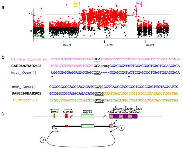
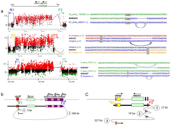
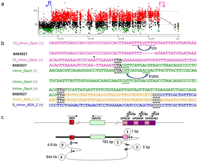
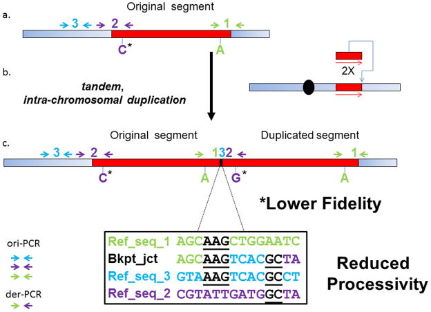
We investigated 67 breakpoint junctions of gene copy number gains in 31 unrelated subjects. We observed a strikingly high frequency of small deletions and insertions (29%) apparently originating from polymerase slippage events, in addition to frameshifts and point mutations in homonucleotide runs (13%), at or flanking the breakpoint junctions of complex copy number variants. These single-nucleotide variants were generated concomitantly with the de novo complex genomic rearrangement (CGR) event. Our findings implicate low-fidelity, error-prone DNA polymerase activity in synthesis associated with DNA repair mechanisms as the cause of local increase in point mutation burden associated with human CGR.


Conflict of interest statement

Competing financial interests

J.R.L. is a paid consultant for Athena Diagnostics, holds stock ownership in 23andMe, Inc. and Ion Torrent Systems, Inc., and is a co-inventor on multiple United States and European patents related to molecular diagnostics. The Department of Molecular and Human Genetics at Baylor College of Medicine derives revenue from molecular genetic testing offered in the Medical Genetics Laboratories (http://www.bcm.edu/geneticlabs/).

Figures

Figure 1
Figure 1. Patient BAB2626 and BAB2628 breakpoint junction mutation load
These patients have at least three mutations at and flanking the CGR breakpoint junction that were likely produced in the same event: two point mutations (transitions) before and after the breakpoint junction, one insertion (AAAG) for which the origin could not be defined, and two long-distance template-switches (1.6 kb and 472.9 kb, respectively). (a) BAB2626/BAB2628 aCGH result and approximate location of the primers (F and R) used to obtain patient specific breakpoint junctions. (b) Breakpoint junction sequence is aligned to the proximal and distal genomic references and color-matched. Strand of alignment (+ or −) is indicated in parenthesis. Microhomology at the breakpoint is indicated by black bold underlined letters. Dashed lines represent nucleotides that did not align to the reference sequence; asterisks indicate point mutations flanking the breakpoint junction. (c) Representation of the genomic structure for the reference genome (top) and for the surmised genomic structure of BAB2626 and BAB2628 (bottom), showing predicted order, origins, and relative orientations of duplicated sequences. Arrows show orientation of DNA sequence relative to the positive strand; filled arrows with circled numbers below represent a template switch that resulted in insertion of segments. The last arrow signifies resumption of replication on the original template that produced the CGR identified by aCGH. Approximate location of primers used to obtain the breakpoint junctions are shown on the bottom.
Figure 2
Figure 2. Short and long template-switches can be observed on either or both sides of CGR breakpoint junctions
(a) For each patient (BAB2623, BAB2991 and BAB3267), the aCGH result along with the breakpoint junction sequences obtained by long-range PCR and Sanger sequencing are shown. Approximate location of the primers (F and R) used to obtain patient-specific breakpoint junctions are represented in the aCGH plot. Breakpoint junction sequence is aligned to the proximal and distal genomic references and color-matched. Strand of alignment (+ or −) is indicated in parenthesis. Microhomology at the breakpoint is indicated by black bold underlined letters. Dashed lines represent deleted nucleotides; blue arrows point to the nucleotides likely involved in the misalignment that generated the deletion. (b) and (c) represent the genomic structure for the reference genome (top) and for the surmised genomic structure (bottom) for patients BAB2623 and BAB2991, respectively, showing predicted order, origins, and relative orientations of duplicated sequences. Arrows show orientation of DNA sequence relative to the positive strand; filled arrows with circled numbers below represent a template switch that resulted in insertion or deletion of segments. Distances between the template-switches are shown in bp or kb. The last arrow signifies resumption of replication on the original template that produced the CGR identified by aCGH. Approximate location of primers used to obtain the breakpoint junctions are shown below the reference genome structure.
Figure 3
Figure 3. Patient BAB3027 breakpoint junction mutational load
Patient BAB3027 presented at least three mutations at and flanking the CGR breakpoint junctions: a frameshift before the breakpoint junction, and multiple template-switch events. (a) BAB3027 aCGH result and approximate location of the primers (F and R) used to obtain patient specific breakpoint junctions. (b) Breakpoint junction sequence is aligned to the proximal and distal genomic references and color-matched. Strand of alignment (+ or −) is indicated in parenthesis. Microhomology at the breakpoint is indicated by black bold underlined letters. Dashed lines represent nucleotides that did not align to the reference sequence; asterisks indicate frameshifts flanking the breakpoint junction. Misalignment and re-annealing of short repeats present in the primer strand and template strand in cis can produce deletion in the newly synthetized strand (forward slippage) or insertion (backward slippage) . In addition, misalignment and re-annealing in trans would produce small inversion at the junctions . (c) Representation of the genomic structure for the reference genome (top) and for the surmised genomic structure (bottom), showing predicted order, origins, and relative orientations of duplicated sequences. Arrows show orientation of DNA sequence relative to the positive strand; filled arrows with circled numbers below represent a template switch that resulted in deletion or insertion of segments. Distance between the template switches are shown in bp or kb. The last arrow signifies resumption of replication on the original template which produced the CGR identified by aCGH.
Figure 4
Figure 4. Representational figure of the types of mutations that can be observed at and flanking the breakpoint junctions of MECP2 duplications
a) Wild type Xq28 segment; b) SNP markers and breakpoint junction analysis indicated that duplications involving MECP2 are frequently intrachromosomal head-to-tail duplications; c) Representational genomic structure of the derivative chromosome and the strategies used to uncover the increased mutational load at the breakpoint junctions such as small templated-insertions, frameshifts and point mutations (ori-PCR and der-PCR, please see main text for further details). Templated insertions suggest reduced processivity whereas presence of SNVs suggests lower fidelity of the replicational process. Blue rectangle represents proximal and distal regions flanking the duplication; red rectangle represents the region that will undergo duplication in (b) #1 and #2 represent proximal and distal breakpoints of the duplication; #3 represents a copy of a short local segment inserted at the breakpoint junction of the duplication. Arrows represent forward and reverse primers used to amplify each one of the involved segments in either original or duplicated copy.

References

    1. Zhang F, Carvalho CM, Lupski JR. Complex human chromosomal and genomic rearrangements. Trends Genet. 2009;25:298–307. - PMC - PubMed
    1. Liu P, Carvalho CM, Hastings PJ, Lupski JR. Mechanisms for recurrent and complex human genomic rearrangements. Curr Opin Genet Dev. 2012;22:211–20. - PMC - PubMed
    1. Conrad DF, et al. Mutation spectrum revealed by breakpoint sequencing of human germline CNVs. Nat Genet. 2010;42:385–91. - PMC - PubMed
    1. Kidd JM, et al. A human genome structural variation sequencing resource reveals insights into mutational mechanisms. Cell. 2010;143:837–47. - PMC - PubMed
    1. Bauters M, et al. Nonrecurrent MECP2 duplications mediated by genomic architecture-driven DNA breaks and break-induced replication repair. Genome Res. 2008;18:847–58. - PMC - PubMed

Publication types

Associated data