

The brain basis of social synchrony
- PMID: 24056729
- PMCID: PMC4127029
- DOI: 10.1093/scan/nst105
The brain basis of social synchrony
Abstract
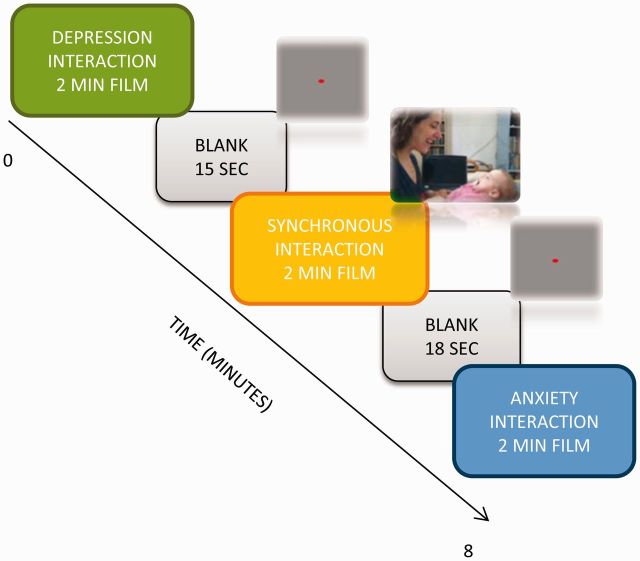
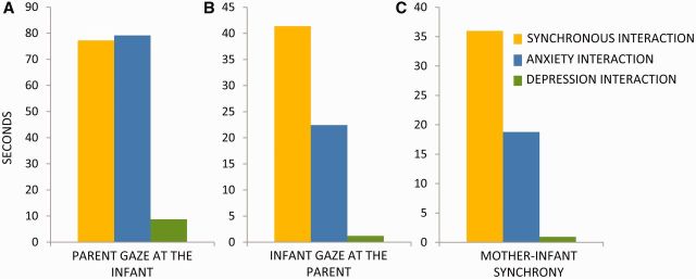
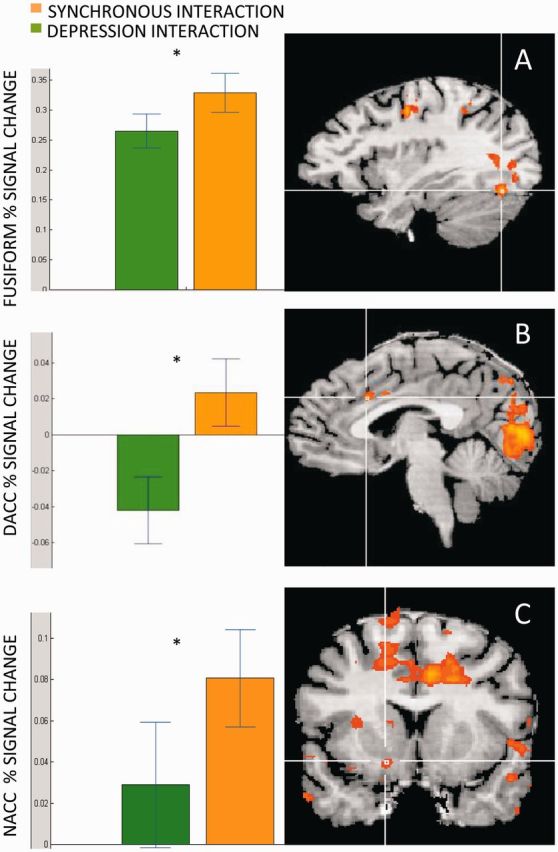
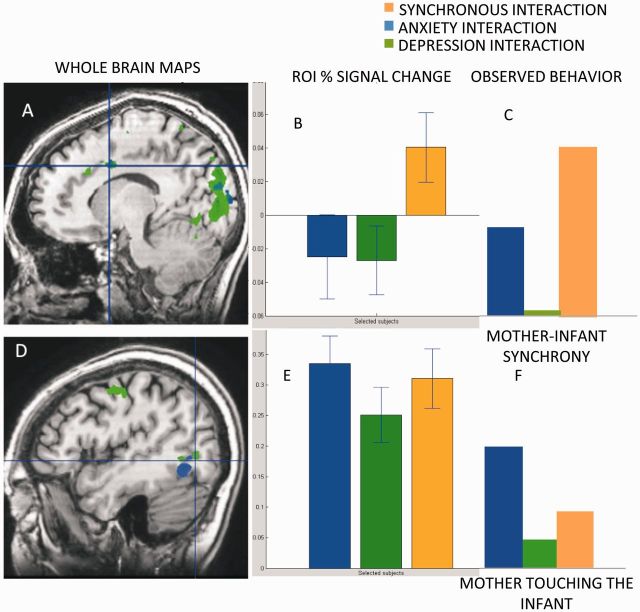
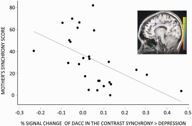
As a social species, humans evolved to detect information from the social behavior of others. Yet, the mechanisms used to evaluate social interactions, the brain networks implicated in such recognition, and whether individual differences in own social behavior determine response to similar behavior in others remain unknown. Here we examined social synchrony as a potentially important mechanism in the evaluation of social behavior and utilized the parenting context, an evolutionarily salient setting of significant consequences for infant survival, to test this issue. The brain response of healthy postpartum mothers to three mother-infant interaction vignettes was assessed. Videos included a typical synchronous interaction and two pathological interactions of mothers diagnosed with postpartum depression and anxiety that showed marked deviations from social synchrony. Mothers' own interactions with their 4- to 6-month-old infants were videotaped and micro-coded for synchrony. Results indicated that the recognition of social synchrony involved activations in the dorsal anterior cingulate cortex (dACC), fusiform, cuneus, inferior parietal lobule, supplementary motor area and NAcc. Mother's own synchrony with her infant correlated with her dACC response to synchrony in others. Findings are consistent with models suggesting that social action underpins social recognition and highlight social synchrony and the mother-infant bond as one prototypical context for studying the brain basis of social understanding.
Keywords: maternal anxiety; maternal depression; mothering; social brain; social synchrony.
© The Author (2013). Published by Oxford University Press. For Permissions, please email: journals.permissions@oup.com.
Figures
References
-
- Atzil S, Hendler T, Zagoory-Sharon O, Winetraub Y, Feldman R. Synchrony and specificity in the maternal and the paternal brain: relations to oxytocin and vasopressin. Journal of the American Academy of Child & Adolescent Psychiatry. 2012;51(8):798–811. - PubMed
-
- Barrier AC, Ruelle E, Haskell MJ, Dwyer CM. Effect of a difficult calving on the vigour of the calf, the onset of maternal behaviour, and some behavioural indicators of pain in the dam. Preventive Veterinary Medicine. 2012;103(4):248–56. - PubMed
-
- Bartels A, Zeki S. The neural correlates of maternal and romantic love. Neuroimage. 2004;21(3):1155–66. - PubMed
Publication types
MeSH terms
LinkOut - more resources
Full Text Sources
Other Literature Sources
Medical
Research Materials
