. 2013 Nov 21;5(22):10894-900.
doi: 10.1039/c3nr03272h. Epub 2013 Sep 20.

Single-cell force spectroscopy of the medically important Staphylococcus epidermidis-Candida albicans interaction

Affiliations

Single-cell force spectroscopy of the medically important Staphylococcus epidermidis-Candida albicans interaction

Audrey Beaussart et al. Nanoscale. .

Abstract

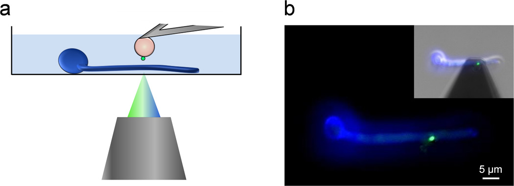
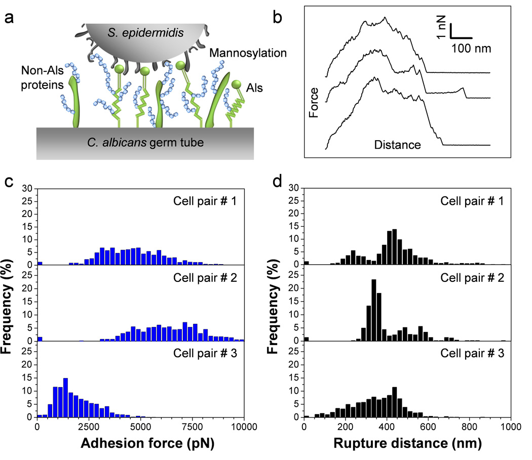
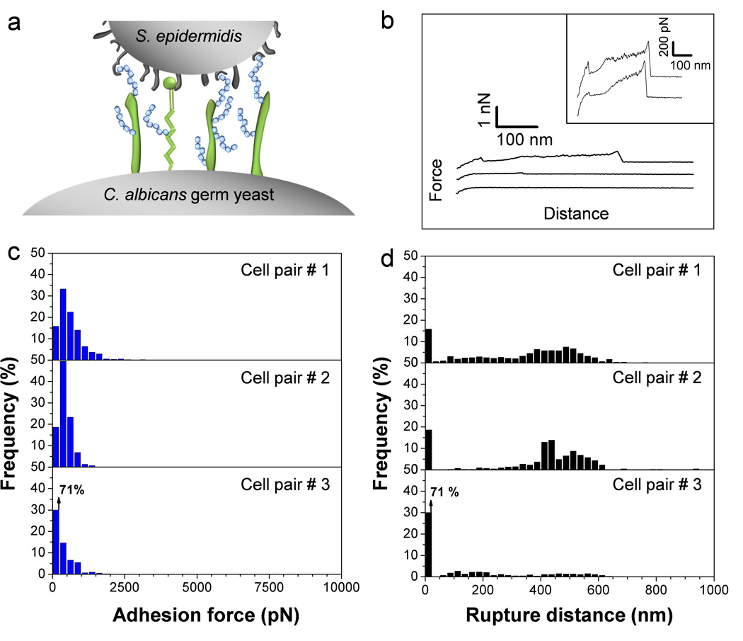
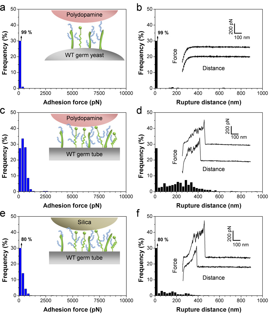
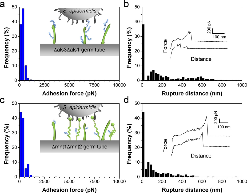
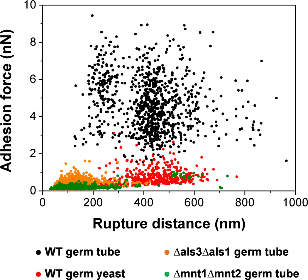
Despite the clinical importance of bacterial-fungal interactions, their molecular details are poorly understood. A hallmark of such medically important interspecies associations is the interaction between the two nosocomial pathogens Staphylococcus aureus and Candida albicans, which can lead to mixed biofilm-associated infections with enhanced antibiotic resistance. Here, we use single-cell force spectroscopy (SCFS) to quantify the forces engaged in bacterial-fungal co-adhesion, focusing on the poorly investigated S. epidermidis-C. albicans interaction. Force curves recorded between single bacterial and fungal germ tubes showed large adhesion forces (~5 nN) with extended rupture lengths (up to 500 nm). By contrast, bacteria poorly adhered to yeast cells, emphasizing the important role of the yeast-to-hyphae transition in mediating adhesion to bacterial cells. Analysis of mutant strains altered in cell wall composition allowed us to distinguish the main fungal components involved in adhesion, i.e. Als proteins and O-mannosylations. We suggest that the measured co-adhesion forces are involved in the formation of mixed biofilms, thus possibly as well in promoting polymicrobial infections. In the future, we anticipate that this SCFS platform will be used in nanomedicine to decipher the molecular mechanisms of a wide variety of pathogen-pathogen interactions and may help in designing novel anti-adhesion agents.


Figures

Fig. 1
Fig. 1
Single-cell force spectroscopy of bacterial-fungal interactions. (a) Schematic of the experimental set-up. (b) Using an integrated AFM-inverted optical microscope, the S. epidermidis probe (green) is approached towards a C. albicans hyphae (blue). The image was obtained using epifluorescence microscopy while the inset shows a merged phase/epifluorescence image.
Fig. 2
Fig. 2
SCFS quantifies the adhesion forces between S. epidermidis and C. albicans germ tubes. (a) Key cell wall components that are involved in C. albicans surface interactions are cell-surface glycoproteins (in green) and mannose-rich glycoconjugates (in blue). (b) Typical force-distance curves recorded in Tris NaCl buffer between S. epidermidis and C. albicans hyphae. (c, d) Adhesion force (c) and rupture length (d) histograms obtained by recording force curves between 3 cell pairs from different cell cultures, and representative of a total of 7 cell pairs (n > 500 force-distances curves for each pair).
Fig. 3
Fig. 3
C. albicans germinating yeasts show much weaker adhesion than germ tubes. (a) Germinating yeasts express fewer Als proteins than germ tubes. (b) Typical force-distance curves recorded in Tris NaCl buffer between S. epidermidis and C. albicans germinating yeasts. (c, d) Adhesion force (c) and rupture length (d) histograms obtained by recording force curves between 3 cell pairs from different cell cultures, and representative of a total of 6 cell pairs (n > 500 force-distances curves for each pair).
Fig. 4
Fig. 4
Control experiments using polydopamine and silica probes. (a–f) Adhesion force (a, c, e) and rupture length (b, d, f) histograms, together with representative force curves, obtained by recording force curves between polydopamine-coated probes and C. albicans germinating yeasts (a, b) or germ tubes (c, d), and between silica probes and C. albicans germ tubes (e, f). For each probe, similar data were obtained in 3 independent experiments.
Fig. 5
Fig. 5
Als proteins and O-mannosylations on the C. albicans surface are required for bacterial adhesion. (a–d) Adhesion force (a, c) and rupture length (b, d) histograms, together with representative force curves, obtained by recording force curves in Tris NaCl buffer between a single S. epidermidis bacterium and a C. albicans germ tube from the mutant als3Δ/als3Δ als1Δ/als1Δ (Δals3Δals1) (a, b) or a C. albicans germ tube from the mutant mnt1Δ/mnt1Δ mnt2Δ/mnt2Δ (Δmnt1Δmnt2) (c, d). For each mutant, similar data were obtained in 3 independent experiments using 3 different cell pairs.
Fig. 6
Fig. 6
Role of cellular morphogenesis, Als proteins and O-mannosylations in S. epidermidis-C. albicans adhesion. Plots of the adhesion forces versus rupture distances measured between S. epidermidis and WT germ tubes (black symbols), WT germinated yeasts (red symbols), als3Δ/als3Δ als1Δ/als1Δ (Δals3Δals1) germ tubes (orange symbols) and mnt1Δ/mnt1Δ mnt2Δ/mnt2Δ (Δmnt1Δmnt2) germ tubes (green symbols). Strong co-adhesion is only observed on germ tubes and involves two types of highly adhesive and extended macromolecules, i.e. Als proteins and O-mannosylations.

References

    1. Peleg AY, Hogan DA, Mylonakis E. Nat. Rev. Microbiol. 2010;8:340–349. - PubMed
    1. Morales DK, Hogan DA. PLoS Pathog. 2010;6:1–4. - PMC - PubMed
    1. Shirtliff ME, Peters BM, Jabra-Rizk MA. FEMS Microbiol. Lett. 2009;299:1–8. - PMC - PubMed
    1. Douglas LJ. Trends Microbiol. 2003;11:30–36. - PubMed
    1. Elias S, Banin E. FEMS Microbiol. Rev. 2012;36:990–1004. - PubMed

Publication types

MeSH terms

LinkOut - more resources