. 2014 Feb 1;23(3):730-48.
doi: 10.1093/hmg/ddt459. Epub 2013 Sep 20.

Reversibility of neuropathology in Tay-Sachs-related diseases

Affiliations

Reversibility of neuropathology in Tay-Sachs-related diseases

María-Begoña Cachón-González et al. Hum Mol Genet. .

Abstract

The GM2 gangliosidoses are progressive neurodegenerative disorders due to defects in the lysosomal β-N-acetylhexosaminidase system. Accumulation of β-hexosaminidases A and B substrates is presumed to cause this fatal condition. An authentic mouse model of Sandhoff disease (SD) with pathological characteristics resembling those noted in infantile GM2 gangliosidosis has been described. We have shown that expression of β-hexosaminidase by intracranial delivery of recombinant adeno-associated viral vectors to young adult SD mice can prevent many features of the disease and extends lifespan. To investigate the nature of the neurological injury in GM2 gangliosidosis and the extent of its reversibility, we have examined the evolution of disease in the SD mouse; we have moreover explored the effects of gene transfer delivered at key times during the course of the illness. Here we report greatly increased survival only when the therapeutic genes are expressed either before the disease is apparent or during its early manifestations. However, irrespective of when treatment was administered, widespread and abundant expression of β-hexosaminidase with consequent clearance of glycoconjugates, α-synuclein and ubiquitinated proteins, and abrogation of inflammatory responses and neuronal loss was observed. We also show that defects in myelination occur in early life and cannot be easily resolved when treatment is given to the adult brain. These results indicate that there is a limited temporal opportunity in which function and survival can be improved-but regardless of resolution of the cardinal pathological features of GM2 gangliosidosis, a point is reached when functional deterioration and death cannot be prevented.


Figures

Figure 1.
Figure 1.
Lifespan of Sandhoff mice after intracranial infusions of rAAVα + rAAVβ is significantly extended. SD mice were injected bilaterally into the striatum and cerebellum at 4 (n = 22), 8 (n = 5), 10 (n = 5) and 12 (n = 12) weeks post-birth (WPB). Control groups: normal controls (n = 22) and SD (UT) mice (n = 37). Kaplan–Meier survival curve, data censored at 2 years (A). One-way ANOVA and Bonferroni multiple post hoc comparisons with mean ± SEM (B).
Figure 2.
Figure 2.
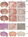
Abundant and wide bio-distribution of enzyme and viral RNA is achieved by intracranial vector delivery. In situ β-hexosaminidase activity staining (hex), red precipitate, of SD mice injected with rAAVα + rAAVβ at 4w (A and B), 10w (D and E) and 12w (G, H and K) age, and killed at their humane end point. Brain parenchyma and choroid plexus (chp) are strongly labelled. Controls were SD (UT) (N), and normal control (O) killed at 4 months of age. Viral mRNA ISH, black stain, of consecutive sections from 4w (C), 10w (F) and 12w (I and L) injected mice is shown. Grey but also white matter (corpus callosum, cc) in brain parenchyma, and choroid plexus were transduced. The substantia nigra (SN) from a 12w injected mouse stained with hex (K) and ISH (L). PAS staining of SN shows absence of glycoconjugate storage (J). Normal control stained with PAS (M). Caudate putamen (CPu), cerebral peduncle (cp), lateral ventricle (LV), medial lemniscus (ml) and primary somatosensory cortex (S1). Scale bars: 2 mm (A, D, G, M, N and O); 500 μm (B, C, E, F, H, I and J–L).
Figure 3.
Figure 3.
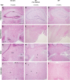
Glycoconjugate storage is reduced throughout the neuraxis. Sections from a representative 12w rAAVα + rAAVβ injected SD mouse killed at the humane end point of 4 months were stained with PAS (B, E, H and K). Controls were 12-week-old SD (UT) (A, D, G and J) and 4-month-old normal control (C, F, I and L). Anatomical regions shown are hippocampus (A–C), hypothalamus (D–F), deep cerebellar nuclei (G–I) and brain stem (J–L). Arrow heads point to PAS-positive neurones. Deep cerebellar nuclei (DCN); fields CA1(CA1) and CA3 (CA3) of hippocampus; fimbria (fi); fourth ventricle (4V); gigantocellular reticular nucleus (Gi); granular layer of the dentate gyrus (GrDG); internal capsule (ic); lateral posterior thalamic nucleus (LP); medial amygdaloidal nucleus (Me); medial vestibular nucleus (MVe); primary somatosensory cortex (S1); pyramidal tract (py); third ventricle (3V); ventromedial hypothalamic nucleus (VMH). Scale bar: 500 μm (A–L).
Figure 4.
Figure 4.
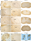
Gene transfer reduces α-synuclein and ubiquitin inclusions. We show representative staining by IHC of hippocampus and thalamus of 4w (C) and 12w (B) injected animals at their humane end points with a monoclonal antibody against α-synuclein. Controls were 12-week-old SD (UT) (A), and 4-month-old normal control (D). Insert in (A) is a magnified view of a cell with characteristic α-synuclein inclusions. Whereas SD (UT) granular layer of the dentate gyrus (GrDG) and lateral posterior thalamic nucleus (LP) show numerous cells with α-synuclein inclusions, treated animals had normalized staining (B and C). In SD (UT), the number of cells containing α-synuclein inclusions and glycoconjugate storage was similar, and localized to the same regions (E), but treated SD and normal controls had neither α-synuclein nor glycoconjugate inclusions (FH). IHC against ubiquitin shows a small number of scattered cells intensely staining in the grey matter of spinal cord of a 4-month-old SD (UT) (I), and absence of inclusions in normal controls (L). While staining intensity in 12w injected mice was only reduced (J), it was fully normalized in the 4w injected group (K). PAS staining demonstrated that a larger number of cells are positive for glycoconjugate storage than for ubiquitin in SD (UT) (M). Treated SD animals had a PAS-staining pattern (N and O) similar to normal controls (P). Inserts in (I) and (M) are magnified views of inclusions-containing cells. Arrowheads point to stained cells. Field CA3 of hippocampus (CA3); granular layer of the dentate gyrus (GrDG); lateral posterior thalamic nucleus (LP). Scale bars: 200 μm (A–H); 100 μm (I–P and inserts).
Figure 5.
Figure 5.
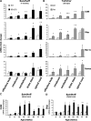
Spatial and temporal up-regulation of markers of inflammation in untreated Sandhoff and Krabbe (Twitcher) mouse models of disease by real-time PCR. Relative mRNA expression of Cd68 and Gfap, markers of activated microglia and astrocytes, respectively, and chemokines Mip-1α and Rantes relative to β-actin were examined in brain and spinal cord at the humane end point of 4 months in Sandhoff (A) and 38 days in twitcher mice (B). Greatest expression occurs in the hindbrain and spinal cord of both models of disease, areas particularly rich in myelin. Temporal expression of Cd68 (C) and Gfap (D) in spinal cord of Sandhoff mice shows largest up-regulation of these markers coincides with the start of the symptomatic phase at around age 13w. Student's t-test; *P < 0.05; **P < 0.01; ***P < 0.001.
Figure 6.
Figure 6.
Gene transfer reduces the number of activated microglia. IHC staining against Cd68 of brain and spinal cord from treated SD at 4w (GI) and 12w (DF) mice was compared with SD (UT) (A–C and M) and normal controls (J–L and N). Treated animals and SD (UT) were killed at their humane end point and normal control at age 4 months. The number of activated microglia was significantly reduced throughout the neuraxis at all transduced ages. Small numbers remain in the VPM/VPL (ventroposterior medial and lateral) nuclei of the thalamus and grey, but not in white matter of spinal cord in 12w injected mice. Whereas most activated microglia in SD (UT) hippocampus had ramified morphology (magnified view in O), those in the stratum lucidum (encircled in A, and magnified in M and P) are amoeboid. Arrowheads and arrows point to ramified and amoeboid microglia, respectively. Field CA3 of hippocampus (CA3). Scale bars: 500 μm (C, F, I and L), 200 μm (A, B, D, E, G, H, J and K), 100 μm (M and N) and 50 μm (O and P).
Figure 7.
Figure 7.
Neuronal degeneration increases with age and is prevented by intracranial gene transfer. SD (UT) (A–D and H) and wild-type mice (E), ages 2w–4 months, were studied by the chemical-development-silver method of Gallyas (21). Silver deposition in the stratum lucidum of the hippocampus (*) is seen at 2w and shows increasing intensity over time. The axons of SD (UT) at the terminal stage of the disease have numerous spheroids (arrowhead) and bulbous ends (arrow) (H). SD mice treated at 4w (F) and 12w (G) have significantly reduced staining. Field CA3 of hippocampus (CA3). Scale bars: 100 μm (A–G) and 25 μm (H).
Figure 8.
Figure 8.
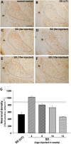
Neuronal cell death is prevented by treatment. NeuN-positive cells were counted in the VPM/VPL thalamic nuclei of SD mice treated at 4w (C), 8w (D), 10w (E) and 12w (F) and killed at their humane end points; SD (UT) (B) and normal controls (A) were killed at 4 months and 4 months–2 years, respectively. Mean ± SEM for each group is represented graphically (G). Horizontal light grey area is NeuN-positive cell numbers in normal controls. 4w injected SD mice (asymptomatic phase) had NeuN-positive cell numbers similar to normal controls. Neuronal density in SD mice injected at age 8–10w (2–3 months, early symptomatic phase) was ∼80–90% that of normal controls; a loss of 15–37% is expected to have already occurred by the time animals were injected (22). P < 0.05 (Bonferroni post hoc test). SD (UT) mice lost 41% of neurones in this study and 50% according to our previous work (22). SD animals injected at 12w had lost ∼60% of neurones by the time they reached their humane end point. Unlike in animals injected at 8–10w, treatment at 12w (late symptomatic phase) did not prevent cell loss (P > 0.05).
Figure 9.
Figure 9.
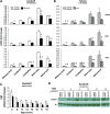
Spatial and temporal expression of myelin markers in untreated Sandhoff and Krabbe (Twitcher) mouse models of disease. Relative mRNA expression of mag, plp and cgt relative to β-actin was examined in brain and spinal cord at the humane end point of 19w in Sandhoff (A) and 38 days in twitcher mice (B). Myelin mRNA expression in SD (UT) is about half that of normal controls in all areas of the brain and spinal cord at the humane end point (A), and reduced expression appears to be an early feature of the disease process (C). Western blot of SD (UT) cerebrum extracts of different ages against the non-compacted myelin marker cnpase also suggests early myelin deficits compared with age-matched controls, while no obvious differences were seen with an antibody against the enzyme th that was probed on the same blot.
Figure 10.
Figure 10.
Compacted myelin proteins are reduced in SD (UT). Amounts of myelin markers Plp (A) and Mbp (C) and neuronal β-tubulin III (E) were analysed relative to β-actin by western blot on cerebrum extracts of SD (UT) and age-matched normal controls. Densitometry analysis of protein species on western blots indicates reduced myelin protein content (B, D), while β-tubulin III composition remains unaltered (F). Student's t-test; *P < 0.05; **P < 0.01.
Figure 11.
Figure 11.
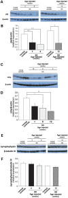
Deficits in myelin protein composition persist after therapeutic gene transfer. Amounts of myelin proteins Plp (A) and Mbp (C) relative to β-actin and neuronal synaptophysin relative to β-tubulin III (E) were analysed by western blot on cerebrum extracts of SD mice treated at 4w, 8w and 12w age and normal controls. Mutant animals were killed at their humane end point and normal controls at a range of ages to cover for the different ages in the treatment groups. Densitometry analysis of protein species on western blots indicates reduced myelin protein content in treated SD (B, D), while synaptophysin relative to β-tubulin III remained largely unchanged (F). *P < 0.05; **P < 0.01 (Bonferroni post hoc test).

Similar articles

Cited by

References

    1. Sandhoff K., Andreae U., Jatzkewitz H. Deficient hexosaminidase activity in an exceptional case of Tay-Sachs disease with additional storage of kidney globoside in visceral organs. Life Sci. 1968;7:283–288. doi:10.1016/0024-3205(68)90024-6. - DOI - PubMed
    1. Okada S., O'Brien J.S. Tay-Sachs disease: generalized absence of a beta-D-N-acetylhexosaminidase component. Science. 1969;165:698–700. doi:10.1126/science.165.3894.698. - DOI - PubMed
    1. Colzelmann E., Sandhoff K. AB variant of infantile GM2 gangliosidosis: deficiency of a factor necessary for stimulation of hexosaminidase A-catalyzed degradation of ganglioside GM2 and glycolipid GA2. Proc. Natl. Acad. Sci. USA. 1978;75:3979–3983. doi:10.1073/pnas.75.8.3979. - DOI - PMC - PubMed
    1. Kolodny E.H., Brady R.O., Volk B.W. Demonstration of an alteration of ganglioside metabolism in Tay-Sachs disease. Biochem. Biophys. Res. Commun. 1969;37:526–531. doi:10.1016/0006-291X(69)90947-4. - DOI - PubMed
    1. Sandhoff K. Variation of beta-N-acetylhexosaminidase-pattern in Tay-Sachs disease. FEBS Lett. 1969;4:351–354. doi:10.1016/0014-5793(69)80274-7. - DOI - PubMed

Publication types

MeSH terms