. 2013 Sep;11(9):e1001652.
doi: 10.1371/journal.pbio.1001652. Epub 2013 Sep 10.

Integrated conformational and lipid-sensing regulation of endosomal ArfGEF BRAG2

Affiliations

Integrated conformational and lipid-sensing regulation of endosomal ArfGEF BRAG2

Kaheina Aizel et al. PLoS Biol. 2013 Sep.

Abstract

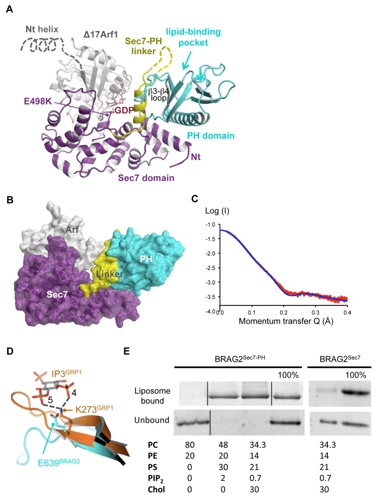
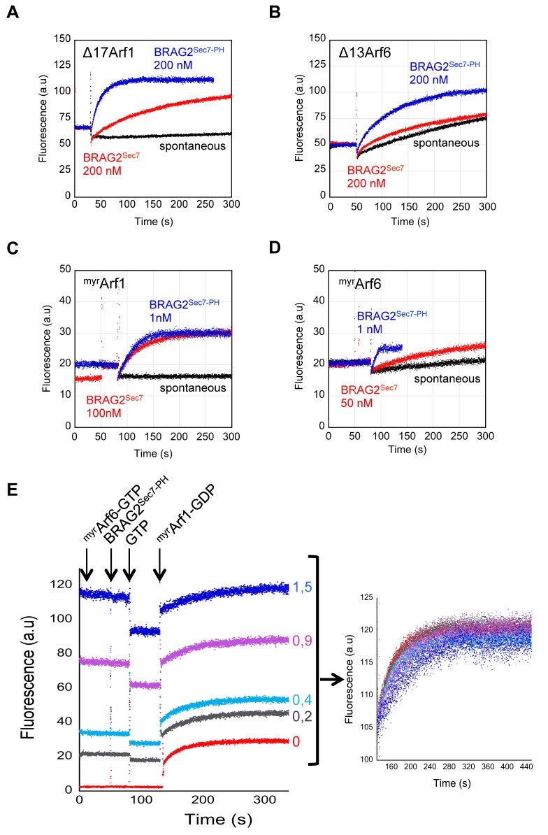
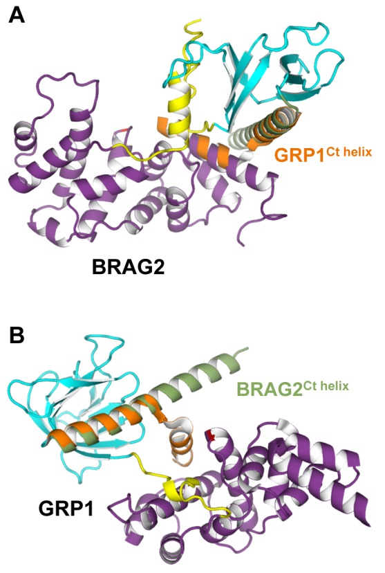
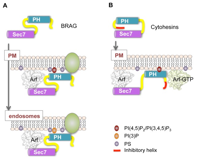
The mechanisms whereby guanine nucleotide exchange factors (GEFs) coordinate their subcellular targeting to their activation of small GTPases remain poorly understood. Here we analyzed how membranes control the efficiency of human BRAG2, an ArfGEF involved in receptor endocytosis, Wnt signaling, and tumor invasion. The crystal structure of an Arf1-BRAG2 complex that mimics a membrane-bound intermediate revealed an atypical PH domain that is constitutively anchored to the catalytic Sec7 domain and interacts with Arf. Combined with the quantitative analysis of BRAG2 exchange activity reconstituted on membranes, we find that this PH domain potentiates nucleotide exchange by about 2,000-fold by cumulative conformational and membrane-targeting contributions. Furthermore, it restricts BRAG2 activity to negatively charged membranes without phosphoinositide specificity, using a positively charged surface peripheral to but excluding the canonical lipid-binding pocket. This suggests a model of BRAG2 regulation along the early endosomal pathway that expands the repertoire of GEF regulatory mechanisms. Notably, it departs from the auto-inhibitory and feedback loop paradigm emerging from studies of SOS and cytohesins. It also uncovers a novel mechanism of unspecific lipid-sensing by PH domains that may allow sustained binding to maturating membranes.


Conflict of interest statement

The authors have declared that no competing interests exist.

Figures

Figure 1
Figure 1. Crystallographic, SAXS, and membrane-binding analysis of BRAG2 reveals an atypical PH domain.
(A) Crystal structure of the Δ17Arf1–GDP/BRAG2Sec7-PH/E498K complex (P2 form, crystallographic statistics in Table S1). Arf1 is in grey, and the domains of BRAG2 are color-coded as indicated. Disordered residues in the linker are indicated by a dotted line. The open end of the β-barrel of the PH domain (arrow), which corresponds to the canonical lipid-binding site of PH domains, aligns with the expected position of the membrane-binding myristoylated N-terminal helix of Arf1 (grey dotted line). (B) Surface representation of the Arf1/BRAG2 complex. The linker and the PH domain form a close-packed structure, which establishes a large intramolecular interface with the N-terminus of the Sec7 domain. Arf1 forms edge contacts with the linker. Residues involved in these interfaces are given in Figures S2A and S3. (C) Synchrotron radiation SAXS analysis of unbound BRAG2Sec7-PH. Fit of the experimental SAXS data of unbound BRAG2Sec7-PH (red) with the scattering curve calculated from the crystal structure of BRAG2Sec7-PH extracted from the complex (blue) is shown. (D) The PH domain of BRAG2 contains a glutamate that replaces a highly conserved phospholipid-binding lysine. A close-up view of the PH domain of BRAG2 (cyan) superposed on the PH domain of GRP1 bound to IP3 (PDB entry code 1U29, orange) is shown. The structure-based sequence alignment of BRAG2 with structures of PH domains with bound phospholipid headgroups is given in Figure S2B. (E) BRAG2Sec7-PH binds to PI(4,5)P2-containing liposomes by its PH domain but not to uncharged liposomes. BRAG2Sec7-PH or BRAG2Sec7 (1 µM) was submitted to flotation assays using liposomes of the indicated composition (% of 1 mM total lipids). The 100% lane corresponds to the theoretical complete recovery of the protein in the fraction.
Figure 2
Figure 2. Quantitative analysis of BRAG2 nucleotide exchange efficiency reveals a dual Arf1/Arf6 specificity and the potentiating role of the PH domain.
(A and B) BRAG2 activates Arf1 and Arf6 and is potentiated by its PH domain in solution. Representative tryptophan fluorescence kinetics curves used to determine kcat/Km given in Table 1 are shown. Exchange reactions were done with 1 µM truncated Arf proteins. SDS-PAGE gels of the proteins are shown in Figure S1A. (C and D) BRAG2 exchange activity towards Arf1 and Arf6 is strongly potentiated by membranes. Representative tryptophan fluorescence kinetics used to determine kcat/Km values given in Table 1 are shown. Exchange reactions were done with 100 µM liposomes (34.3% PC, 14% PE, 21% PS, 0,7%PI(4,5)P2, 30% cholesterol) and with 0.4 µM myrArf proteins. The detailed analysis of Arf6 activation using experimental initial velocities is given in Figure S4. (E) BRAG2 is not regulated by a feedback loop. Activation of myrArf1 by BRAG2Sec7-PH was analyzed by tryptophan fluorescence kinetics using the same liposomes as in Figure 2C. Liposomes were pre-incubated with increasing amounts of myrArf6–GTP as indicated. The right panel shows the kinetics associated with the formation of myrArf1–GTP corrected for the intrinsic fluorescence of myrArf6–GTP.
Figure 3
Figure 3. Unspecific sensitivity of the atypical PH domain of BRAG2 to negatively charged membranes.
(A) BRAG2 is activated by negatively charged membranes but does not discriminate between phosphoinositides. The histogram shows nucleotide exchange rates of BRAG2Sec7-PH (1 nM) towards myrArf1–GDP (0.4 µM) using 100 µM of liposomes containing 2% PI and 30% PS complemented with 48% PC and 20% PE, except for uncharged liposomes containing 80% PC and 20% PE. Reactions were initiated by addition of 100 µM GTP. k obs values are means of at least three experiments and are given ±S.D. (B) The proposed membrane-binding surface of the PH domain of BRAG2. Positively charged residues are shown in dark blue. Residues mutated in the canonical lipid-binding pocket are shown. (C) BRAG2 does not use the lipid-binding pocket of its PH domain to recognize negatively charged membranes. Nucleotide exchange activity of BRAG2Sec7-PH mutants carrying the E639A and E639K mutation in the PH pocket, using myrArf1 and liposomes of the indicated compositions. k obs are expressed as a percentage of the exchange rate of wild-type BRAG2Sec7-PH. Nucleotide exchange in solution using Δ17Arf1–GDP is shown on the left. (D) The proposed membrane-facing surface of the PH domain has a strong positive electrostatic potential. The electrostatic potential map is contoured at –5 kT/e (in red) and 5 kT/e (blue). The view as in Figure 1A.
Figure 4
Figure 4. Structural basis for the diverging regulatory mechanisms of BRAG2 and cytohesins.
(A) Superposition of the PH domain of GRP1 to that of BRAG2 shows that the kinked auto-inhibitory C-terminal helix of GRP1 (in orange) would conflict with the Sec7 domain of BRAG2. (B) Superposition of the PH domain of BRAG2 to the auto-inhibited structure of GRP1 shows that the straight C-terminal helix of BRAG2 (in green) would not be auto-inhibitory in GRP1.
Figure 5
Figure 5. Diverging regulatory models of cytohesin and BRAG ArfGEFs on cellular membranes.
(A) BRAG2 is constitutively active in solution (top panel), but strongly potentiated by negatively charged membranes such as those found at the plasma membrane (middle panel) and early endosomes (bottom panel). The PH domain interacts nonspecifically with PS- and PI-containing membranes outside the canonical lipid-binding pocket. Additional specificity may be achieved by interaction with receptors (shown in green). (B) Cytohesins are autoinhibited in solution (top panel) and activated by specific binding of their PH domain to PI(4,5)P2 or PI(3,4,5)P3 and to Arf–GTP at the plasma membrane (bottom panel).

Comment in

  • Expanding the regulatory repertoire of GEFs.
    Robinson R. Robinson R. PLoS Biol. 2013 Sep;11(9):e1001654. doi: 10.1371/journal.pbio.1001654. Epub 2013 Sep 10. PLoS Biol. 2013. PMID: 24058296 Free PMC article. No abstract available.

References

    1. D'Souza-Schorey C, Chavrier P (2006) ARF proteins: roles in membrane traffic and beyond. Nat Rev Mol Cell Biol 7: 347–358. - PubMed
    1. Cherfils J, Zeghouf M (2013) Regulation of small GTPases by GEFs, GAPs, and GDIs. Physiol Rev 93: 269–309. - PubMed
    1. Pasqualato S, Renault L, Cherfils J (2002) Arf, Arl, Arp and Sar proteins: a family of GTP-binding proteins with a structural device for ‘front-back’ communication. EMBO Rep 3: 1035–1041. - PMC - PubMed
    1. Renault L, Guibert B, Cherfils J (2003) Structural snapshots of the mechanism and inhibition of a guanine nucleotide exchange factor. Nature 426: 525–530. - PubMed
    1. DiNitto JP, Delprato A, Gabe Lee MT, Cronin TC, Huang S, et al. (2007) Structural basis and mechanism of autoregulation in 3-phosphoinositide-dependent Grp1 family Arf GTPase exchange factors. Mol Cell 28: 569–583. - PMC - PubMed

Publication types

MeSH terms