

Cardiac myocyte diversity and a fibroblast network in the junctional region of the zebrafish heart revealed by transmission and serial block-face scanning electron microscopy
- PMID: 24058412
- PMCID: PMC3751930
- DOI: 10.1371/journal.pone.0072388
Cardiac myocyte diversity and a fibroblast network in the junctional region of the zebrafish heart revealed by transmission and serial block-face scanning electron microscopy
Abstract
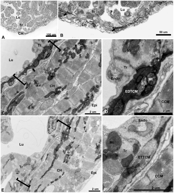
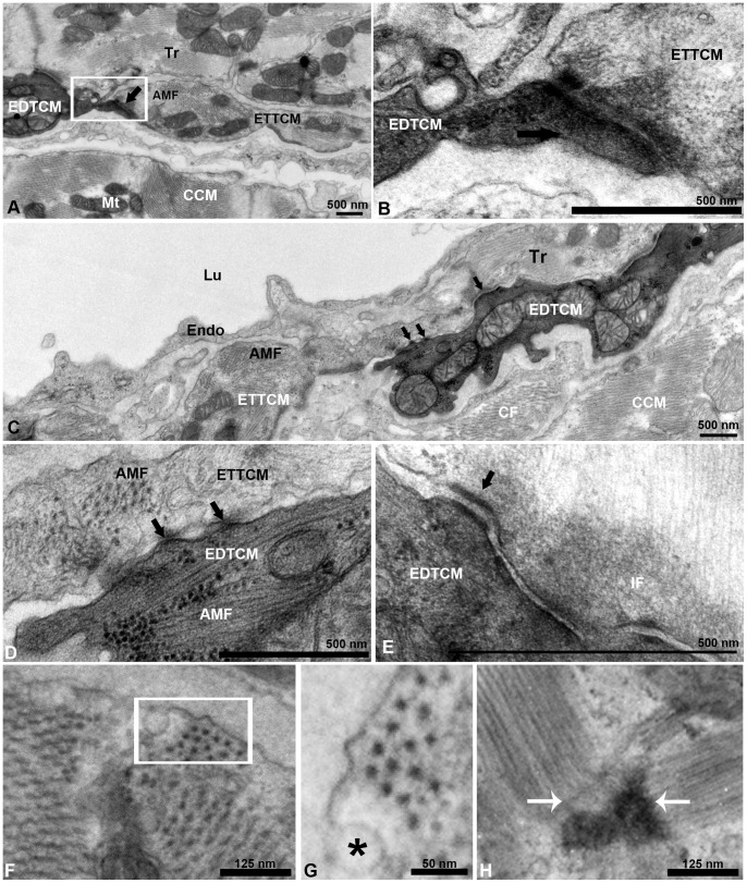
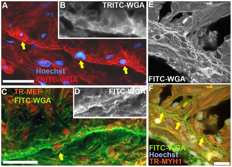
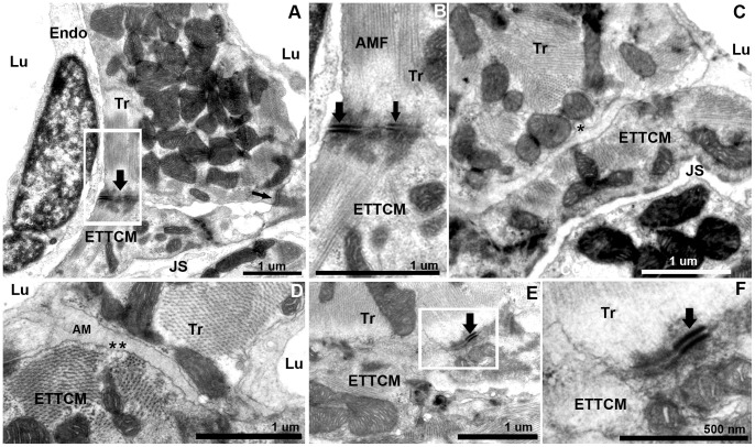
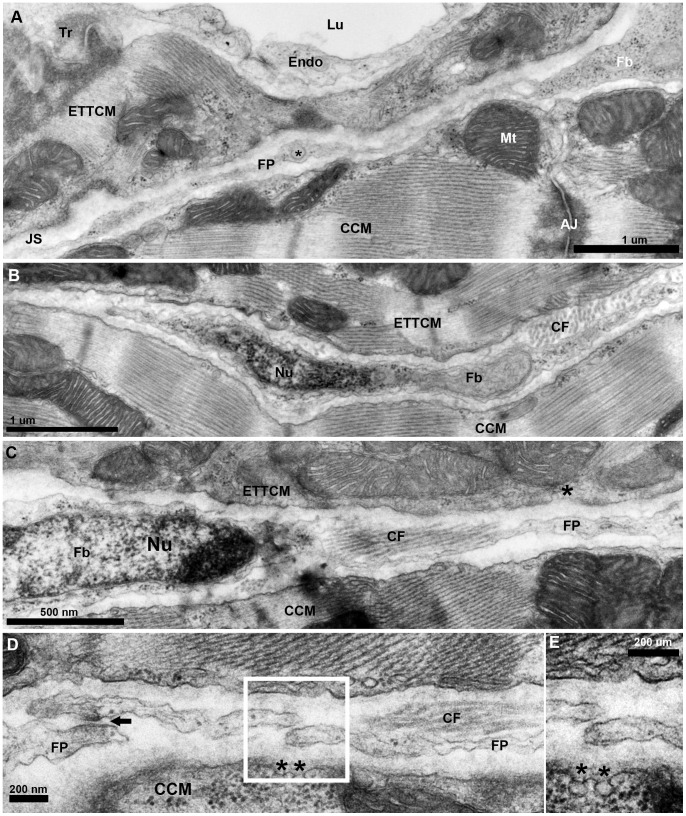
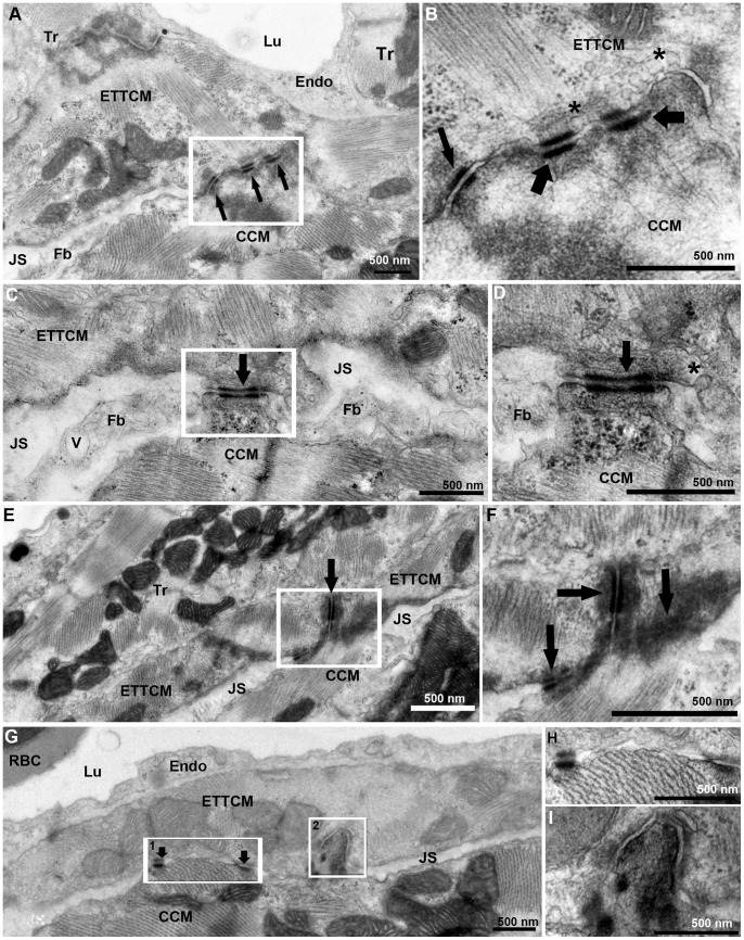
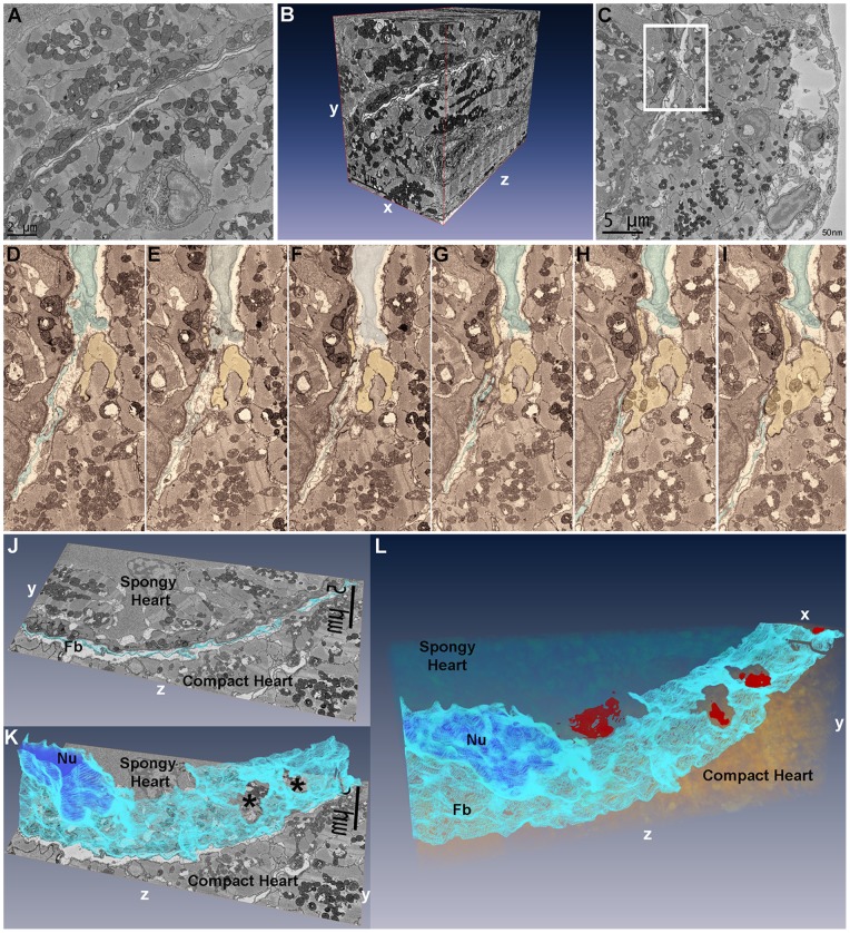
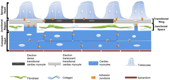
The zebrafish has emerged as an important model of heart development and regeneration. While the structural characteristics of the developing and adult zebrafish ventricle have been previously studied, little attention has been paid to the nature of the interface between the compact and spongy myocardium. Here we describe how these two distinct layers are structurally and functionally integrated. We demonstrate by transmission electron microscopy that this interface is complex and composed primarily of a junctional region occupied by collagen, as well as a population of fibroblasts that form a highly complex network. We also describe a continuum of uniquely flattened transitional cardiac myocytes that form a circumferential plate upon which the radially-oriented luminal trabeculae are anchored. In addition, we have uncovered within the transitional ring a subpopulation of markedly electron dense cardiac myocytes. At discrete intervals the transitional cardiac myocytes form contact bridges across the junctional space that are stabilized through localized desmosomes and fascia adherentes junctions with adjacent compact cardiac myocytes. Finally using serial block-face scanning electron microscopy, segmentation and volume reconstruction, we confirm the three-dimensional nature of the junctional region as well as the presence of the sheet-like fibroblast network. These ultrastructural studies demonstrate the previously unrecognized complexity with which the compact and spongy layers are structurally integrated, and provide a new basis for understanding development and regeneration in the zebrafish heart.
Conflict of interest statement
Figures
Similar articles
-
The intercellular organization of the two muscular systems in the adult salmonid heart, the compact and the spongy myocardium.J Anat. 2009 Nov;215(5):536-47. doi: 10.1111/j.1469-7580.2009.01129.x. Epub 2009 Jul 22. J Anat. 2009. PMID: 19627390 Free PMC article.
-
Unravelling the ultrastructural details of αT-catenin-deficient cell-cell contacts between heart muscle cells by the use of FIB-SEM.J Microsc. 2020 Sep;279(3):189-196. doi: 10.1111/jmi.12855. Epub 2019 Dec 22. J Microsc. 2020. PMID: 31828778
-
Distribution and three-dimensional structure of intercellular junctions in canine myocardium.Circ Res. 1989 Mar;64(3):563-74. doi: 10.1161/01.res.64.3.563. Circ Res. 1989. PMID: 2645060
-
Cytoarchitecture and intercalated disks of the working myocardium and the conduction system in the mammalian heart.Anat Rec A Discov Mol Cell Evol Biol. 2004 Oct;280(2):940-51. doi: 10.1002/ar.a.20109. Anat Rec A Discov Mol Cell Evol Biol. 2004. PMID: 15368339 Review.
-
Serial block face scanning electron microscopy for the study of cardiac muscle ultrastructure at nanoscale resolutions.J Mol Cell Cardiol. 2014 Nov;76:1-11. doi: 10.1016/j.yjmcc.2014.08.010. Epub 2014 Aug 20. J Mol Cell Cardiol. 2014. PMID: 25149127 Review.
Cited by
-
Proteomic Analysis of Exosomes during Cardiogenic Differentiation of Human Pluripotent Stem Cells.Cells. 2021 Oct 1;10(10):2622. doi: 10.3390/cells10102622. Cells. 2021. PMID: 34685602 Free PMC article.
-
Reactive oxygen species during heart regeneration in zebrafish: Lessons for future clinical therapies.Wound Repair Regen. 2021 Mar;29(2):211-224. doi: 10.1111/wrr.12892. Epub 2021 Jan 20. Wound Repair Regen. 2021. PMID: 33471940 Free PMC article. Review.
-
Exercise, programmed cell death and exhaustion of cardiomyocyte proliferation in aging zebrafish.Dis Model Mech. 2021 Jul 1;14(7):dmm049013. doi: 10.1242/dmm.049013. Epub 2021 Jul 22. Dis Model Mech. 2021. PMID: 34296752 Free PMC article.
-
Serial Block-Face Scanning Electron Microscopy (SBF-SEM) of Biological Tissue Samples.J Vis Exp. 2021 Mar 26;(169):10.3791/62045. doi: 10.3791/62045. J Vis Exp. 2021. PMID: 33843931 Free PMC article.
-
The careg element reveals a common regulation of regeneration in the zebrafish myocardium and fin.Nat Commun. 2017 May 3;8:15151. doi: 10.1038/ncomms15151. Nat Commun. 2017. PMID: 28466843 Free PMC article.
References
-
- Santer RM (1980) Morphological studies on the ventricle of teleost and elasmobranch hearts. J Zool, Lond 190: 259–272.
-
- Sanchez-Quintana D, Garcia-Martinez V, Climent V, Hurle JM (1995) Morphological analysis of the fish heart ventricle: myocardial and connective tissue architecture in teleost species. Ann Anat 177: 267–274. - PubMed
-
- Sanchez-Quintana D, Hurle JM (1987) Ventricular myocardial architecture in marine fishes. Anat Rec 217: 263–273. - PubMed
-
- Santer RM (1985) Morphology and innervation of the fish heart. Adv Anat Embryol Cell Biol 89: 1–102. - PubMed
-
- Reifers F, Walsh EC, Leger S, Stainier DY, Brand M (2000) Induction and differentiation of the zebrafish heart requires fibroblast growth factor 8 (fgf8/acerebellar). Development 127: 225–235. - PubMed
Publication types
MeSH terms
Substances
Grants and funding
LinkOut - more resources
Full Text Sources
Other Literature Sources
