. 2013 Sep 18;8(9):e74523.
doi: 10.1371/journal.pone.0074523. eCollection 2013.

Point mutations in the transmembrane region of the clic1 ion channel selectively modify its biophysical properties

Affiliations

Point mutations in the transmembrane region of the clic1 ion channel selectively modify its biophysical properties

Stefania Averaimo et al. PLoS One. .

Abstract

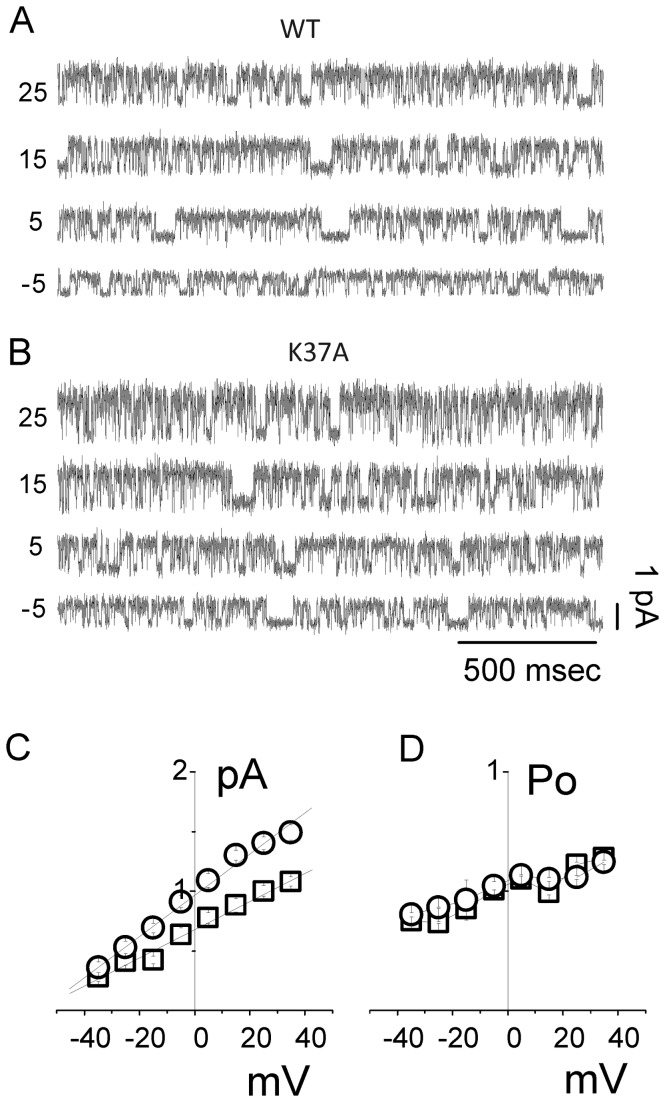
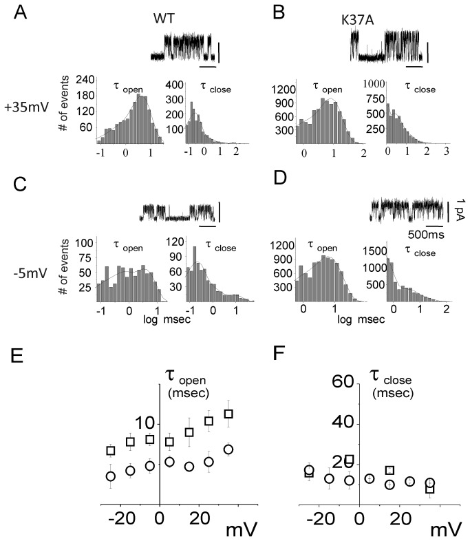
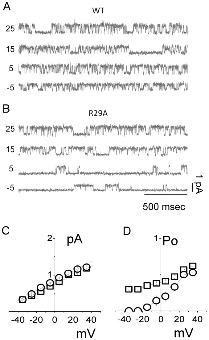
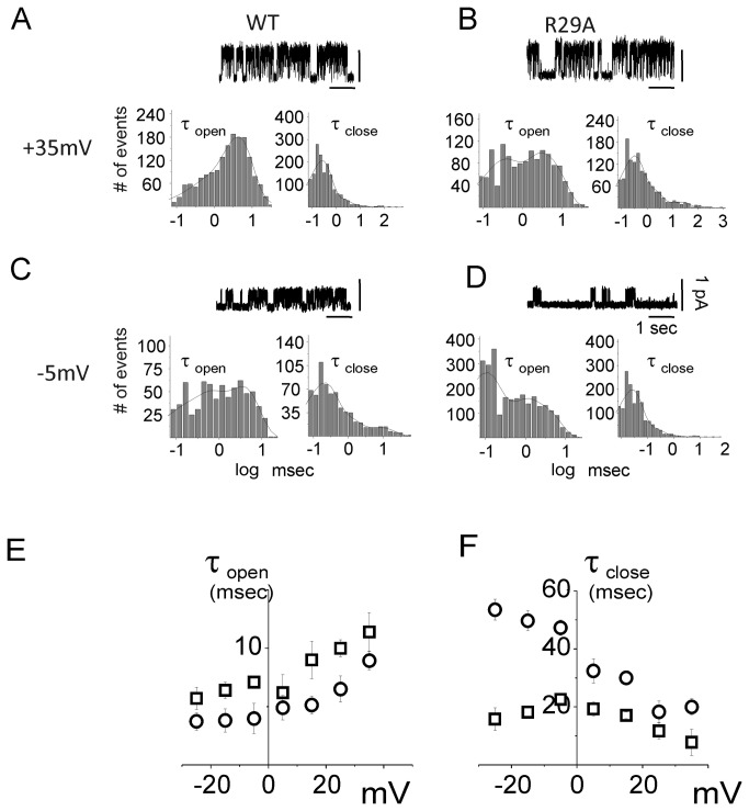
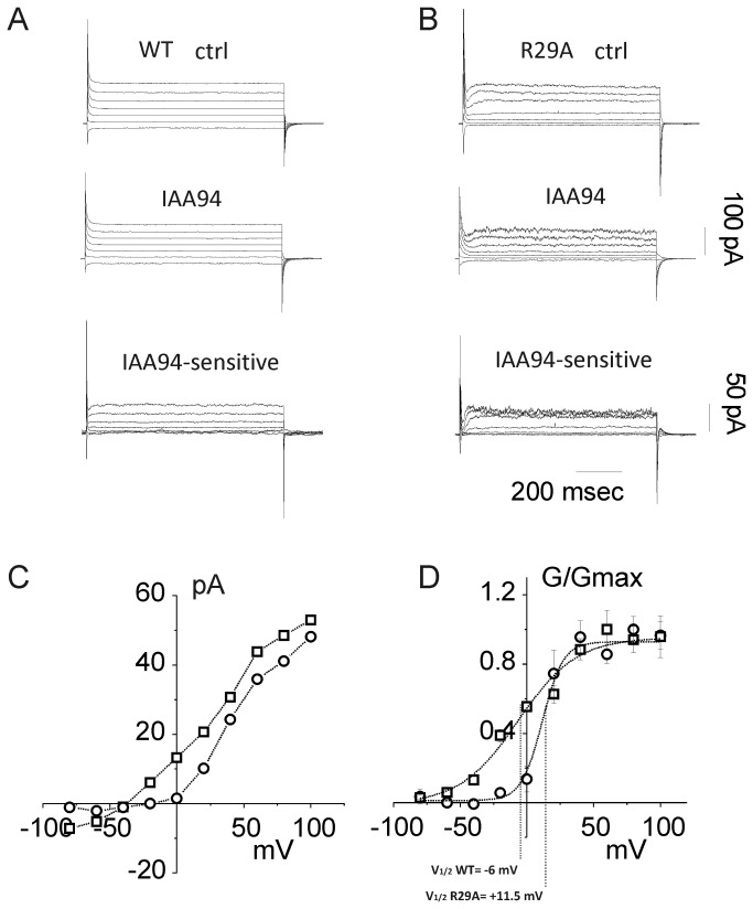
Chloride intracellular Channel 1 (CLIC1) is a metamorphic protein that changes from a soluble cytoplasmic protein into a transmembrane protein. Once inserted into membranes, CLIC1 multimerises and is able to form chloride selective ion channels. Whilst CLIC1 behaves as an ion channel both in cells and in artificial lipid bilayers, its structure in the soluble form has led to some uncertainty as to whether it really is an ion channel protein. CLIC1 has a single putative transmembrane region that contains only two charged residues: arginine 29 (Arg29) and lysine 37 (Lys37). As charged residues are likely to have a key role in ion channel function, we hypothesized that mutating them to neutral alanine to generate K37A and R29A CLIC1 would alter the electrophysiological characteristics of CLIC1. By using three different electrophysiological approaches: i) single channel Tip-Dip in artificial bilayers using soluble recombinant CLIC1, ii) cell-attached and iii) whole-cell patch clamp recordings in transiently transfected HEK cells, we determined that the K37A mutation altered the single-channel conductance while the R29A mutation affected the single-channel open probability in response to variation in membrane potential. Our results show that mutation of the two charged amino acids (K37 and R29) in the putative transmembrane region of CLIC1 alters the biophysical properties of the ion channel in both artificial bilayers and cells. Hence these charged residues are directly involved in regulating its ion channel activity. This strongly suggests that, despite its unusual structure, CLIC1 itself is able to form a chloride ion channel.


Conflict of interest statement

Competing Interests: The authors have declared that no competing interests exist.

Figures

Figure 1
Figure 1. CLIC1 ion channel activity from Tip-Dip bilayer experiments.
Current recordings from -80 to +80 mV, 20 mV interval, are shown in (A) for wild type CLIC1 (left), K37A (center) and R29A (right) CLIC1 protein. The upper panel of Figure 1B depicts the single-channel current-voltage data for WT (□) and K37A (○) CLIC1 proteins. The average single-channel conductance differs between WT and K37A, calculated as 30.1 ± 0.2 and 42.4 ± 0.2 pS, respectively (n = 5, p < 0.001). In contrast, the channel open probability of the K37A mutation is very similar to the WT (Figure 1B, lower panel). The current/voltage relationships for both WT (□) and R29A CLIC1 (○) are shown in (C). The single-channel conductance (upper panel), is 30.1 ± 0.2 and 29 ± 0.2 pS for WT and R29A, respectively. The average open probability for WT (□) and R29A mutated protein (○) is shown in the lower panel.
Figure 2
Figure 2. Endogenous CLIC1 is not expressed on the plasma membrane of untransfected HEK cells.
(A) Family of currents for an untransfected HEK293 cell at varying applied voltages from -60 to +60 mV with 20 mV increment. Top panel: whole cell current in resting conditions; middle panel: after IAA94 perfusion; lower panel: IAA94-sensitive (CLIC1-mediated) current obtained from subtraction. (B) i/V curve of an IAA94-sensitive current in WT CLIC1 transfected HEK293 cell (□), and in an untransfected HEK293 cell (○). (C) Plot of the average of the IAA94-sensitive current as a percentage of the control current of untransfected HEK cells (n=5). Data are shown as mean ± SEM. CLIC1-mediated current is completely absent in untransfected HEK cells.
Figure 3
Figure 3. Cell-attached recordings of HEK cells transiently transfected with WT and K37A CLIC1 protein.
Single-channel current traces are shown for WT (A) and K37A (B) CLIC1 transfected cells. The membrane voltage was clamped at different values (indicated on the left of each trace) in 10 mV step increments. Single-channel current/voltage plots are shown in (C) for WT (□) and K37A (○). Average single-channel conductance of 12.1 ± 0.6 pS for WT and 17.4 ± 0.8 pS for K37A with an extrapolated reversal potential of -58 ± 0.7 mV and -56 ± 1.2 mV, respectively, was calculated. The open probability obtained at each membrane potential is shown in (D) for WT (□) and K37A (○).
Figure 4
Figure 4. Open (τopen) and close (τclose) time constants of the WT and K37A CLIC1 ion channel.
In panels (A) and (B), membrane potentials were held at +35 mV while panels (C) and (D) concern cell membrane potentials held at -5 mV for WT (A, C) and K37A (B, D) CLIC1 transfected HEK cells. (A) to (D) shows the open (left) and close (right) time distributions for each condition. Four seconds of single channel recordings appear as inserts in the corresponding panels for each condition. The open and close time distribution histograms were fitted by a double exponential decay function and plotted on a semi-logarithmic scale. Panels (E) and (F) depict open and close time distributions as a function of membrane potential for WT (□) and K37A (○) transfected HEK cells.
Figure 5
Figure 5. Cell-attached recordings of HEK cells transiently transfected with WT CLIC1 and R29A CLIC1 protein.
Single-channel current traces are shown for WT (A) and R29A (B) transfected cells. The voltage steps are from -5 to + 25 mV (10 mV step increment). Single-channel current/voltage plots are shown in (C) for WT (□) and R29A (○). Average single-channel conductance of 12.3 ± 0.1 pS for WT and 13.1 ± 0.3 pS for R29A and an extrapolated reversal potential of -63 ± 0.4 mV and -61 ± 0.6 mV, respectively. The open probability obtained at each membrane potential is shown in (D) for WT (□) and R29A (○).
Figure 6
Figure 6. Open (τopen) and close (τclose) time constants of the WT and R29A CLIC1 ion channel.
In panels (A) and (B), membrane potentials were held at +35 mV while panels (C) and (D) concern cell membrane potentials held at -5 mV. The Figure shows open (left) and close (right) time distribution for WT (A, C) and R29A (B, D) CLIC1 transfected HEK cells. Four seconds of single channel recordings appear as inserts in the corresponding panels for each condition. The open and close time distribution histograms were fitted by a double exponential decay function and plotted on a semi-logarithmic scale. Panels (E) and (F) depict open and close time distributions as a function of membrane potential for WT (□) and R29A (○) transfected HEK cells.
Figure 7
Figure 7. Whole-cell current in R29A transfected HEK cells.
Family of whole-currents for WT (A) and R29A (B) transfected HEK cells (top panels). Voltage steps lasting 800 ms from holding potential of -30 mV to membrane potential of -80 to +100 mV (20 mV step increment). The middle panels depict the whole-cell currents after perfusion of 50 µM of IAA94. The bottom panels represent the IAA94-sensitive currents obtained by subtraction of the middle panel current from the upper panel current. Note that the IAA94-sensitive currents are plotted on a different scale with the scale bars on the right hand side of the figure. (C) an example of current/voltage relationship of the IAA94-sensitive current from a WT (□) and a R29A (○) transfected HEK cell. (D) Averaged G/V plots from IAA94-sensitive current of WT (□) and R29A mutant (○), from 5 independent experiments.

References

    1. Valenzuela SM, Martin DK, Por SB, Robbins JM, Warton K et al. (1997) Molecular cloning and expression of a chloride ion channel of cell nuclei. J Biol Chem 272: 12575-12582. doi:10.1074/jbc.272.19.12575. PubMed: 9139710. - DOI - PubMed
    1. Yang SG, Wang WY, Ling TJ, Feng Y, Du XT et al. (2010) alpha-Tocopherol quinone inhibits beta-amyloid aggregation and cytotoxicity, disaggregates preformed fibrils and decreases the production of reactive oxygen species, NO and inflammatory cytokines. Neurochem Int 57: 914-922. doi:10.1016/j.neuint.2010.09.011. PubMed: 20933033. - DOI - PubMed
    1. Littler DR, Harrop SJ, Goodchild SC, Phang JM, Mynott AV et al. (2010) The enigma of the CLIC proteins: Ion channels, redox proteins, enzymes, scaffolding proteins? FEBS Lett 584: 2093-2101. doi:10.1016/j.febslet.2010.01.027. PubMed: 20085760. - DOI - PubMed
    1. Averaimo S, Milton RH, Duchen MR, Mazzanti M (2010) Chloride intracellular channel 1 (CLIC1): Sensor and effector during oxidative stress. FEBS Lett 584: 2076-2084. doi:10.1016/j.febslet.2010.02.073. PubMed: 20385134. - DOI - PubMed
    1. Edwards JC, Kahl CR (2010) Chloride channels of intracellular membranes. FEBS Lett 584: 2102-2111. doi:10.1016/j.febslet.2010.01.037. PubMed: 20100480. - DOI - PMC - PubMed

Publication types