

Vascular surgery, microsurgery and supramicrosurgery for treatment of chronic diabetic foot ulcers to prevent amputations
- PMID: 24058622
- PMCID: PMC3772888
- DOI: 10.1371/journal.pone.0074704
Vascular surgery, microsurgery and supramicrosurgery for treatment of chronic diabetic foot ulcers to prevent amputations
Abstract
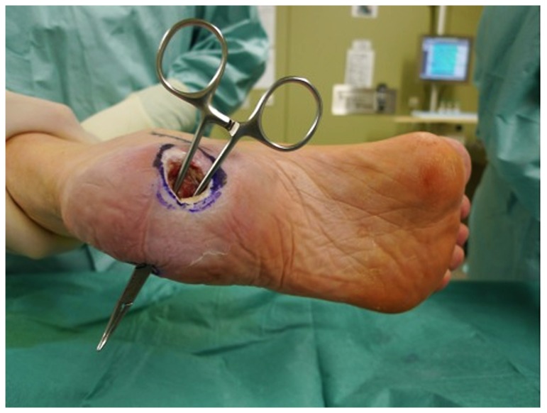
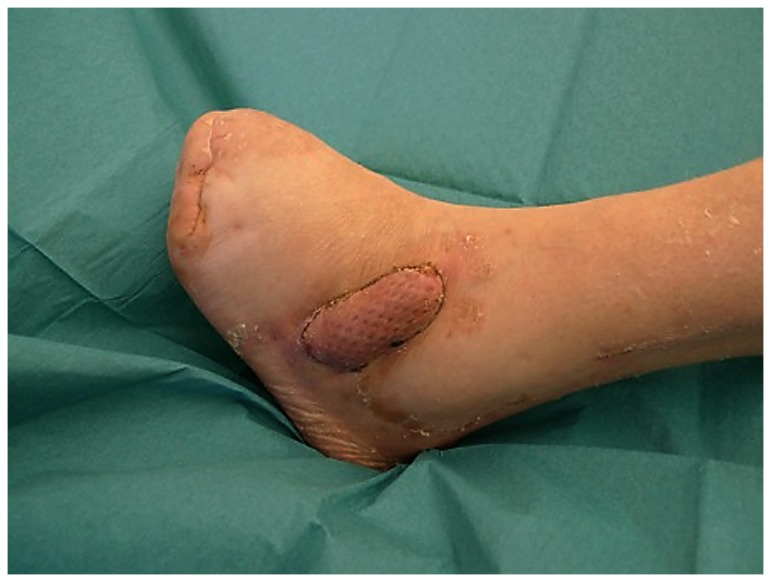
Introduction: Diabetic foot ulcers occur in approximately 2,5% of patients suffering from diabetes and may lead to major infections and amputation. Such ulcers are responsible for a prolonged period of hospitalization and co- morbidities caused by infected diabetic foot ulcers. Small, superficial ulcers can be treated by special conservative means. However, exposed bones or tendons require surgical intervention in order to prevent osteomyelitis. In many cases reconstructive surgery is necessary, sometimes in combination with revascularization of the foot. There are studies on non surgical treatment of the diabetic foot ulcer. Most of them include patients, classified Wagner 1-2 without infection. Patients presenting Wagner 3D and 4D however are at a higher risk of amputation. The evolution of microsurgery has extended the possibilities of limb salvage. Perforator based flaps can minimize the donorsite morbidity.
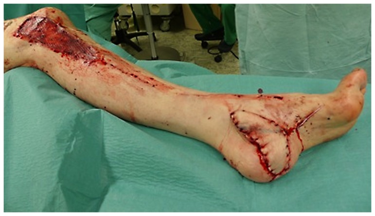
Patients and methods: 41 patients were treated with free tissue transfer for diabetic foot syndrome and chronic defects. 44 microvascular flaps were needed. The average age of patients was 64.3 years. 18 patients needed revascularization. 3 patients needed 2 microvascular flaps. In 6 cases supramicrosurgical technique was used.
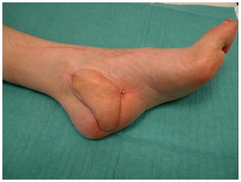
Results: There were 2 flap losses leading to amputation. 4 other patients required amputation within 6 months postoperatively due to severe infection or bypass failure. Another 4 patients died within one year after reconstruction. The remaining patients were ambulated.
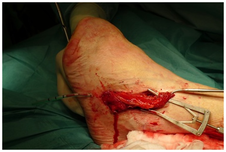
Discussion: Large defects of the foot can be treated by free microvascular myocutaneous or fasciocutaneous tissue transfer. If however, small defects, exposing bones or tendons, are not eligible for local flaps, small free microvascular flaps can be applied. These flaps cause a very low donor site morbidity. Arterialized venous flaps are another option for defect closure. Amputation means reduction of quality of life and can lead to an increased mortality postoperatively.
Conflict of interest statement
Figures
References
-
- World Health Organization and International Diabetes Federation, Europe (1990) Diabetes care and Research in Europe. The Saint Vincent Declaration. Diabet Med 7: 360-363. doi:10.1111/j.1464-5491.1990.tb01405.x. PubMed: 2140091. - DOI - PubMed
-
- Armstrong DG, Lipsky BA (2004) Diabetic foot infections: stepwise medical and surgical management. Int Wound J 1: 123-132. doi:10.1111/j.1742-4801.2004.00035.x. PubMed: 16722884. - DOI - PMC - PubMed
-
- Dellon AL, Muse VL, Nickerson DS, Akre T, Anderson SR et al. (2012) Prevention of ulceration, amputation, and reduction of hospitalization: outcomes of a prospective multicenter trial of tibial neurolysis in patients with diabetic neuropathy. J Reconstr Microsurg 28: 241-246. doi:10.1055/s-0032-1306372. PubMed: 22411624. - DOI - PubMed
-
- Scherer SS, Tobalem M, Vigato E, Heit Y, Modarressi A (2012) Nonactivated versus Thrombin-ActivatedPlatelets on Wound Healing and Fibroblast-to-Myofibroblast Differentiation In Vivo and In Vitro. Plast Reconstr Surg 129(1): 46e-54e. doi:10.1097/PRS.0b013e3182362010. PubMed: 22186584. - DOI - PubMed
MeSH terms
LinkOut - more resources
Full Text Sources
Other Literature Sources
Medical
