. 2013 Sep 13;8(9):e74898.
doi: 10.1371/journal.pone.0074898. eCollection 2013.

Fine structure of the male reproductive system and reproductive behavior of Lutzomyia longipalpis sandflies (Diptera: Psychodidae: Phlebotominae)

Affiliations

Fine structure of the male reproductive system and reproductive behavior of Lutzomyia longipalpis sandflies (Diptera: Psychodidae: Phlebotominae)

Carolina N Spiegel et al. PLoS One. .

Erratum in

  • PLoS One. 2014;9(1). doi:10.1371/annotation/7d6a463d-41b1-4225-b09b-edecc565071c

Abstract

Background: The male reproductive system of insects can have several tissues responsible for the secretion of seminal fluid proteins (SFPs), such as testes, accessory glands, seminal vesicles, ejaculatory duct and ejaculatory bulb. The SFPs are transferred during mating and can induce several physiological and behavioral changes in females, such as increase in oviposition and decrease in sexual receptivity after copulation. The phlebotomine Lutzomyia longipalpis is the main vector of visceral leishmaniasis. Despite its medical importance, little is known about its reproductive biology. Here we present morphological aspects of the male L. longipalpis reproductive system by light, scanning and transmission electron microscopy, and compare the mating frequency of both virgin and previously mated females.

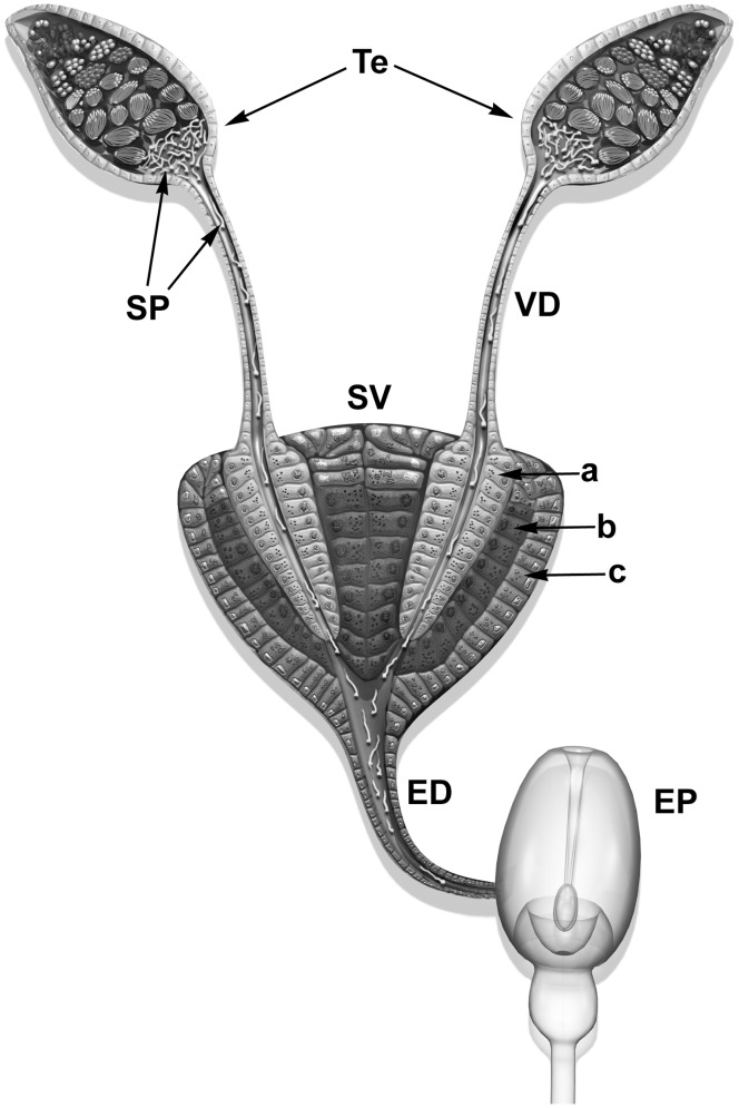
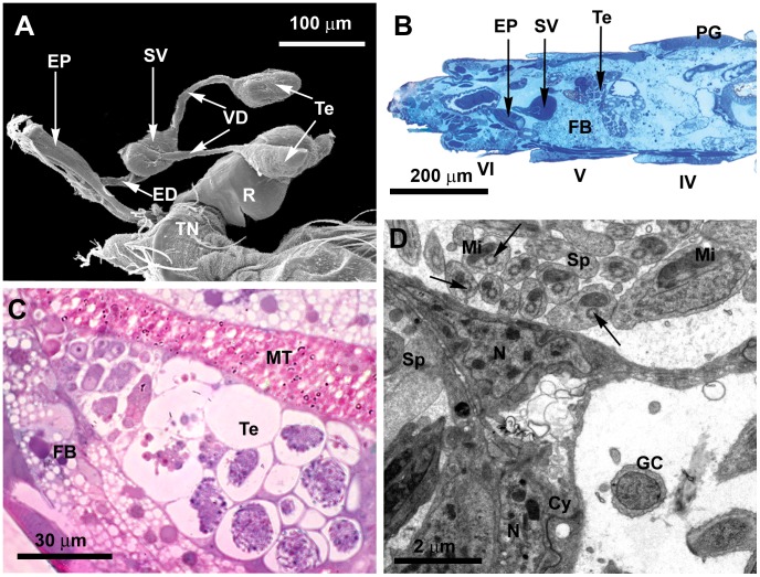
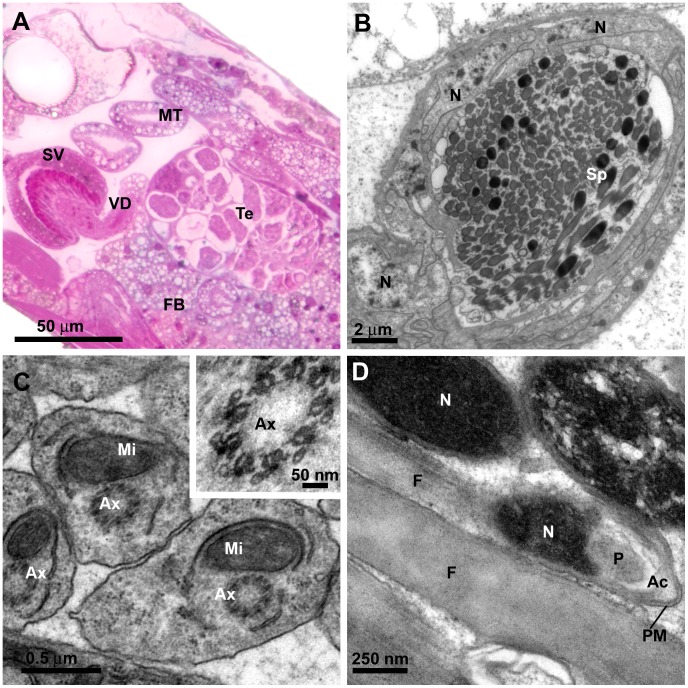
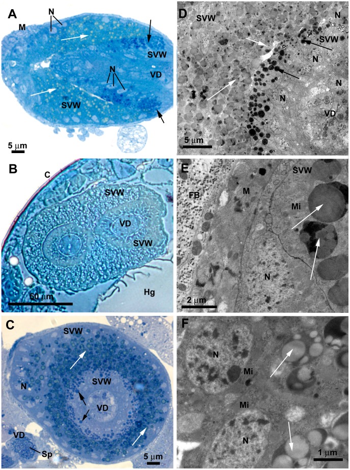
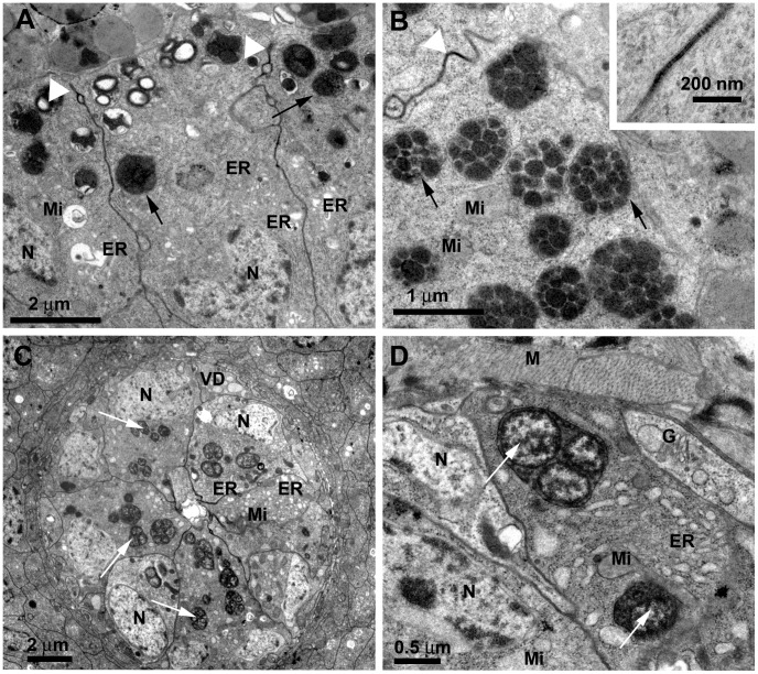
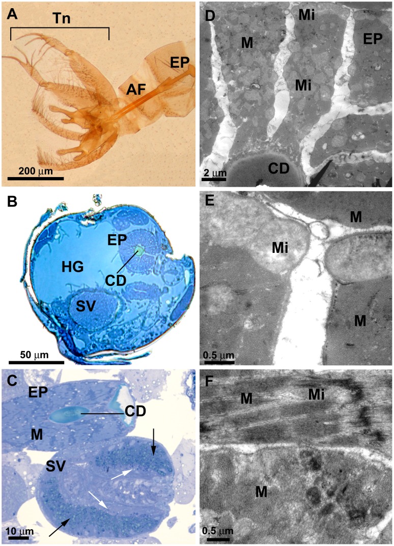
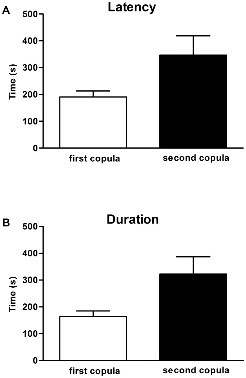
Results: The male L. longipalpis reproductive system is comprised by a pair of oval-shaped testes linked to a seminal vesicle by vasa deferentia. It follows an ejaculatory duct with an ejaculatory pump (a large bulb enveloped by muscles and associated to tracheas). The terminal endings of the vasa deferentia are inserted into the seminal vesicle by invaginations of the seminal vesicle wall, which is composed by a single layer of gland cells, with well-developed endoplasmic reticulum profiles and secretion granules. Our data suggest that the seminal vesicle acts both as a spermatozoa reservoir and as an accessory gland. Mating experiments support this hypothesis, revealing a decrease in mating frequency after copulation that indicates the effect of putative SFPs.

Conclusion: Ultrastructural features of the L. longipalpis male seminal vesicle indicated its possible role as an accessory gland. Behavioral observations revealed a reduction in mating frequency of copulated females. Together with transcriptome analyses from male sandfly reproductive organs identifying ESTs encoding orthologs of SFPs, these data indicate the presence of putative L. longipalpis SFPs reducing sexual mating frequency of copulated females.


Conflict of interest statement

Competing Interests: The authors have declared that no competing interests exist.

Figures

Figure 1
Figure 1. Schematic view of the male reproductive system of Lutzomia longipalpis.
A pair of testes (Te) is linked via vasa deferentia (VD) to a pear-shaped seminal vesicle (SV). An ejaculatory duct (ED) then connects the seminal vesicle to the ejaculatory pump (EP). The vasa deferentia are inserted (a) into invaginations (b) of the seminal vesicle wall (c). Cells at layers “a”, “b” and “c” present different types of cytoplasmic granules; SP: mature spermatozoa. Modified from Valdez (2001) and Fausto et al. 2000.
Figure 2
Figure 2. General aspect of the L. longipalpis male reproductive system and the testes.
A) SEM of an isolated male reproductive system, showing the pair of testes (Te), the vasa deferentia (VD), the seminal vesicle (SV), the ejaculatory duct (ED), the ejaculatory pump (EP) and the external genitalia or terminalia (TN). Also note the rectum (R). B) Light microscopy (LM) section showing the localization of the reproductive organs in the abdomen. Testes (Te) and the seminal vesicle (SV) are located in the fifth abdominal segment (V). Note pheromone gland cells (PG) in the fourth tergite (IV), and the ejaculatory pump (EP) in the sixth abdominal segment (VI). Note the well-developed fat body (FB) surrounding the testis and the seminal vesicle. C) LM section of a testis (Te) showing the organization of germ cells with different stages of development along the testis. Note the proximity of Malpighi tubules (MT) and the fat body (FB). D) Transmission electron microscopy (TEM) of germ cells (GC) and spermatozoa (Sp) surrounded by cystocytes (Cy), forming the cyst. In the spermatozoa, note the axoneme (arrow) lacking the central pair of microtubules and the mitochondria (Mi) close to it; N: nucleus of the cystocytes.
Figure 3
Figure 3. L. longipalpis male reproductive system: vas deferens and spermatozoa.
A) LM section of the fifth abdominal segment, showing the testis (Te) and the vas deferens (VD) that conducts the sperm from the testis to the seminal vesicle (SV). Note the germ cells inside the testis with different shapes. A well-developed fat body (FB) surrounds the testis and the seminal vesicle; MT: Malpighian tubule. B) TEM of the tubular vas deferens packed with mature spermatozoa (Sp). It is enveloped by a thin basal membrane and a layer of interdigitated epithelial cells; N: nucleus of the epithelial cell. C) Detail of spermatozoa by TEM showing the flagelar axoneme (Ax), which presents a 9+0 array of microtubules; Mi: mitochondrion. A higher magnification of the axoneme is also shown in the inset. D) Longitudinal section by TEM of spermatozoa showing flagellum (F), capping perforatorium (P) and the thin acrosomal complex (Ac); PM: plasma membrane; N: meshwork of compact DNA at the spermatozoon head.
Figure 4
Figure 4. L. longipalpis male reproductive system: seminal vesicle, general aspects and the external epithelium.
A) LM longitudinal section through the seminal vesicle shows that it is surrounded by muscle cells (M) and formed by three consecutive distinct layers of cells: the outer epithelial seminal vesicle wall (SVW), the denser, invaginated epithelial wall of the seminal vesicle and the inner circular epithelial layer of the vas deferens (VD). Note the basal localization of the nuclei (N) and the different granule types inside the cells at the inner and outer seminal vesicle wall (black and white arrows, respectively). B) LM transversal section of the seminal vesicle showing the insertion of the terminal endings of the vasa deferentia (VD), forming two lobes. Note the folded seminal vesicle wall (SVW) around the vasa deferentia; C: Cuticle; Hg: Hindgut. C) LM section showing the different spherical granules (arrows) in the cytoplasm of the cells of the seminal vesicle wall (SVW), and the basal location of the nuclei (N); Sp: spermatozoon; VD: vas deferens. D) TEM showing the cells at the outer seminal vesicle wall (SVW) with several cytoplasmic granules (white arrows) with different electron-density as compared to that of granules in the invaginated seminal vesicle wall (black arrows). Note the basal location of the nuclei (N) and the inner vas deferens (VD). E) TEM image showing the muscle fibers (M) and the fat body (FB) that envelope the seminal vesicle. The arrows indicate the dense granules; N: nucleus; Mi: mitochondria; SVW seminal vesicle wall. F) Detail by TEM showing the large nucleus (N) and mitochondria (Mi) located at the basal portion of cells at the peripheral epithelium of the seminal vesicle wall. The cytoplasmic granules are not membrane bound and appear to contain lipid droplets of different densities (arrows).
Figure 5
Figure 5. L. longipalpis male reproductive system: inner seminal vesicle wall and vas deferens.
A) TEM image showing cells from the inner seminal vesicle wall. They present well-developed endoplasmic reticulum (ER) and the cytoplasmic granules (arrows) are more compact than those of cells from the outer layer; the cells are joined by septate junctions (white arrowhead); N: Nucleus; Mi: Mitochondria. B) Detail by TEM showing the dense granules (arrows) of cells from the inner seminal vesicle wall: they are membrane bound and contain several electron dense vesicles; Mi: mitochondria. A detail of a septate junction (white arrowhead) is shown in the inset. C) TEM image showing cells at the inner vas deferens (VD). They present well-developed endoplasmic reticulum (ER), cytoplasmic granules (arrows) and a large ovoid nucleus (N) at the basal portion; Mi: Mitochondria. D) Detail by TEM of cells at the inner vas deferens: They contain mitochondria (Mi), Golgi complex (G), large amount of swollen endoplasmic reticulum cisternae (ER) and present membrane-bound cytoplasmic granules that contain one to several vesicles, each presenting peripheral electron-density (arrows); the vas deferens wall is enveloped by a thin layer of muscle cells (M); N: nucleus.
Figure 6
Figure 6. L. longipalpis male reproductive system: the ejaculatory pump.
A) LM detail of the cuticular ejaculatory pump (EP) and the two aedeagal filaments (AF). Note the external genitalia, or terminalia (Tn), formed by modification of the eighth, ninth and tenth abdominal segments. B) LM transversal section of the ejaculatory pump (EP) showing the central cuticular duct (CD); HG: Hind gut; SV: seminal vesicle. C) LM longitudinal section of the ejaculatory pump (EP) showing well-developed muscle fibers (M) surrounding a central cuticular duct (CD). Below is a longitudinal section through the seminal vesicle (SV) at the point of insertion of a vas deferens (to the right), showing the two consecutive seminal vesicle cell layers with different granule types (black and white arrows). D) TEM transversal section of the ejaculatory pump (EP) showing the muscle fibers (M) with mitochondria (Mi) surrounding the cuticular duct (CD) E) TEM detail of the muscle fibers (M) with mitochondria (Mi). F) TEM detail showing the disposition of the muscle fibers (M); The thin outer sheet is disposed transversally. Mi: mitochondria.
Figure 7
Figure 7. Behavior analysis.
(A) Latency to start copula in virgin females (first copula) and already-mated females (second copula). Two-tailed t test, p<0.01. (B) Duration of copula of virgin females (first copula) and already-mated females (second copula). Two-tailed t test, p<0.01.

References

    1. Araki AS, Vigoder FM, Bauzer LGSR, Ferreira GEM, Souza NA, et al.. (2009) Molecular and behavioral differentiation among Brazilian populations of Lutzomyia longipalpis (Diptera: Psychodidae: Phlebotominae). PLoS Neglect Trop Dis 3: e365. Available: http://www.pubmedcentral.nih.gov/articlerender.fcgi?artid=2628317&tool=p.... Accessed 2013 Aug 16. - PMC - PubMed
    1. Lainson R, Rangel EF (2005) Lutzomyia longipalpis and the eco-epidemiology of American visceral leishmaniasis, with particular reference to Brazil: a review. Mem Inst Oswaldo Cruz 100: 811–827. Available: http://www.bioline.org.br/pdf?oc05169. Accessed 2013 Aug 16. - PubMed
    1. Kelly DW, Dye C (1997) Pheromones, kairomones and the aggregation dynamics of the sandfly Lutzomyia longipalpis Anim Behav 53: 721–731. Available: http://www.sciencedirect.com/science/article/B6W9W-45MFNR4-13/2/b70fce00.... Accessed 2013 Aug 16.
    1. Ward R, Phillips A, Burnet B, Marcondes C (1988) The Lutzomyia longipalpis complex: reprodution and distribution. MW Service, Biosystematics of Haematophagous Insects. Oxford: Oxford University Press. 258–269.
    1. Spiegel CN, Jeanbourquin P, Guerin PM, Hooper AM, Claude S, et al.. (2005) (1S,3S,7R)-3-methyl-alpha-himachalene from the male sandfly Lutzomyia longipalpis (Diptera: Psychodidae) induces neurophysiological responses and attracts both males and females. J Insect Physiol 51: 1366–1375. Available: http://www.sciencedirect.com/science/article/pii/S0022191005001812. Accessed 2013 Aug 16. - PubMed

Publication types

Substances

LinkOut - more resources