. 2013 Sep 18;8(9):e75018.
doi: 10.1371/journal.pone.0075018. eCollection 2013.

Mechanistic insight into the relationship between N-terminal acetylation of α-synuclein and fibril formation rates by NMR and fluorescence

Affiliations

Mechanistic insight into the relationship between N-terminal acetylation of α-synuclein and fibril formation rates by NMR and fluorescence

Lijuan Kang et al. PLoS One. .

Abstract

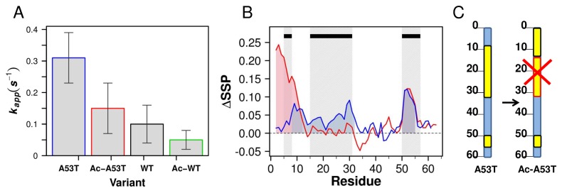
Aggregation of α-synuclein (αSyn), the primary protein component in Lewy body inclusions of patients with Parkinson's disease, arises when the normally soluble intrinsically disordered protein converts to amyloid fibrils. In this work, we provide a mechanistic view of the role of N-terminal acetylation on fibrillation by first establishing a quantitative relationship between monomer secondary structural propensity and fibril assembly kinetics, and secondly by demonstrating in the N-terminal acetylated form of the early onset A53T mutation, that N-terminal transient helices formed and/or inhibited by N-terminal acetylation modulate the fibril assembly rates. Using NMR chemical shifts and fluorescence experiments, we report that secondary structural propensity in residues 5-8, 14-31, and 50-57 are highly correlated to fibril growth rate. A four-way comparison of secondary structure propensity and fibril growth rates of N-terminally acetylated A53T and WT αSyn with non-acetylated A53T and WT αSyn present novel mechanistic insight into the role of N-terminal acetylation in amyloid fibril formation. We show that N-terminal acetylation inhibits the formation of the "fibrillation promoting" transient helix at residues 14-31 resulting from the A53T mutation in the non-acetylated variant and supports the formation of the "fibrillation inhibiting" transient helix in residues 1-12 thereby resulting in slower fibrillation rates relative to the previously studied non-acetylated A53T variant. Our results highlight the critical interplay of the region-specific transient secondary structure of the N-terminal region with fibrillation, and the inhibitory role of the N-terminal acetyl group in fibril formation.


Conflict of interest statement

Competing Interests: The authors have declared that no competing interests exist.

Figures

Figure 1
Figure 1. Quantitative correlation of SSP values to fibril kinetics in the non-acetylated human-mouse chimera αSyn set.
A. The N-, NAC, and C-terminal regions are shown in the schematic representation of αSyn. The black dots represent seven possible substitutions between human and mouse WT αSyn. B. Boxplot diagram representing the dispersion of SSP values for each human-mouse chimera as a function of residue, where each boxplot is a five-number summary of the SSP distribution of the eight variants. Residues are shaded based on their correlation with the growth rate (kapp), data from a previous study [39]. Residues with high positive correlation are shaded in red (r > 0.7), high negative correlation in blue (r < -0.7) and no correlation are shown as grey (-0.7 < r < 0.7). C. The representation is the same as in Figure 1B, however, the boxplots are shaded based on correlation to lag times. Residues with highest positive correlation are shaded in light red (r > 0.5), with highest negative correlation are shaded in light blue (r < -0.5) and no correlation with grey (-0.5 < r < 0.5). D. Correlation between the average SSP in regions that have a strong correlation with k app determined by Figure 1B (residues 5–8, 14–31, and 50–57) and k app(variant)/k app(HHH) for all eight variants, of which details of naming and BMRB accession numbers are indicated in Table 1. The correlation coefficient is r = 0.93. The correlation function is Y = (2.32±0.2)+ (0.57±0.9)* X.E. Correlation between the average SSP in regions that have strong correlation with lag times determined by Figure 1C (residues 51–55 and 84–87) and lag times (HHH)/lag times (variant) for all eight variants. The correlation coefficient is r = 0.75. The correlation function is Y = (2.46±0.66) + (0.53±0.19)* X.
Figure 2
Figure 2. NMR and fibril morphology comparison of Ac-WT and Ac-A53T.
A. Overlay of 15N-1H HSQC of Ac-A53T (black) and Ac-WT(magenta) at 15 °C in PBS buffer at pH 7.4. B. SSP analysis of Ac-WT (magenta) and Ac-A53T (black) plotted for the N-terminal region. The overlay of the rest of the proteins can be seen in Figure S2. C. Negatively stained electron micrographs of the end products of fibril formation of Ac-A53T fibril. The scale bar is 200 nm.
Figure 3
Figure 3. The effect of the A53T mutation upon the acetylated mutant.
A. Histogram plot of the apparent rate of fibril growth, k app of A53T (blue), Ac-A53T (red), WT (black) and Ac-WT (green) calculated by sigmoidal fitting of ThT fluorescence curves. Due to the different purification approach from the previously published one, the absolute k app values are different, however the ratio relative to WT is the same. B. Differences of SSP shown for the N terminal residues 1 to 60 for Ac-A53T vs. WT (red) and A53T (blue) vs. WT; ΔSSP = SSP (Variant) -SSP (WT). The red and blue shading in the positive regions of the SSP curves correspond to increased transient helix observed for Ac-A53T and A53T relative the WT. The black rectangles at the top of the plot represent the regions of increased transient helix that are quantitatively correlated with accelerated growth rates from the chimera set (Figure 1B). The overlay of the shaded regions and the black rectangles highlights the overlapping boundaries between these data sets. These data in combination with the fibril kinetics suggest that increased helicity in residues 1-12 is fibril inhibiting, while increased helicity in residues14-31, 50-57 are fibril accelerating, and increased transient helix at 5-8 may depend on the overall context. C. A schematic representation of the secondary structure propensity differences in regions in A53T and Ac-A53T, based on the ∆SSP of Figure 3B. The blue rectangle represents the N-terminal sequence, the black scale the residue number, and the yellow blocks the increase of SSP values of the variants relative to WT. The red outline and an X in the block represents the removal, or inability to sample increased SSP in this region by N-terminal acetylation highlighting that transient helix supported by the A53T mutation in the non-acetylated protein is not supported alongside acetylation.
Figure 4
Figure 4. Schematic diagram of a conformational selection and population shift mechanism for A53T and acetylated proteins.
The representation of the population distribution is based on a description by Ma et al. The populations of the various monomer species in the fibrillation process are represented by blue curves. Monomer conformations exist in a heterogeneous ensemble and are shown by schematic drawings with the red cylinder representing α-helix. The disordered monomers sample large heterogeneous ensembles that are differentially populated; both fibrillation prone and non-fibrillation prone monomers exist within the heterogeneous ensemble. Sequence modifications shift the population to favored conformations, which are boxed. The increase in transient helix propensity of the modified protein relative to the WT αSyn is presented. Secondary structure propensity correlations with fibril growth rates suggest that the fibrillation prone conformation consists of increased transient helix in residues 14–31 and 50–57 relative to WT, while transient helix in residues 1–12 arising from N-terminal acetylation is fibrillation inhibiting. By definition, the WT protein does not have fibrillation prone regions, and Ac-A53T which contains both aggregation promoting (residues 50–57) and fibrillation inhibiting conformations (residues 1–12) has similar fibrillation behavior to WT. Ac-WT, which contains only the fibrillation inhibiting conformation (residues 1–12) fibrillates the slowest.

References

    1. Lansbury PT Jr. (1999) Evolution of amyloid: what normal protein folding may tell us about fibrillogenesis and disease. Proc Natl Acad Sci U S A 96: 3342-3344. doi:10.1073/pnas.96.7.3342. PubMed: 10097040. - DOI - PMC - PubMed
    1. Perutz MF (1999) Glutamine repeats and neurodegenerative diseases: molecular aspects. Trends Biochem Sci 24: 58-63. doi:10.1016/S0968-0004(98)01350-4. PubMed: 10098399. - DOI - PubMed
    1. Dobson CM (1999) Protein misfolding, evolution and disease. Trends Biochem Sci 24: 329-332. doi:10.1016/S0968-0004(99)01445-0. PubMed: 10470028. - DOI - PubMed
    1. Chiti F, Dobson CM (2006) Protein misfolding, functional amyloid, and human disease. Annu Rev Biochem 75: 333-366. doi:10.1146/annurev.biochem.75.101304.123901. PubMed: 16756495. - DOI - PubMed
    1. Ross CA, Poirier MA (2004) Protein aggregation and neurodegenerative disease. Nat Med 10 Suppl: S10-S17. doi:10.1038/nm1066. PubMed: 15272267. - DOI - PubMed

Publication types

MeSH terms

LinkOut - more resources