. 2013;2(3):107-129.
doi: 10.3978/j.issn.2218-676X.2013.04.01.

DNA damage tolerance: a double-edged sword guarding the genome

Affiliations

DNA damage tolerance: a double-edged sword guarding the genome

Gargi Ghosal et al. Transl Cancer Res. 2013.

Abstract

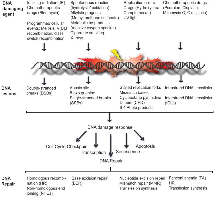
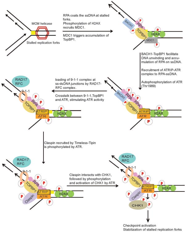
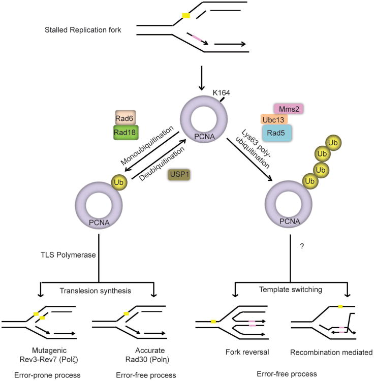
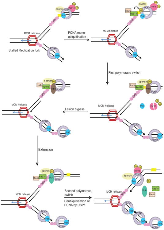
Preservation of genome integrity is an essential process for cell homeostasis. During the course of life of a single cell, the genome is constantly damaged by endogenous and exogenous agents. To ensure genome stability, cells use a global signaling network, namely the DNA damage response (DDR) to sense and repair DNA damage. DDR senses different types of DNA damage and coordinates a response that includes activation of transcription, cell cycle control, DNA repair pathways, apoptosis, senescence, and cell death. Despite several repair mechanisms that repair different types of DNA lesions, it is likely that the replication machinery would still encounter lesions that are mis-repaired or not repaired. Replication of damaged genome would result in high frequency of fork collapse and genome instability. In this scenario, the cells employ the DNA damage tolerance (DDT) pathway that recruits a specialized low fidelity translesion synthesis (TLS) polymerase to bypass the lesions for repair at a later time point. Thus, DDT is not a repair pathway per se, but provides a mechanism to tolerate DNA lesions during replication thereby increasing survival and preventing genome instability. Paradoxically, DDT process is also associated with increased mutagenesis, which can in turn drive the cell to cancer development. Thus, DDT process functions as a double-edged sword guarding the genome. In this review, we will discuss the replication stress induced DNA damage-signaling cascade, the stabilization and rescue of stalled replication forks by the DDT pathway and the effect of the DDT pathway on cancer.

Keywords: DNA damage tolerance (DDT); proliferating cell nuclear antigen (PCNA); replicative DNA polymerase; stalled replication forks; translesion polymerase; translesion synthesis (TLS).


Conflict of interest statement

Disclosure: The authors declare no conflict of interest.

Figures

Figure 1
Figure 1
DNA damage response network. Endogenous and environmental sources of DNA damaging agent infict damage to the DNA that range from modified bases, intra- and inter-strand crosslinks, cyclobutane pyrimidine dimers, 6-4 photoproducts, single- and double-stranded DNA breaks. Upon sensing DNA damage the cells activate the DDR network which activates cellular processes such as cell-cycle checkpoint control, transcription, DNA repair machinery, senescence and/or cell death. DNA repair pathways act independently or coordinate to repair DNA lesions
Figure 2
Figure 2
DNA damage signaling at replication forks. During DNA replication, lesions (yellow square) in the DNA template block the progression of DNA polymerases and result in the stalling and uncoupling of replicative polymerases with helicase activities of the replication machinery at the lesion site, generating tracts of single-strand DNA (ssDNA) due to continuous unwinding of DNA by helicases. ssDNA is subsequently bound by RPA. RPA-ssDNA then serves to signal the recruitment of ATR through its interacting partner, ATRIP, where it phosphorylates and activates Chk1. ATR-Chk1 activation requires intricate crosstalk between TopBP1, BACH1, 9-1-1 complex, Timeless, Tipin and Claspin mediator proteins at the replication forks. ATR-Chk1 pathway serves to activate cell cycle checkpoint and DNA repair machinery to repair the DNA lesion
Figure 3
Figure 3
DNA damage tolerance pathway (DDT): lesions (yellow Square) in the DNA template blocks progression of high-fidelity replicative polymerase resulting in stalled replication forks. DNA damage tolerance mechanism mediates bypass of lesions by replicating over damaged DNA by low-fidelity DNA polymerases (translesion synthesis) or using the undamaged sister chromatid as a template (template switching). Template switching is mediated by structural rearrangement of the replication fork either by recombination or fork reversal. The key regulator of DDT pathway is the modification of PCNA. Under undamaged conditions replicative polymerase binds to unmodified PCNA during DNA replication. Upon genotoxic stress, PCNA is ubiquitinated at K164 to initiate DNA damage tolerance pathways. Monoubiquitination of PCNA promotes translesion synthesis, while polyubiquitination facilitates template switching. PCNA is monoubiquitinated by RAD18-RAD6 E3-ligase and polyubiquitinated by Rad5 (human homologue, SHPRH or HLTF). Following lesion bypass Usp1 deubiquitinates PCNA, thereby facilitating loading of the replicative polymerase to resume DNA synthesis
Figure 4
Figure 4
Proposed model for TLS pathway. Replication fork stalling uncouples the replicative helicase from normal high-fidelity DNA polymerases resulting in DNA unwinding and generation of tracts of ssDNA, which is coated by RPA. RPA-ssDNA serves to initiate the ATR-Chk1 pathway to activate cell cycle checkpoint control. RPA-ssDNA also recruits RAD18 E3-ligase to activate DNA damage tolerance pathway. PCNA monoubiquitinated at K164 (ub-PCNA) by RAD18-RAD6 operates as a molecular switch from normal DNA replication to the TLS. Under normal conditions ubiquitinated PAF15 is bound to PCNA. Upon DNA damage PAF15 is degraded by the proteasome and this facilitates the binding of TLS polymerase to ub-PCNA. Additionally, Spartan is recruited to DNA damage sites by ub-PCNA and is required to stabilize RAD18 and ub-PCNA on the chromatin. TLS polymerase Polη (Pol eta) bound to ub-PCNA, inserts a nucleotide directly opposite the lesion and requires an additional TLS polymerase, such as Polζ (Pol zeta), to extend beyond the insertion. Following extention, the second polymerase switch is initiated where the TLS polymerase is replaced by high fidelity replicative DNA polymerase. USP1 deubiquitinates PCNA and DNA synthesis is resumed by high-fidelity replicative DNA polymerase. The precise mechanism of polymerase switching and regulation of TLS by Spartan, PAF15 and USP1 is still unclear
Figure 5
Figure 5
TLS synthesis in ICL repair by Fanconi anemia (FA) pathway: replicative DNA polymerase stalls upon encounter of an interstrand crosslink (ICL) in DNA. FA core complex (FANC-A, B, C, E, F, G, L and M) and associated proteins (FAAP20, FAAP24, FAAP100) are activated and recruited to the ICL site where, FANCL serves as E3-ligase, and monoubiquitinates FANCI-D2 heterodimer. Monoubiquitinated FANCI-D2 complex is recruited to the chromatin and recruits FAN1 nuclease to sites of damage. ICLs are incised and unhooked by the action of several structure specific endonucleases; MUS81-EME1, SNM1A, FAN1, XPF-ERCC1 and SLX1-SLX4. Complete repair of ICL is mediated by co-ordinated action of TLS and HR processes. FAAP20 interacts with the FA core complex and binds to monoubiquitinated REV1. FAAP20 may direct REV1 and Pol ζ to the unhooked crosslink to catalyze lesion bypass across the ICL adduct creating a suitable substrate for repair by the HR pathway

References

    1. Kasparek TR, Humphrey TC. DNA double-strand break repair pathways, chromosomal rearrangements and cancer. Semin Cell Dev Biol. 2011;22:886–97. - PubMed
    1. Bartek J, Bartkova J, Lukas J. DNA damage signalling guards against activated oncogenes and tumour progression. Oncogene. 2007;26:7773–9. - PubMed
    1. Moraes MC, Neto JB, Menck CF. DNA repair mechanisms protect our genome from carcinogenesis. Front Biosci. 2012;17:1362–88. - PubMed
    1. Hoeijmakers JH. DNA damage, aging, and cancer. N Engl J Med. 2009;361:1475–85. - PubMed
    1. Lindahl T. Instability and decay of the primary structure of DNA. Nature. 1993;362:709–15. - PubMed