. 2013 Dec 15;456(3):409-15.
doi: 10.1042/BJ20130860.

Positive and negative phosphorylation regulates RIP1- and RIP3-induced programmed necrosis

Affiliations

Positive and negative phosphorylation regulates RIP1- and RIP3-induced programmed necrosis

Thomas McQuade et al. Biochem J. .

Abstract

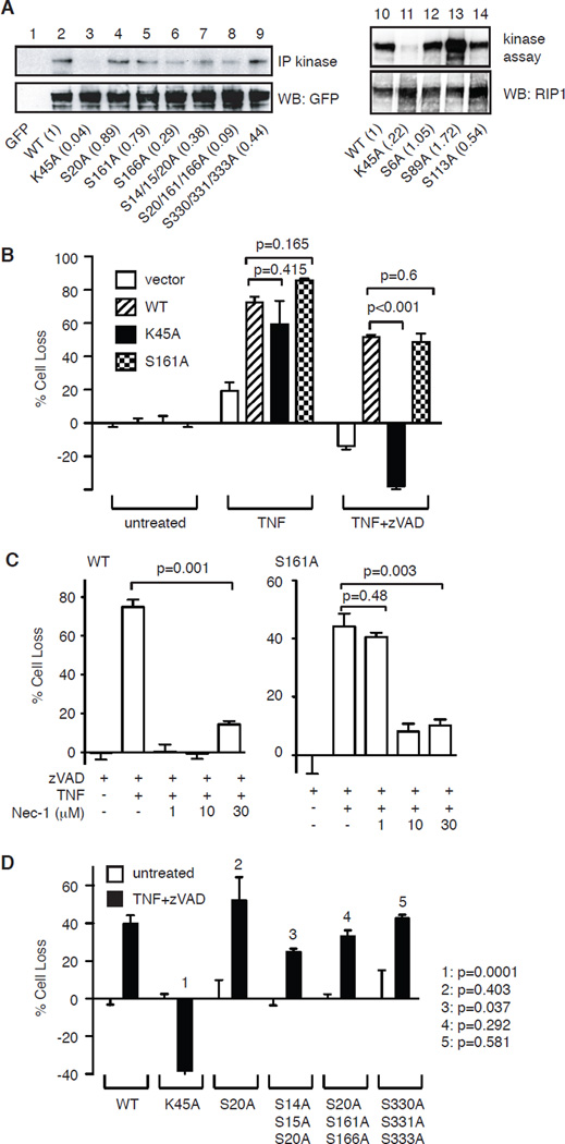
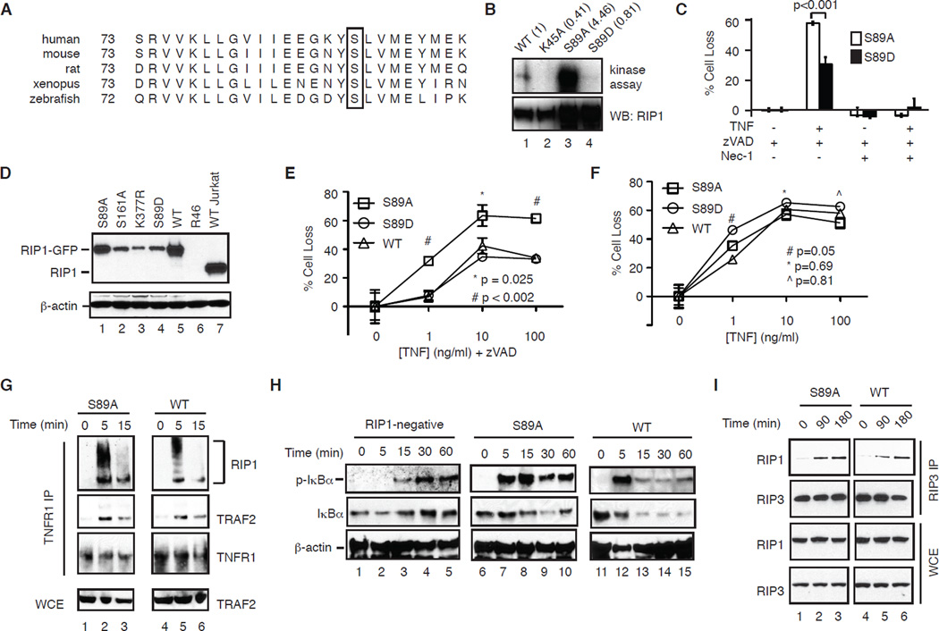
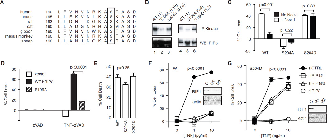
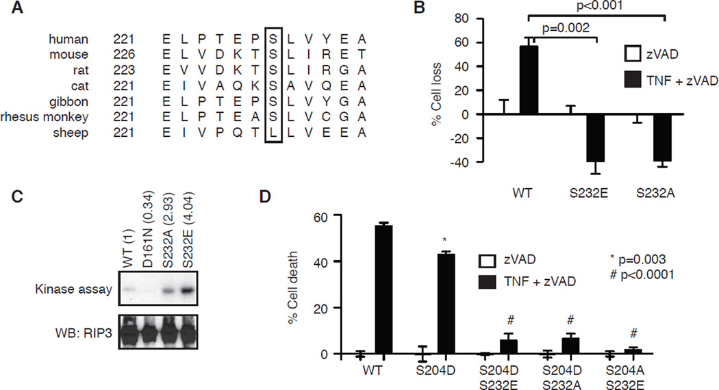
Programmed necrosis or necroptosis is controlled by the action of two serine/threonine kinases, RIP1 (receptor-interacting serine/threonine protein kinase 1; also known as RIPK1) and RIP3. The phosphorylation of RIP1 and RIP3 is critical for assembly of the necrosome, an amyloid-like complex that initiates transmission of the pro-necrotic signal. In the present study, we used site-directed mutagenesis to systematically examine the effects of putative phosphoacceptor sites on RIP1 and RIP3 on TNF (tumour necrosis factor)-induced programmed necrosis. We found that mutation of individual serine residues in the kinase domain of RIP1 had little effect on RIP1 kinase activity and TNF-induced programmed necrosis. Surprisingly, an alanine residue substitution for Ser(89) enhanced RIP1 kinase activity and TNF-induced programmed necrosis without affecting RIP1-RIP3 necrosome formation. This indicates that Ser(89) is an inhibitory phosphoacceptor site that can dampen the pro-necrotic function of RIP1. In addition, we show that a phosphomimetic mutant of RIP3, S204D, led to programmed necrosis that was refractory to RIP1 siRNA and insensitive to necrostatin-1 inhibition. Our results show that programmed necrosis is regulated by positive and inhibitory phosphorylation events.


Figures

Figure 1
Figure 1. The effect of serine phosphorylation on RIP1 kinase activity
(A) Effect of alanine substitutions of reported phosphorylation sites on RIP1 kinase activity. HEK293T cells were transfected with the indicated GFP-tagged RIP1 plasmids. The in vitro kinase activity was determined using RIP1 autophosphorylation as readout. The numbers in parentheses represent kinase activity normalized to expression level of RIP1-GFP on bottom panel. (B) Ser161 does not control the pro-necrotic function of RIP1. RIP1-deficient Jurkat cells were transfected with the indicated RIP1-GFP plasmids or empty GFP vector. Transfected GFP+ cells were tested for TNF-induced apoptosis or TNF and zVAD-induced programmed necrosis. (C) Ser161 controls sensitivity to Nec-1 inhibition. RIP1-deficient Jurkat cells stably expressing wild type RIP1-GFP or S161A-RIP1-GFP were treated with TNF, zVAD, and the indicated doses of Nec-1. (D) RIP1-deficient Jurkat cells were transfected with the indicated RIP1-GFP plasmids. Transfected GFP+ cells were tested for TNF and zVAD-induced programmed necrosis. p values were calculated by comparing WT transfectants with the indicated mutants using Student’s t test.
Figure 2
Figure 2. S89A-RIP1 promotes necrosome assembly and programmed necrosis
(A) Sequence alignment showing conservation of Ser89 in RIP1 from different species. (B) The indicated RIP1-GFP mutants were expressed and purified from HEK293T cells and subjected to in vitro kinase. (C) RIP1-deficient Jurkat cells were transiently transfected with the indicated RIP1-GFP plasmids and tested for TNF and zVAD-induced programmed necrosis. (D) Expression of RIP1-GFP in different clones of RIP1-deficient Jurkat cells. (E) S89A-RIP1 cells exhibited higher level of TNF and zVAD-induced programmed necrosis. (F) TNF-induced apoptosis in Jurkat cells expressing S89A, S89D or WT RIP1. (G) Similar assembly of TNFR-1 signaling complex in WT and S89A-RIP1 Jurkat cells. (H) TNF-induced phosphorylation of IκBα in parental RIP1-deficient Jurkat and clones expressing different RIP1. (I) Assembly of the RIP1-RIP3 necrosome is not affected in the S89A-RIP1 Jurkat cells.
Figure 3
Figure 3. Ser204 critically regulates RIP3 function
(A) Sequence alignment of RIP3 reveals conservation of Ser204 (in mouse RIP3) among different species (box). (B) Effects of alanine and aspartic acid substitution of Ser204 on mRIP3 and Ser199 on hRIP3 kinase activity. The indicated RIP3-GFP fusion proteins were expressed in 293T cells were subjected to in vitro kinase assay. The numbers in parentheses represent the normalized kinase activity compared to wild type RIP3. (C) RIP3−/− fibroblasts stably expressing the indicated RIP3-GFP fusion proteins were treated with TNF and zVAD to induce programmed necrosis, with or without 1 µM Nec-1. (D) The indicated RIP3-GFP plasmids were transfected into RIP3−/− fibroblasts and tested for TNF and zVAD-induced programmed necrosis. (E) RIP3−/− fibroblasts stably transfected with the indicated RIP3-GFP plasmids were tested for TNF and cycloheximide (CHX) induced apoptosis. One-way ANOVA analysis was performed to compare the three different groups. (F-G) RIP3−/− fibroblasts stably expressing the indicated WT or S204D RIP3-GFP fusion protein were transfected with the indicated siRNAs. TNF and zVAD-induced programmed necrosis was determined. Western blots show the level of silencing by the RIP siRNAs. Two-way ANOVA analysis was performed.
Figure 4
Figure 4. Ser232 does not regulate RIP3 function in programmed necrosis
(A) Sequence alignment of RIP3 reveals that Ser232 (in mouse RIP3) is not conserved in sheep RIP3 (box). (B) RIP3−/− fibroblasts were transiently transfected with the indicated GFP-tagged RIP3. Programmed necrosis was induced by TNF and zVAD. Cell death was determined in the GFP+ population. (C) The indicated RIP3-GFP fusion proteins were expressed in 293T cells and subjected to in vitro kinase assay. Autophosphorylation of RIP3 was revealed. (D) RIP3−/− fibroblasts stably expressing the indicated RIP3-GFP. Programmed necrosis was induced by TNF and zVAD. p values represent comparison between WT and mutant clones using Student’s t test.

References

    1. Moriwaki K, Chan FK. RIP3: a molecular switch for necrosis and inflammation. Genes Dev. 2013;27:1640–1649. - PMC - PubMed
    1. Li J, McQuade T, Siemer AB, Napetschnig J, Moriwaki K, Hsiao YS, Damko E, Moquin D, Walz T, McDermott A, Chan FK, Wu H. The RIP1/RIP3 necrosome forms a functional amyloid signaling complex required for programmed necrosis. Cell. 2012;150:339–350. - PMC - PubMed
    1. Sun L, Wang H, Wang Z, He S, Chen S, Liao D, Wang L, Yan J, Liu W, Lei X, Wang X. Mixed lineage kinase domain-like protein mediates necrosis signaling downstream of RIP3 kinase. Cell. 2012;148:213–227. - PubMed
    1. Zhao J, Jitkaew S, Cai Z, Choksi S, Li Q, Luo J, Liu ZG. Mixed lineage kinase domain-like is a key receptor interacting protein 3 downstream component of TNF-induced necrosis. Proc Natl Acad Sci U S A. 2012;109:5322–5327. - PMC - PubMed
    1. Wang Z, Jiang H, Chen S, Du F, Wang X. The mitochondrial phosphatase PGAM5 functions at the convergence point of multiple necrotic death pathways. Cell. 2012;148:228–243. - PubMed

Publication types

MeSH terms