

The role of hepatocyte nuclear factor 4alpha in metastatic tumor formation of hepatocellular carcinoma and its close relationship with the mesenchymal-epithelial transition markers
- PMID: 24059685
- PMCID: PMC3852538
- DOI: 10.1186/1471-2407-13-432
The role of hepatocyte nuclear factor 4alpha in metastatic tumor formation of hepatocellular carcinoma and its close relationship with the mesenchymal-epithelial transition markers
Abstract
Background: Mesenchymal-epithelial transition (MET) is now suggested to participate in the process of metastatic tumor formation. However, in hepatocellular carcinoma (HCC) the process is still not well revealed.
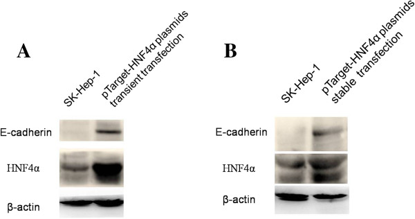
Methods: Paraffin-embedded tissue samples were obtained from 13 patients with HCC in Shengjing Hospital of China Medical University. The expression of E-cadherin, Fibronectin, N-cadherin, Vimentin, Hepatocyte nuclear factor 4alpha (HNF4alpha), Snail and Slug was assessed in primary tumors and their corresponding metastases by immunohistochemical staining. Next, the expression of HNF4alpha and E-cadherin in four HCC cell lines was examined. Furthermore, SK-Hep-1 cells were transfected with human HNF4alpha expression vector, and the change of E-cadherin expression was assessed.
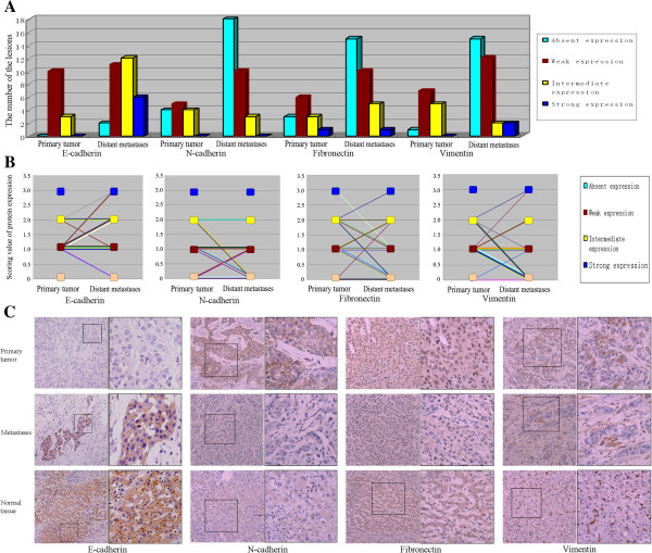
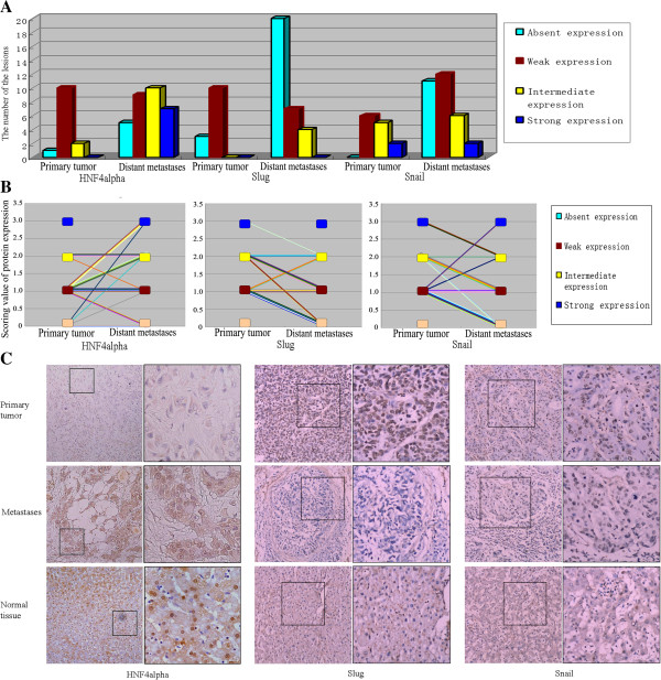
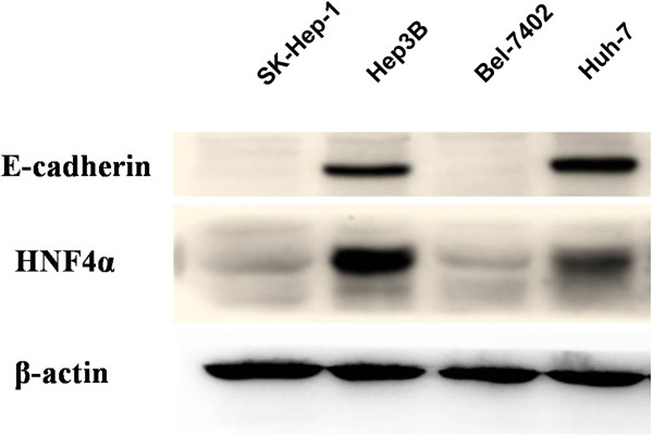
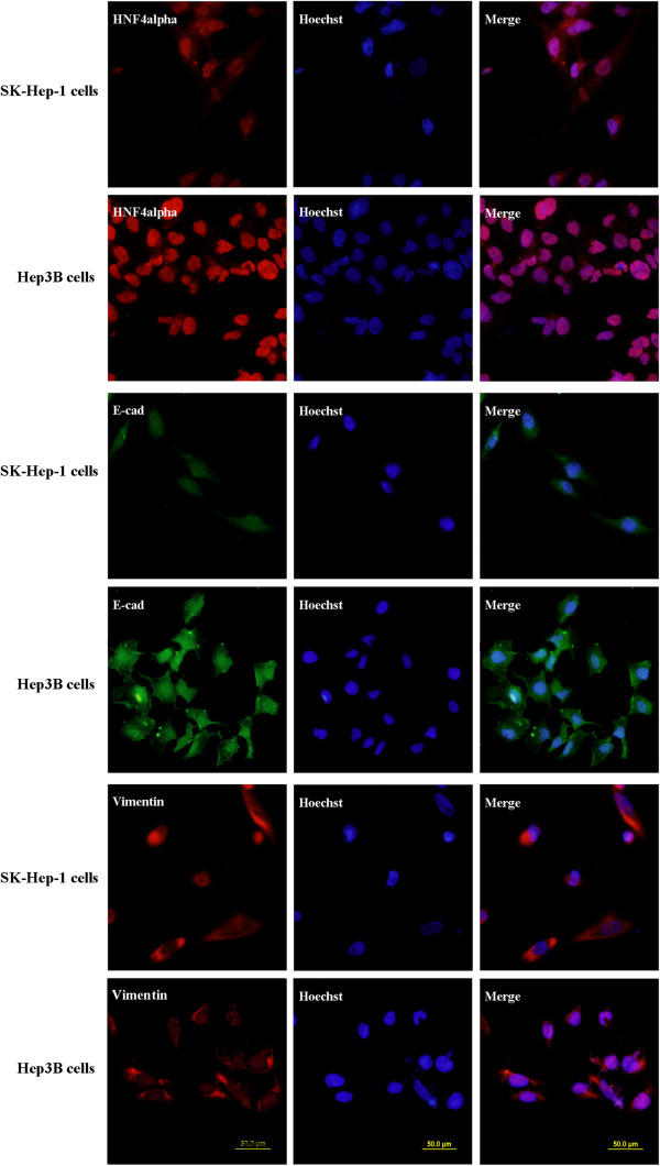
Results: 45.2% (14/31) of the lesions in the metastases showed increased E-cadherin expression compared with the primaries, suggesting the possible occurrence of MET in metastatic tumor formation of HCC, as re-expression of E-cadherin is proposed to be the important hallmark of MET. The occurrence of MET was also confirmed by the reduced expression of Fibronectin (54.8%, 17/31), N-cadherin (38.7%, 12/31) and Vimentin (61.3%, 19/31) in the metastases. 45.2% (14/31) of the lesions in the metastases also showed increased HNF4alpha expression, and 67.7% (21/31) and 48.4% (15/31) of metastases showed decreased Snail and Slug expression respectively. Statistical results showed that the expression of HNF4alpha was positively related with that of E-cadherin, and negatively correlated with that of Snail, Slug and Fibronectin, suggesting that the expression change of the MET markers in the metastatic lesions might be associated with HNF4alpha. Among the four HCC cell lines, both HNF4alpha and E-cadherin expressed high in Hep3B and Huh-7 cells, but low in SK-Hep-1 and Bel-7402 cells. Furthermore, the expression of E-cadherin increased accordingly when SK-Hep-1 cells were transfected with human HNF4alpha expression vector, further confirming the role of HNF4alpha in the regulation of E-cadherin expression.
Conclusions: Our clinical observations and experimental data indicate that HNF4alpha might play a crucial role in the metastatic tumor formation of HCC, and the mechanism may be related with the process of phenotype transition.
Figures
Similar articles
-
PRRX1 deficiency induces mesenchymal-epithelial transition through PITX2/miR-200-dependent SLUG/CTNNB1 regulation in hepatocellular carcinoma.Cancer Sci. 2021 Jun;112(6):2158-2172. doi: 10.1111/cas.14853. Epub 2021 Apr 8. Cancer Sci. 2021. PMID: 33587761 Free PMC article.
-
FoxM1 overexpression promotes epithelial-mesenchymal transition and metastasis of hepatocellular carcinoma.World J Gastroenterol. 2015 Jan 7;21(1):196-213. doi: 10.3748/wjg.v21.i1.196. World J Gastroenterol. 2015. PMID: 25574092 Free PMC article.
-
MicroRNA-122 triggers mesenchymal-epithelial transition and suppresses hepatocellular carcinoma cell motility and invasion by targeting RhoA.PLoS One. 2014 Jul 3;9(7):e101330. doi: 10.1371/journal.pone.0101330. eCollection 2014. PLoS One. 2014. PMID: 24992599 Free PMC article.
-
Role of Hepatocyte Nuclear Factor 4 Alpha in Liver Cancer.Semin Liver Dis. 2024 Aug;44(3):383-393. doi: 10.1055/a-2349-7236. Epub 2024 Jun 20. Semin Liver Dis. 2024. PMID: 38901435 Review.
-
Deregulation of hepatocyte nuclear factor 4 (HNF4)as a marker of epithelial tumors progression.Exp Oncol. 2010 Sep;32(3):167-71. Exp Oncol. 2010. PMID: 21403612 Review.
Cited by
-
Liver resection versus radiofrequency ablation in octogenarian patients for hepatocellular carcinoma: a propensity score multicenter analysis.Surg Endosc. 2023 Apr;37(4):3029-3036. doi: 10.1007/s00464-022-09826-2. Epub 2022 Dec 19. Surg Endosc. 2023. PMID: 36534162
-
Single-cell multi-modal chromatin profiles revealing epigenetic regulations of cells in hepatocellular carcinoma.Clin Transl Med. 2024 Sep;14(9):e70000. doi: 10.1002/ctm2.70000. Clin Transl Med. 2024. PMID: 39210544 Free PMC article.
-
Evaluating markers of epithelial-mesenchymal transition to identify cancer patients at risk for metastatic disease.Clin Exp Metastasis. 2016 Jan;33(1):53-62. doi: 10.1007/s10585-015-9757-7. Epub 2015 Oct 27. Clin Exp Metastasis. 2016. PMID: 26507436 Free PMC article.
-
A multidimensional integration analysis reveals potential bridging targets in the process of colorectal cancer liver metastasis.PLoS One. 2017 Jun 19;12(6):e0178760. doi: 10.1371/journal.pone.0178760. eCollection 2017. PLoS One. 2017. PMID: 28628609 Free PMC article.
-
Hepatocyte nuclear factor 4α and cancer-related cell signaling pathways: a promising insight into cancer treatment.Exp Mol Med. 2021 Jan;53(1):8-18. doi: 10.1038/s12276-020-00551-1. Epub 2021 Jan 18. Exp Mol Med. 2021. PMID: 33462379 Free PMC article. Review.
References
-
- Herszényi L, Tulassay Z. Epidemiology of gastrointestinal and liver tumors. Eur Rev Med Pharmacol Sci. 2010;14:249–258. - PubMed
MeSH terms
Substances
LinkOut - more resources
Full Text Sources
Other Literature Sources
Medical
Research Materials
Miscellaneous
