

Tumor cell response to bevacizumab single agent therapy in vitro
- PMID: 24059699
- PMCID: PMC3849065
- DOI: 10.1186/1475-2867-13-94
Tumor cell response to bevacizumab single agent therapy in vitro
Abstract
Background: Angiogenesis represents a highly multi-factorial and multi-cellular complex (patho-) physiologic event involving endothelial cells, tumor cells in malignant conditions, as well as bone marrow derived cells and stromal cells. One main driver is vascular endothelial growth factor (VEGFA), which is known to interact with endothelial cells as a survival and mitogenic signal. The role of VEGFA on tumor cells and /or tumor stromal cell interaction is less clear. Condition specific (e.g. hypoxia) or tumor specific expression of VEGFA, VEGF receptors and co-receptors on tumor cells has been reported, in addition to the expression on the endothelium. This suggests a potential paracrine/autocrine loop that could affect changes specific to tumor cells.
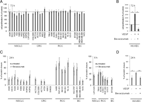
Methods: We used the monoclonal antibody against VEGFA, bevacizumab, in various in vitro experiments using cell lines derived from different tumor entities (non small cell lung cancer (NSCLC), colorectal cancer (CRC), breast cancer (BC) and renal cell carcinoma (RCC)) in order to determine if potential VEGFA signaling could be blocked in tumor cells. The experiments were done under hypoxia, a major inducer of VEGFA and angiogenesis, in an attempt to mimic the physiological tumor condition. Known VEGFA induced endothelial biological responses such as proliferation, migration, survival and gene expression changes were evaluated.
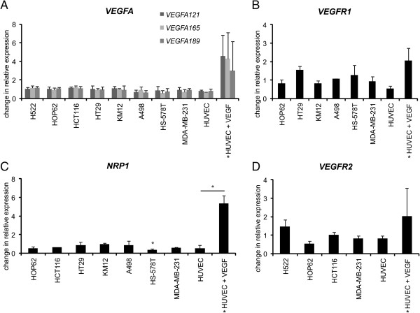
Results: Our study was able to demonstrate expression of VEGF receptors on tumor cells as well as hypoxia regulated angiogenic gene expression. In addition, there was a cell line specific effect in tumor cells by VEGFA blockade with bevacizumab in terms of proliferation; however overall, there was a limited measurable consequence of bevacizumab therapy detected by migration and survival.
Conclusion: The present study showed in a variety of in vitro experiments with several tumor cell lines from different tumor origins, that by blocking VEGFA with bevacizumab, there was a limited autocrine or cell-autonomous function of VEGFA signaling in tumor cells, when evaluating VEGFA induced downstream outputs known in endothelial cells.
Figures


Similar articles
-
FOXF1 promotes angiogenesis and accelerates bevacizumab resistance in colorectal cancer by transcriptionally activating VEGFA.Cancer Lett. 2018 Dec 28;439:78-90. doi: 10.1016/j.canlet.2018.09.026. Epub 2018 Sep 22. Cancer Lett. 2018. PMID: 30253191
-
Vascular Endothelial Growth Factor A Regulates the Secretion of Different Angiogenic Factors in Lung Cancer Cells.J Cell Physiol. 2016 Jul;231(7):1514-21. doi: 10.1002/jcp.25243. Epub 2015 Nov 24. J Cell Physiol. 2016. PMID: 26542886
-
Targeting tumor cell-derived CCL2 as a strategy to overcome Bevacizumab resistance in ETV5+ colorectal cancer.Cell Death Dis. 2020 Oct 24;11(10):916. doi: 10.1038/s41419-020-03111-7. Cell Death Dis. 2020. PMID: 33099574 Free PMC article.
-
Vascular endothelial growth factor (VEGF) as a target of bevacizumab in cancer: from the biology to the clinic.Curr Med Chem. 2006;13(16):1845-57. doi: 10.2174/092986706777585059. Curr Med Chem. 2006. PMID: 16842197 Review.
-
Monoclonal antibodies targeting vascular endothelial growth factor: current status and future challenges in cancer therapy.BioDrugs. 2009;23(5):289-304. doi: 10.2165/11317600-000000000-00000. BioDrugs. 2009. PMID: 19754219 Review.
Cited by
-
Immunomodulation on tumor immune microenvironment in acquired targeted therapy resistance and implication for immunotherapy resistance.Transl Oncol. 2025 Apr;54:102353. doi: 10.1016/j.tranon.2025.102353. Epub 2025 Mar 8. Transl Oncol. 2025. PMID: 40058234 Free PMC article. Review.
-
Vascularized tumor on a microfluidic chip to study mechanisms promoting tumor neovascularization and vascular targeted therapies.Theranostics. 2025 Jan 1;15(3):766-783. doi: 10.7150/thno.95334. eCollection 2025. Theranostics. 2025. PMID: 39776800 Free PMC article.
-
Combined effect of propranolol, vincristine and bevacizumab on HUVECs and BJ cells.Exp Ther Med. 2019 Jan;17(1):307-315. doi: 10.3892/etm.2018.6925. Epub 2018 Nov 2. Exp Ther Med. 2019. PMID: 30651796 Free PMC article.
-
In vitro vascularized immunocompetent patient-derived model to test cancer therapies.iScience. 2023 Sep 29;26(10):108094. doi: 10.1016/j.isci.2023.108094. eCollection 2023 Oct 20. iScience. 2023. PMID: 37860774 Free PMC article.
-
Candidate SNP Markers of Gender-Biased Autoimmune Complications of Monogenic Diseases Are Predicted by a Significant Change in the Affinity of TATA-Binding Protein for Human Gene Promoters.Front Immunol. 2016 Apr 4;7:130. doi: 10.3389/fimmu.2016.00130. eCollection 2016. Front Immunol. 2016. PMID: 27092142 Free PMC article.
References
LinkOut - more resources
Full Text Sources
Other Literature Sources
