

Diesel exhaust particle induction of IL-17A contributes to severe asthma
- PMID: 24060272
- PMCID: PMC3845500
- DOI: 10.1016/j.jaci.2013.06.048
Diesel exhaust particle induction of IL-17A contributes to severe asthma
Abstract
Background: IL-17A has been implicated in severe forms of asthma. However, the factors that promote IL-17A production during the pathogenesis of severe asthma remain undefined. Diesel exhaust particles (DEPs) are a major component of traffic-related air pollution and are implicated in asthma pathogenesis and exacerbation.
Objective: We sought to determine the mechanism by which DEP exposure affects asthma severity using human and mouse studies.
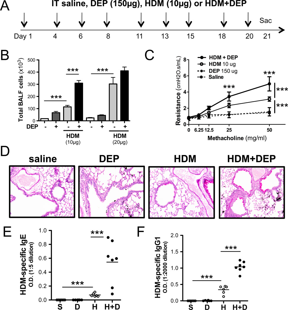
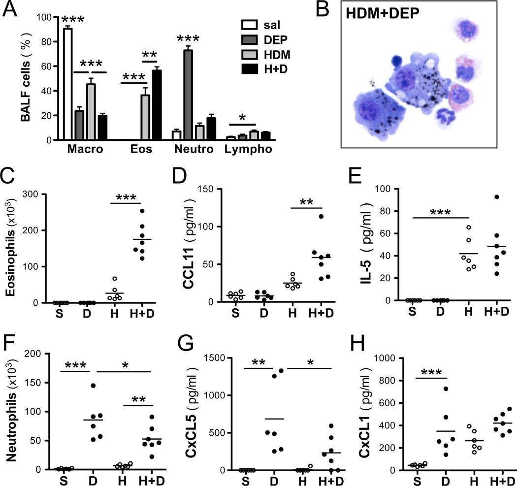
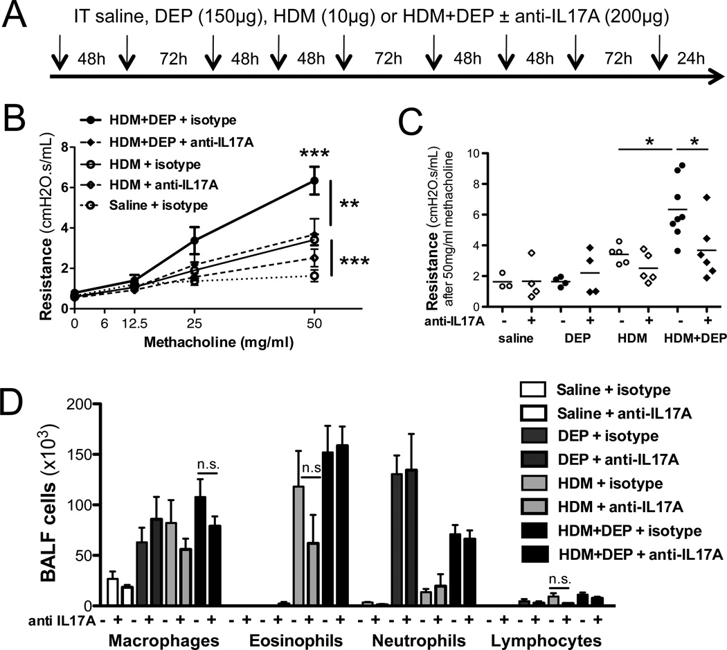
Methods: BALB/c mice were challenged with DEPs with or without house dust mite (HDM) extract. Airway inflammation and function, bronchoalveolar lavage fluid cytokine levels, and flow cytometry of lung T cells were assessed. The effect of DEP exposure on the frequency of asthma symptoms and serum cytokine levels was determined in children with allergic asthma.
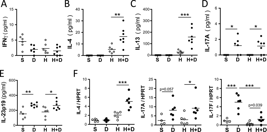
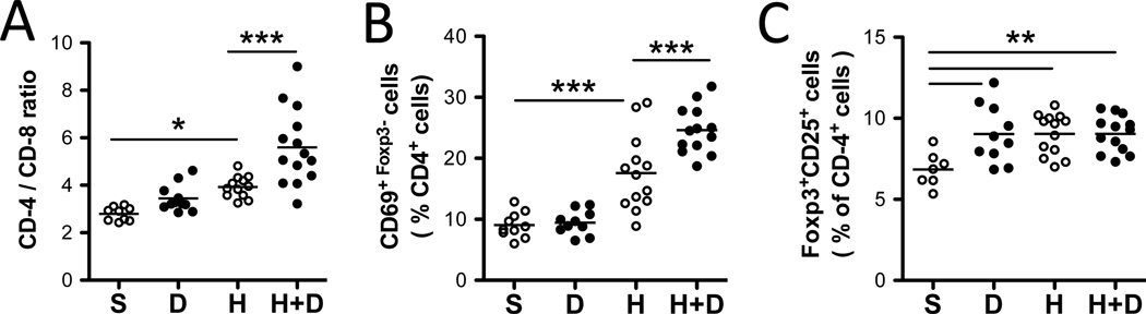
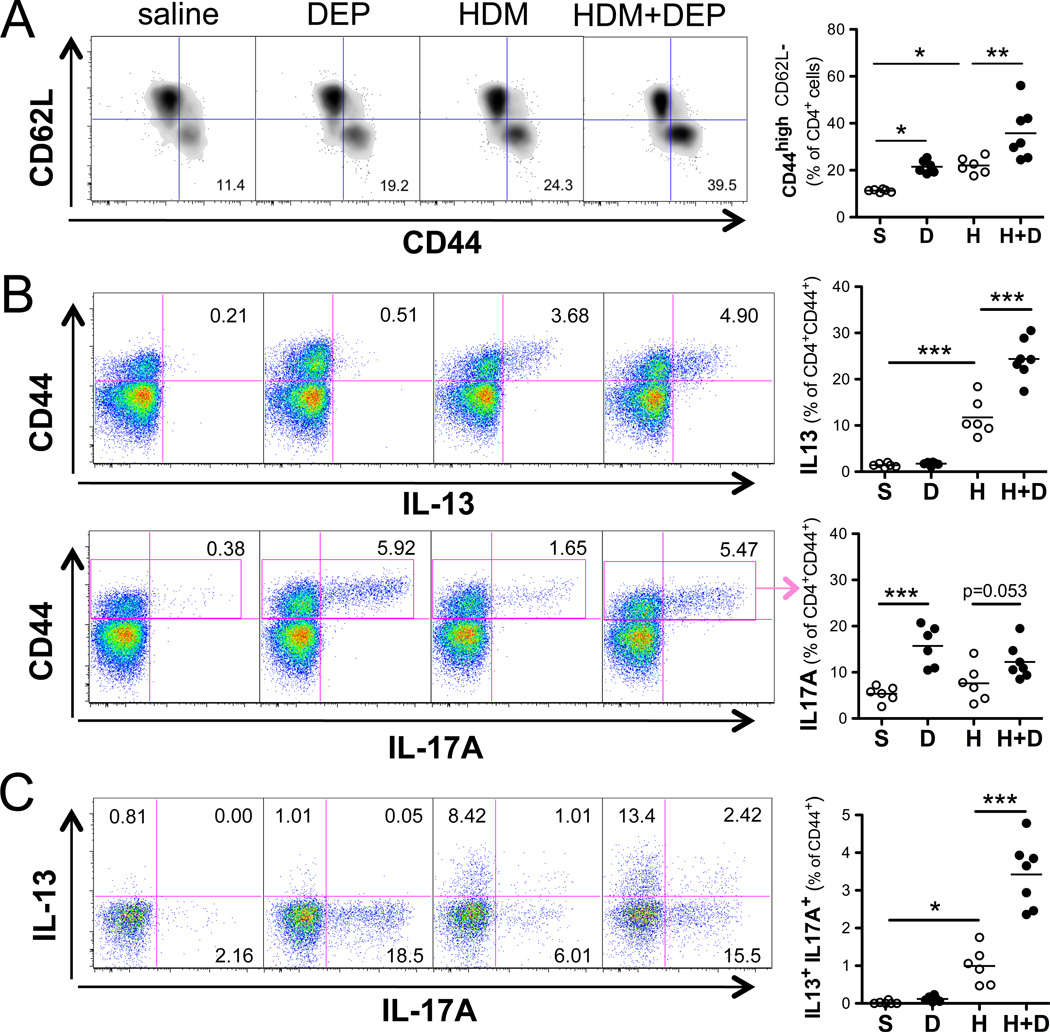
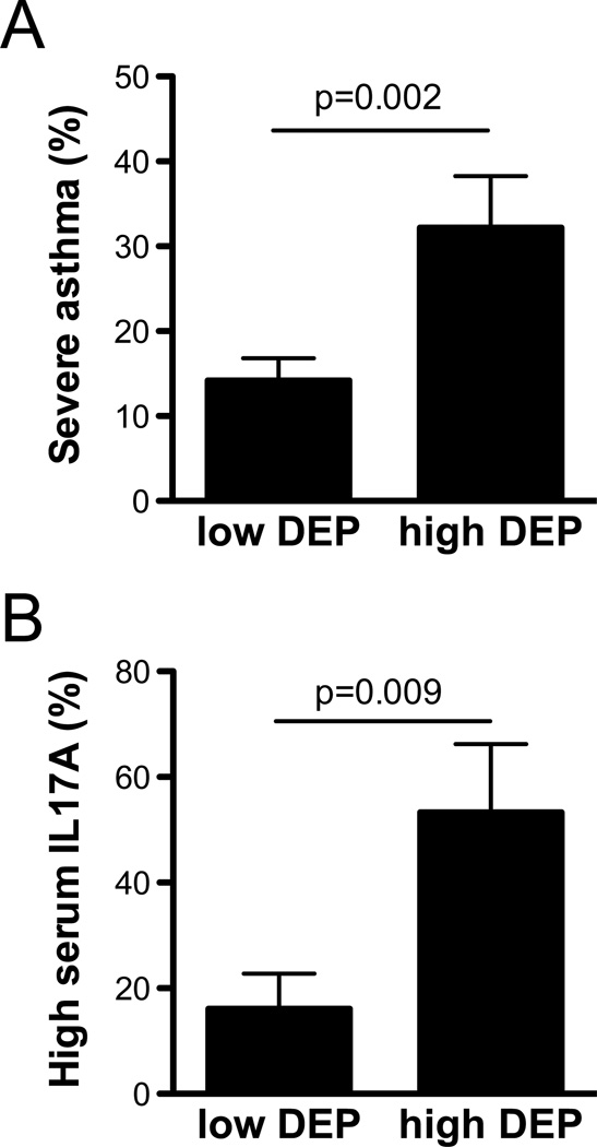
Results: In mice exposure to DEPs alone did not induce asthma. DEP and HDM coexposure markedly enhanced airway hyperresponsiveness compared with HDM exposure alone and generated a mixed T(H)2 and T(H)17 response, including IL-13(+)IL-17A(+) double-producing T cells. IL-17A neutralization prevented DEP-induced exacerbation of airway hyperresponsiveness. Among 235 high DEP-exposed children with allergic asthma, 32.2% had more frequent asthma symptoms over a 12-month period compared with only 14.2% in the low DEP-exposed group (P = .002). Additionally, high DEP-exposed children with allergic asthma had nearly 6 times higher serum IL-17A levels compared with low DEP-exposed children.
Conclusions: Expansion of T(H)17 cells contributes to DEP-mediated exacerbation of allergic asthma. Neutralization of IL-17A might be a useful potential therapeutic strategy to counteract the asthma-promoting effects of traffic-related air pollution, especially in highly exposed patients with severe allergic asthma.
Keywords: AHR; Airway hyperresponsiveness; Allergic asthma; BALF; Bronchoalveolar lavage fluid; DEP; Diesel exhaust particle; Forkhead box protein 3; Foxp3; GCPCR; Greater Cincinnati Pediatric Clinic Repository; HDM; House dust mite; IL-13 receptor; IL-13R; IL-17A; OR; Odds ratio; PE; PEES; Pediatric Environmental Exposures Study; Phycoerythrin; Regulatory T; SPT; Skin prick test; Treg; diesel exhaust particle; house dust mite; regulatory T cell.
Copyright © 2013 American Academy of Allergy, Asthma & Immunology. Published by Mosby, Inc. All rights reserved.
Conflict of interest statement
Conflict of interest: The authors have declared that there are no conflicts of interest. AL Budelsky is a paid employee and stockholder in Amgen, Inc.
Figures
Comment in
-
Reply: To PMID 24060272.J Allergy Clin Immunol. 2014 May;133(5):1496-7. doi: 10.1016/j.jaci.2013.12.1097. Epub 2014 Mar 15. J Allergy Clin Immunol. 2014. PMID: 24636093 No abstract available.
-
DEP-induced T(H)17 response in asthmatic subjects.J Allergy Clin Immunol. 2014 May;133(5):1495-6, 1496.e1. doi: 10.1016/j.jaci.2013.12.1095. Epub 2014 Mar 15. J Allergy Clin Immunol. 2014. PMID: 24636096 No abstract available.
References
-
- Simpson JL, Scott R, Boyle MJ, Gibson PG. Inflammatory subtypes in asthma: assessment and identification using induced sputum. Respirology. 2006;11:54–61. - PubMed
-
- Al-Ramli W, Prefontaine D, Chouiali F, Martin JG, Olivenstein R, Lemiere C, Hamid Q. T(H)17-associated cytokines (IL-17A and IL-17F) in severe asthma. J Allergy Clin Immunol. 2009;123:1185–1187. - PubMed
-
- Linden A, Laan M, Anderson GP. Neutrophils, interleukin-17A and lung disease. Eur Respir J. 2005;25:159–172. - PubMed
Publication types
MeSH terms
Substances
Grants and funding
LinkOut - more resources
Full Text Sources
Other Literature Sources
Medical
