Comparative Study
. 2013 Oct 22;52(42):7512-21.
doi: 10.1021/bi401176d. Epub 2013 Oct 9.

Divergent evolution of ligand binding in the o-succinylbenzoate synthase family

Affiliations
Comparative Study

Divergent evolution of ligand binding in the o-succinylbenzoate synthase family

Denis Odokonyero et al. Biochemistry. .

Abstract

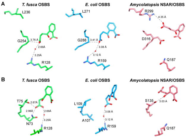
Thermobifida fusca o-succinylbenzoate synthase (OSBS), a member of the enolase superfamily that catalyzes a step in menaquinone biosynthesis, has an amino acid sequence that is 22 and 28% identical with those of two previously characterized OSBS enzymes from Escherichia coli and Amycolatopsis sp. T-1-60, respectively. These values are considerably lower than typical levels of sequence identity among homologous proteins that have the same function. To determine how such divergent enzymes catalyze the same reaction, we determined the structure of T. fusca OSBS and identified amino acids that are important for ligand binding. We discovered significant differences in structure and conformational flexibility between T. fusca OSBS and other members of the enolase superfamily. In particular, the 20s loop, a flexible loop in the active site that permits ligand binding and release in most enolase superfamily proteins, has a four-amino acid deletion and is well-ordered in T. fusca OSBS. Instead, the flexibility of a different region allows the substrate to enter from the other side of the active site. T. fusca OSBS was more tolerant of mutations at residues that were critical for activity in E. coli OSBS. Also, replacing active site amino acids found in one protein with the amino acids that occur at the same place in the other protein reduces the catalytic efficiency. Thus, the extraordinary divergence between these proteins does not appear to reflect a higher tolerance of mutations. Instead, large deletions outside the active site were accompanied by alteration of active site size and electrostatic interactions, resulting in small but significant differences in ligand binding.


Figures

Figure 1
Figure 1
The OSBS family. A) The o-succinylbenzoate synthase (OSBS) and N-succinylamino acid racemase (NSAR) reactions. The enolate anion intermediate that occurs in all reactions in the enolase superfamily is red; blue atoms are lost or rearranged in the reactions. R = hydrophobic amino acid side chain. B) Phylogenetic tree showing the division of the OSBS family into eight major subfamilies. The structure of T. fusca OSBS (PDB: 2OPJ and 2 QVH) from the Actinobacteria subfamily is reported in this work. It is compared to E. coli OSBS (PDB: 1FHV, 1FHU, and 1R6W) from the γ-Proteobacteria 1 subfamily and Amycolatopsis NSAR/OSBS (PDB: 1SJB) from the NSAR/OSBS subfamily.
Figure 2
Figure 2
Comparison of OSBS enzymes from Thermobifida fusca (2QVH), Escherichia coli (1FHV), and Amycolatopsis sp. T-1-60 (1SJB)., A) Insertions and deletions. Regions in red are deleted in one of the other enzymes. Regions in blue are insertions in that enzyme. Regions in orange have insertions and deletions in all three enzymes and cannot be aligned to each other. OSB is shown in black, and the magnesium ion is shown in lime green. The 20s loops and 50s loops are marked with a star and a circle, respectively. The three α-helices of the capping domain are labeled. B) Active site accessibility. Space-filling models have been rotated 90° toward the right (top) or left (bottom) relative to panel A. The 20s loop is cyan, the 50s loop is pink, the rest of the capping domain is light gray, and the barrel domain is green. Another subunit of the Amycolatopsis NSAR/OSBS octamer is shown in dark grey to illustrate subunit interactions. OSB is shown in black.
Figure 3
Figure 3
Comparison of conformation changes upon ligand binding. Apo- and ligand-bound structures were aligned by superimposing the conserved catalytic motifs in the barrel domain. The apo-structure is in dark grey, and the ligand-bound structure is light grey. Regions that become ordered upon ligand binding are red, and residues whose alpha-carbons shift by >2 Å are in yellow. A) T. fusca OSBS in the presence and absence of OSB (2QVH and 2OPJ, respectively). B) E. coli OSBS in the absence (1FHU) and presence of OSB (1FHV) or SHCHC (1R6W).,
Figure 4
Figure 4
Conformation changes in the active site. A) In T. fusca OSBS, R17 forms a salt bridge with one of the metal-binding residues in the absence of OSB and Mg2+ (dark grey; 2OPJ). When OSB and Mg2+ are bound, R17 interacts with three residues from the fifties loop instead (light grey; 2QVH). B) In T. fusca OSBS bound to OSB (2QVH; grey), R17, F39, V230, and A229 form a pocket for binding the top of OSB (black). The structure is rotated ~90° toward the left relative to panel A. In E. coli OSBS bound to OSB (1FHV), the 20s and 50s loops interact via R20 and F51. The Mg2+ ion is shown as a lime sphere. C) In E. coli OSBS, L109 and S262 form a slot where the succinyl tail of the substrate and product bind. The Apo-structure (1FHU) is dark grey, the product-bound structure is light blue (1FHV), and the substrate-bound structure is light grey (1R6W)., OSB and SHCHC are black.
Figure 5
Figure 5
Comparison of ligand contacts in the active site. A) Position of the ligand relative to R128, L236, and G254 of T. fusca OSBS and related enzymes., B) Interaction between the ligand and β-strand 1 of the barrel domain. The images are rotated ~90° toward the left relative to panel A, so that L236 and G254 would be behind the ligand.

References

    1. Gerlt JA, Babbitt PC, Rayment I. Divergent evolution in the enolase superfamily: the interplay of mechanism and specificity. Arch Biochem Biophys. 2005;433:59–70. - PubMed
    1. Rakus JF, Kalyanaraman C, Fedorov AA, Fedorov EV, Mills-Groninger FP, Toro R, Bonanno J, Bain K, Sauder JM, Burley SK, Almo SC, Jacobson MP, Gerlt JA. Computation-facilitated assignment of the function in the enolase superfamily: a regiochemically distinct galactarate dehydratase from Oceanobacillus iheyensis. Biochemistry. 2009;48:11546–11558. - PMC - PubMed
    1. Song L, Kalyanaraman C, Fedorov AA, Fedorov EV, Glasner ME, Brown S, Imker HJ, Babbitt PC, Almo SC, Jacobson MP, Gerlt JA. Prediction and assignment of function for a divergent N-succinyl amino acid racemase. Nat Chem Biol. 2007;3:486–491. - PubMed
    1. Kalyanaraman C, Imker HJ, Fedorov AA, Fedorov EV, Glasner ME, Babbitt PC, Almo SC, Gerlt JA, Jacobson MP. Discovery of a dipeptide epimerase enzymatic function guided by homology modeling and virtual screening. Structure. 2008;16:1668–1677. - PMC - PubMed
    1. Lukk T, Sakai A, Kalyanaraman C, Brown SD, Imker HJ, Song L, Fedorov AA, Fedorov EV, Toro R, Hillerich B, Seidel R, Patskovsky Y, Vetting MW, Nair SK, Babbitt PC, Almo SC, Gerlt JA, Jacobson MP. Homology models guide discovery of diverse enzyme specificities among dipeptide epimerases in the enolase superfamily. Proc Natl Acad Sci U S A. 2012;109:4122–4127. - PMC - PubMed

Publication types

LinkOut - more resources