. 2013 Dec;34(38):9877-85.
doi: 10.1016/j.biomaterials.2013.08.082. Epub 2013 Sep 20.

Human progenitor cell recruitment via SDF-1α coacervate-laden PGS vascular grafts

Affiliations

Human progenitor cell recruitment via SDF-1α coacervate-laden PGS vascular grafts

Kee-Won Lee et al. Biomaterials. 2013 Dec.

Abstract

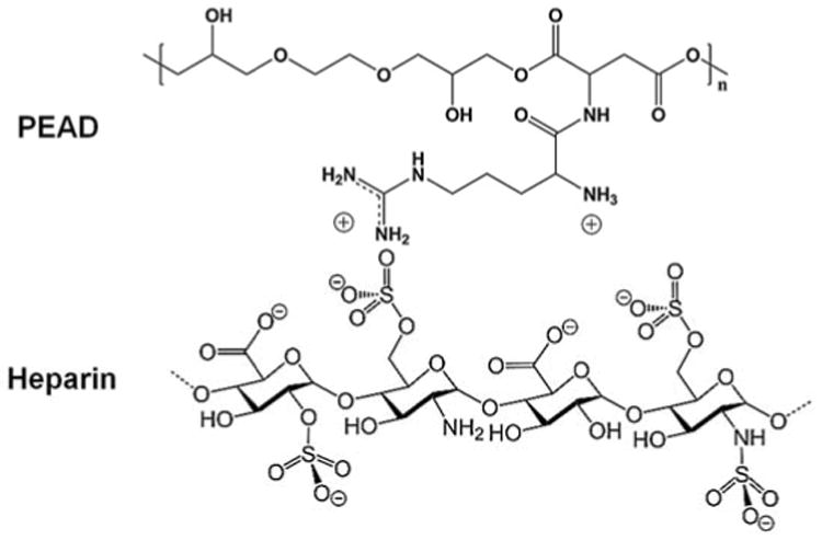
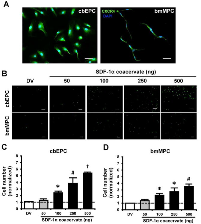
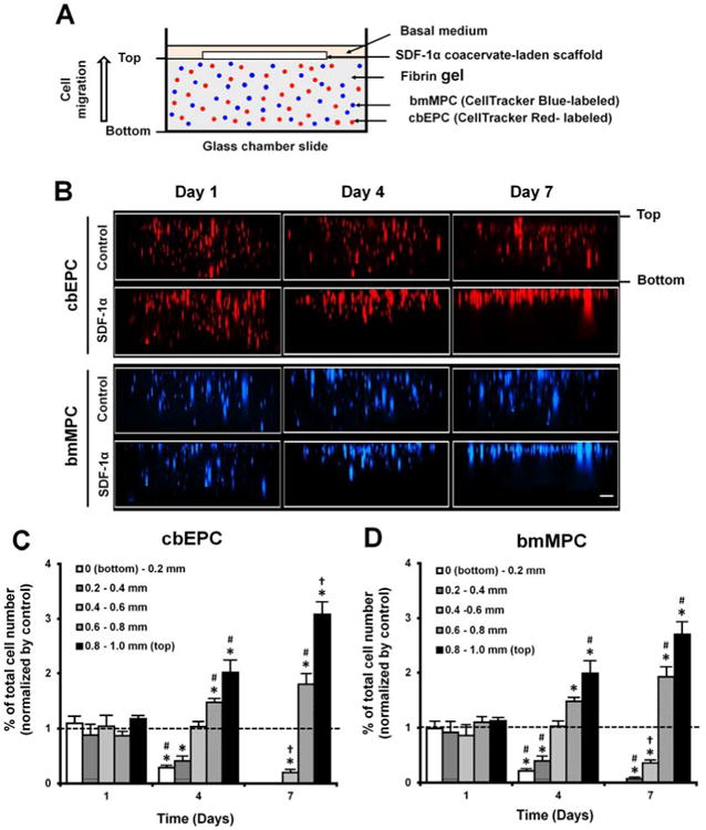
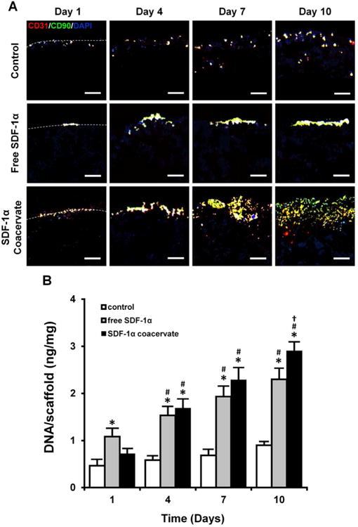
Host cell recruitment is crucial for vascular graft remodeling and integration into the native blood vessel; it is especially important for cell-free strategies which rely on host remodeling. Controlled release of growth factors from vascular grafts may enhance host cell recruitment. Stromal cell-derived factor (SDF)-1α has been shown to induce host progenitor cell migration and recruitment; however, its potential in regenerative therapies is often limited due to its short half-life in vivo. This report describes a coacervate drug delivery system for enhancing progenitor cell recruitment into an elastomeric vascular graft by conferring protection of SDF-1α. Heparin and a synthetic polycation are used to form a coacervate, which is incorporated into poly(glycerol sebacate) (PGS) scaffolds. In addition to protecting SDF-1α, the coacervate facilitates uniform scaffold coating. Coacervate-laden scaffolds have high SDF-1α loading efficiency and provide sustained release under static and physiologically-relevant flow conditions with minimal initial burst release. In vitro assays showed that coacervate-laden scaffolds enhance migration and infiltration of human endothelial and mesenchymal progenitor cells by maintaining a stable SDF-1α gradient. These results suggest that SDF-1α coacervate-laden scaffolds show great promise for in situ vascular regeneration.

Keywords: Coacervate; Human progenitor cells; Poly(glycerol sebacate); Polycation; Stromal cell-derived factor (SDF)-1α; Tissue engineering.


Figures

Figure 1
Figure 1
Composition of a self-assembled coacervate.
Figure 2
Figure 2
Characterization and chemotaxis of progenitor cells. (A) CXCR4 expression of cbEPCs and bmMPCs, (B) Fluorescent images of migrated progenitor cells on the bottom of transwell insert membrane. Scale bar = 200 μm. (C) and (D) Quantification of migrated cells. Cell numbers in each group were normalized by those in basal medium group. *p < 0.05 (compared to DV and 50), #p < 0.05 (compared to DV, 50, and 100), p < 0.05 (compared to all other groups) (n = 4).
Figure 3
Figure 3
Incorporation and distribution of SDF-1α coacervate in PGS scaffolds. (A) Scanning electron micrographs of empty (control) and SDF-1α coacervate-coated scaffolds. Bottom row represents partial magnification of the box shown in top row. Scale bar = 100 μm (top row) and 10 μm (bottom row). (B) and (C) distribution of the fluorescence-labeled SDF-1α coacervate in the flat and tubular scaffolds. Scale bar = 500 μm (all). L: lumen of the scaffold.
Figure 4
Figure 4
In vitro release profiles of SDF-1α from the coacervate-coated scaffolds in static and flow conditions.
Figure 5
Figure 5
Migration of progenitor cells from SDF-1α coacervate-laden scaffolds. (A) Schematic of the cell migration assay. (B) Cross-sectional images of fibrin gels. The difference in migration of fluorescence-labeled progenitor cells in response to empty scaffold (Control) and SDF-1α coacervate-laden scaffold (SDF-1α). (C) Quantification of vertically migrated cells in the fibrin gel of SDF-1α coacervate-laden scaffolds. Cell numbers at each depth in both groups were normalized by total number of cells in the entire gel. Final normalized cell numbers in SDF-1α group were divided into those in control group and represented as a fold difference vs. control. Dashed line represents the normalized cell numbers in control group. *p < 0.05 (compared to control), #p < 0.05 (compared to day 1), p < 0.05 (compared to days 1 and 4).
Figure 6
Figure 6
Recruitment of progenitor cells into the SDF-1α coacervate-laden scaffolds. (A) Immunofluorescent staining for CD31 (cbEPC marker, red), CD90 (bmMPC marker, green), and DAPI (nuclei, blue). Dashed lines represent the border between the outer region (top) and the scaffolds (bottom). Scale bar = 100 μm (all). (B) Quantification of the number of cells in scaffolds. The number of cells was normalized by the wet weight of scaffolds. *p < 0.05 (compared to control), #p < 0.05 (compared to day 1), p < 0.05 (compared to day 4) (n = 4).

References

    1. Weinberg CB, Bell E. A blood vessel model constructed from collagen and cultured vascular cells. Science. 1986;231:397–400. - PubMed
    1. Niklason LE, Gao J, Abbott WM, Hirschi KK, Houser S, Marini R, et al. Functional arteries grown in vitro. Science. 1999;284:489–93. - PubMed
    1. L'Heureux N, Paquet S, Labbe R, Germain L, Auger FA. A completely biological tissue-engineered human blood vessel. Faseb J. 1998;12:47–56. - PubMed
    1. Dahl SL, Kypson AP, Lawson JH, Blum JL, Strader JT, Li Y, et al. Readily available tissue-engineered vascular grafts. Sci Transl Med. 2011;3:68ra9. - PubMed
    1. Quint C, Kondo Y, Manson RJ, Lawson JH, Dardik A, Niklason LE. Decellularized tissue-engineered blood vessel as an arterial conduit. Proc Natl Acad Sci U S A. 2011;108:9214–9. - PMC - PubMed

Publication types