. 2013 Nov;112(7):1395-407.
doi: 10.1093/aob/mct215. Epub 2013 Sep 22.

Auxin and cytokinin control formation of the quiescent centre in the adventitious root apex of Arabidopsis

Affiliations

Auxin and cytokinin control formation of the quiescent centre in the adventitious root apex of Arabidopsis

F Della Rovere et al. Ann Bot. 2013 Nov.

Abstract

Background and aims: Adventitious roots (ARs) are part of the root system in numerous plants, and are required for successful micropropagation. In the Arabidopsis thaliana primary root (PR) and lateral roots (LRs), the quiescent centre (QC) in the stem cell niche of the meristem controls apical growth with the involvement of auxin and cytokinin. In arabidopsis, ARs emerge in planta from the hypocotyl pericycle, and from different tissues in in vitro cultured explants, e.g. from the stem endodermis in thin cell layer (TCL) explants. The aim of this study was to investigate the establishment and maintenance of the QC in arabidopsis ARs, in planta and in TCL explants, because information about this process is still lacking, and it has potential use for biotechnological applications.

Methods: Expression of PR/LR QC markers and auxin influx (LAX3)/efflux (PIN1) genes was investigated in the presence/absence of exogenous auxin and cytokinin. Auxin was monitored by the DR5::GUS system and cytokinin by immunolocalization. The expression of the auxin-biosynthetic YUCCA6 gene was also investigated by in situ hybridization in planta and in AR-forming TCLs from the indole acetic acid (IAA)-overproducing superroot2-1 mutant and its wild type.

Key results: The accumulation of auxin and the expression of the QC marker WOX5 characterized the early derivatives of the AR founder cells, in planta and in in vitro cultured TCLs. By determination of PIN1 auxin efflux carrier and LAX3 auxin influx carrier activities, an auxin maximum was determined to occur at the AR tip, to which WOX5 expression was restricted, establishing the positioning of the QC. Cytokinin caused a restriction of LAX3 and PIN1 expression domains, and concomitantly the auxin biosynthesis YUCCA6 gene was expressed in the apex.

Conclusions: In ARs formed in planta and TCLs, the QC is established in a similar way, and auxin transport and biosynthesis are involved through cytokinin tuning.

Keywords: Adventitious root apex; Arabidopsis thaliana; WOX5; auxin biosynthesis; auxin transport; cytokinin localization; quiescent centre; root meristemoids; stem endodermis; thin cell layers.


Figures
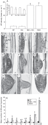
Fig. 1.
Fig. 1.
(A) AR mean density (± s.e.) in the hypocotyl of 14-day-old wild-type seedlings grown on HF, Kin (0·1 µm), IBA (10 µm) + Kin (0·1 µm) and NAA (2 µm) media, and hypocotyl mean length (inset). (B–J) Developmental stages of ARs in wild-type seedlings grown on HF medium. (B) First anticlinal divisions in the hypocotyl pericycle (arrows). (C) Outer (OL) and inner (IL) layers formed by periclinal divisions in the cells originated by the first anticlinal divisions. (D) OL periclinal doubling. (E) IL doubling, leading to a four-layered ARP. (F) ARP dome establishment. (G) ARP dome with protoderm specification (arrow). (H) Stage VII ARP showing cells with LR QC morphology (asterisks). (I) Developed ARP emerging from the hypocotyl (QC shown by the asterisks). (J) Apex of a mature AR (QC shown by the asterisks). (B–J) Histological longitudinal radial sections stained with toluidine blue (Ws ecotype). (K) Mean number (± s.e.) of ARs at different stages in wild-type seedlings grown on HF, IBA (10 µm) + Kin (0·1 µm) and NAA (2 µm) media. (A and K) a,cP < 0·01 differences from other treatments; bP < 0·05 difference between IBA + Kin and NAA. Columns with the same letter are not significantly different. n = 30 (Col ecotype). Scale bars: (B–G, J) = 10 µm; (H) = 20 µm; (I) = 30 µm. I–VII, developmental ARP stages, p, hypocotyl pericycle; x, protoxylem; c1–c2, cortex; end endodermis; e, epidermis.
Fig. 2.
Fig. 2.
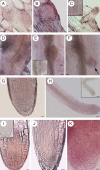
Expression of PR/LR QC markers during AR formation in Col seedlings grown for 14 d under various hormonal treatments. (A–G; HF) QC25::GUS (A), pAGL42::GFP (B) and pWOX5::GFP (D) in the QC at stage VII, and pWOX5::GFP at stage II (C). QC25::GUS (E), pAGL42::GFP (F) and pWOX5::GFP (G) in the QC, and lateral initials (G) of emerged ARs. (H–L; NAA) pAGL42::GFP (H) and QC25::GUS (I) in the QC of stage VII ARPs. pWOX5::GFP at stage II (J), QC25::GUS at the tip of a regular AR (K) and pAGL42::GFP in the twin tip of a fasciated AR (L). (M–R; IBA + Kin) pAGL42::GFP in the QC of not yet emerged (M) and emerged (N) ARPs. pWOX5::GFP at stage II (O), and in the QC, lateral initials and pericycle cells forming LRs (arrows) in emerged ARPs (P). QC25::GUS in the QC at stage VII (Q), and in emerged ARPs (R). Insets in fluorescence pictures show corresponding bright-field images. Scale bars: = (A, B and inset, E, F, H–K, M–O, R) 10 µm; (C, D, G, L, P–Q, and insets in C, F, J, N–O) = 20 µm; (insets in D, G, H) = 30 µm; (inset in P) = 40 µm; (inset in L) = 100 µm.
Fig. 3.
Fig. 3.
DR5::GUS, monitoring IAA presence in developing ARs from 14-day-old Col seedlings grown under different hormonal treatments. (A–C; HF) DR5 in a stage IV ARP located at the hypocotyl transition zone (A), in the tip of a stage VII ARP (B) and in the QC, flanking initials, columella and developing vasculature of a mature AR (C). (D–F; NAA) Strong DR5 at stage III (arrow) and in surrounding hypocotyl cells (D), at stage VII (E) and in the QC, surrounding initials, cap and differentiated tissues (arrow) of a mature AR (F). (G–I; IBA + Kin) DR5 at stage II (G), at the base and tip of a stage VII ARP (H) and in the QC, surrounding initials, columella and developing vasculature of a mature AR (I). (J–L; Kin) Faint DR5 signal in a stage III ARP located at the transition zone (J), stronger expression at the tip of a stage VII ARP (K) and in the QC, surrounding initials and columella of a mature AR (L). Scale bars: (A, B, D, E, G–L) = 10 µm; (C, F) = 20 µm.
Fig. 4.
Fig. 4.
PIN1::GUS expression in developing ARs from 14-day-old Col seedlings grown under different hormonal treatments. (A–C; HF) PIN1 expression at stage III (A), at the base and in the middle of a stage VII ARP (B) and in the niche, cap and procambium of a mature AR (C). (D–F; NAA) High PIN1 expression at stage III (D), at stage VII (E) and in the whole apex of a mature AR (F). (G–I; IBA + Kin) High PIN1 expression at stage III (G) and VII (H), and in a mature AR tip, except for the differentiating cortex and epidermis (I). (J–L; Kin) Very weak PIN1 expression at stage I (J), higher expression at the base and middle part of a stage VII ARP (K) and faint expression in the niche and procambium of a mature AR apex (L). Scale bars: (A,B) = 10 µm; (C–L) = 20 µm.
Fig. 5.
Fig. 5.
LAX3::GUS expression during AR development in 14-day-old Col seedlings grown under different hormonal conditions. (A–F; HF) Expression at stage III (A) and IV (B), at the base of a stage VII ARP (C), in the forming vasculature of emerging (D) and elongating ARPs (E) and in a few cap cells, and the differentiating vasculature of a mature AR (F). (G–K; NAA) High and uniform expression at stage IV (G) and VI (H), in the ARP basal half, before (I) and after (J) protrusion, and in cap cells and precociously differentiated tissues in a mature AR (K). (L–P; IBA + Kin) High expression at stage III (L) and V (M), in the basal half of an emerging ARP (N), in the vasculature of an elongating ARP (O) and a mature AR (P), and in some cap cells in the latter (P). (Q–U; Kin, hypocotyl transition zone) LAX3 expression at stage II (Q) and IV (R), at the base of a stage VII ARP (S), in the developing vasculature of elongating ARPs (T) and mature ARs (U), and faintly in the AR cap (U). Scale bars: (A, B, H, I, K, M, Q–S) = 10 µm; (D, G, L, N–P, U) = 20 µm; (C, E, F, J, T) = 30 µm.
Fig. 6.
Fig. 6.
Expression of QC markers (A–J), auxin monitoring (K–O), and auxin efflux (P–R) and influx (S–W) gene expression during AR formation in IBA + Kin-cultured TCLs. (A, B) WOX5 expressed in meristematic cell clusters (A) and root meristemoids (B). Corresponding light microscopy images are shown in the insets. (C, D) Emerging ARPs (C) and apices of elongated ARs (D) showing WOX5 in the QC and lateral initials. Corresponding light microscopy images are shown in the insets. (E) Early-domed ARPs showing the appearance of pAGL42. (F) Emerging ARPs with pAGL42 expression in the QC. (G) Confocal microscopy image of mature AR apices with pAGL42::GFP signal in the QC. (H) No expression in early-forming ARPs of QC25::GUS TCLs, but expression in the QC of emerged ARPs (I) and elongated ARs (J). (K) Meristematic cell clusters with DR5::GUS expression. (L) Detail of a meristemoid showing signal. (M, N) Not yet protruded ARPs showing DR5 expression in the tip (arrows in N). (O) DR5::GUS expression in the niche and cap of mature ARs. The inset shows expression in the forming vasculature. (P) PIN1 expression in early-domed ARPs, corresponding to stage VII in planta, and endodermis-derived cells at the base. (Q) Not yet emerged ARPs showing PIN1 signal, mainly in the tip and forming vasculature. (R) Protruded ARP with PIN1 expression in the vasculature, procambium and apex. (S) LAX3 expression in forming meristemoids (square). (T) Not yet protruded ARPs with LAX3 expression at the base. (U, V) Elongating ARPs after protrusion, with LAX3 expression in the vasculature, but not in the apex. (W) Mature AR with LAX3 signal in the developing vasculature and some cap cells (arrow). Insets in A–D, toluidine blue section staining. Scale bars: (insets in A and C, G) = 10 µm; (A, C–F, H, J, O, inset in O, V, W) = 20 µm; (inset in B, I, T) = 30 µm; (inset in D, K–M, S, U) = 40 µm; (N, P–R) = 50 µm; (B) = 100 µm.
Fig. 7.
Fig. 7.
Localization of trans-zeatin riboside (A–C) and YUCCA6 transcription (D–H) during AR development in planta [14-day-old Col (A–F) and Ws (G, H) seedlings grown under HF treatment], and YUCCA6 transcription in ARs from TCLs cultured with/without IBA + Kin (I–K). (A) Stage IV (arrow) and stage VI ARPs showing a diffuse cytokinin immunostaining. (B) ARP just before protrusion showing high cytokinin immunostaining in the protoderm, in particular. (C) Elongating ARP with extensive cytokinin signal at the tip. Differentiating epidermis with staining (arrow) magnified in the inset (longitudinal tangential section). (D–E) In situ hybridizations showing YUCCA6 transcription at stage IV (D) and VI (E). Sense probe control shown in the inset of E. (F–H) YUCCA6 transcription at the tip (arrow) of protruded ARPs (F), all over the AR apex (G), and in the AR procambium and differentiating vasculature (H). Sense probe control shown in the inset of H. (I) YUCCA6 transcription in AR apices from IBA + Kin-cultured Ws TCLs. The sense probe control is shown in the inset. (J, K) Low (J) and high (K) YUCCA6 transcription in the apices of ARs formed by sur2-1 TCLs cultured without hormones and with IBA + Kin, respectively. (D–F, H, K) Whole-mount RNA in situ hybridizations; (G, I, J) RNA in situ hybridizations on longitudinal sections of resin-embedded ARs. Scale bars: (inset in C, G, I–K) = 10 µm; (A–C, inset in I) = 20 µm; (D–F, inset in E, H) = 30 µm; (inset in H) = 40 µm.
Fig. 8.
Fig. 8.
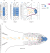
Model of auxin flow, gene expression and cytokinin localization during AR formation in planta. At stage I, auxin (IAA) is diverted from the basipetal flow along the vascular parenchyma cells (vp) adjacent to the protoxylem (x) of the hypocotyl (Hy) towards the pericycle (P) cells by PIN1, activating LAX3 and auxin accumulation (blue colour) in the founder cells. At stage II, auxin is maintained in the first-formed inner and outer AR layers by PIN1 (yellow arrow) and LAX3 (light-blue arrows), and WOX5 is expressed. At stage VII, PIN1 drives auxin flow towards the ARP tip throughout the middle cell files, because cytokinin (pink colour) downregulates PIN1 in the peripheral ARP layers. Cytokinin also downregulates LAX3, limiting the carrier activity at the ARP base (up to the dotted line). The auxin flow driven by PIN1 towards the tip results in an apical auxin maximum, limiting WOX5 expression at the distal tip, and here establishing the position of the QC. Auxin biosynthesis by YUCCA6 (green diamonds) contributes to auxin maximum positioning in the tip. LAX3 is also active in the Hy endodermis (End), cortex (C) and epidermis (E) around the ARP, possibly favouring protrusion. In the mature AR, the auxin maximum (blue colour) encompasses the QC, flanking initials and cap cells (columella, in particular), and WOX5 QC expression is maintained. Auxin biosynthesis by YUCCA6 is also maintained (green diamonds), contributing to the persistence of apical auxin accumulation. Also cytokinin is present at the AR tip (pink stars), contributing to the maintenance of auxin homeostasis by a downregulation of PIN1 in the forming epidermis/cortex, and of LAX3 in the entire tip, except the cap (light-blue dots). PIN1 and LAX3 are expressed in the AR vasculature, and LAX3 expression stops at the elongation zone border (dotted line).

Similar articles

Cited by

References

    1. Airoldi CA, Della Rovere F, Falasca G, et al. The Arabidopsis BET bromodomain factor GTE4 is involved in maintenance of the mitotic cell cycle during plant development. Plant Physiology. 2010;152:1320–1334. - PMC - PubMed
    1. Aloni R, Langhans M, Aloni E, Dreieicher E, Ullrich CI. Root-synthesized cytokinin in Arabidopsis is distributed in the shoot by the transpiration stream. Journal of Experimental Botany. 2005;56:1535–1544. - PubMed
    1. Barlier I, Kowalczyk M, Marchant A, et al. The SUR2 gene of Arabidopsis thaliana encodes the cytochrome P450 CYP83B1, a modulator of auxin homeostasis. Proceedings of the National Academy of Sciences, USA. 2000;97:14819–14824. - PMC - PubMed
    1. Benková E, Michniewicz M, Sauer M, et al. Local, efflux-dependent auxin gradients as a common module for plant organ formation. Cell. 2003;115:591–602. - PubMed
    1. van den Berg C, Willemsen V, Hendriks G, Weisbeek P, Scheres B. Short-range control of cell differentiation in the Arabidopsis root meristem. Nature. 1997;390:287–289. - PubMed

Publication types

MeSH terms