Review
. 2013:9:691.
doi: 10.1038/msb.2013.48.

Biomedically relevant circuit-design strategies in mammalian synthetic biology

Affiliations
Review

Biomedically relevant circuit-design strategies in mammalian synthetic biology

William Bacchus et al. Mol Syst Biol. 2013.

Abstract

The development and progress in synthetic biology has been remarkable. Although still in its infancy, synthetic biology has achieved much during the past decade. Improvements in genetic circuit design have increased the potential for clinical applicability of synthetic biology research. What began as simple transcriptional gene switches has rapidly developed into a variety of complex regulatory circuits based on the transcriptional, translational and post-translational regulation. Instead of compounds with potential pharmacologic side effects, the inducer molecules now used are metabolites of the human body and even members of native cell signaling pathways. In this review, we address recent progress in mammalian synthetic biology circuit design and focus on how novel designs push synthetic biology toward clinical implementation. Groundbreaking research on the implementation of optogenetics and intercellular communications is addressed, as particularly optogenetics provides unprecedented opportunities for clinical application. Along with an increase in synthetic network complexity, multicellular systems are now being used to provide a platform for next-generation circuit design.


Conflict of interest statement

The authors declare that they have no conflict of interest.

Figures

Figure 1
Figure 1
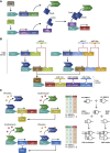
Synthetic circuits based on the rewired cell-signaling pathways. (A) Guanabenz-induced synthetic circuit for the treatment of metabolic syndrome. Cells engineered to express the chimeric trace amine-associated receptor (cTAAR1) respond to Guanabenz by activating endogenous cAMP signaling. Increased levels of cAMP activate PCRE-driven transgene expression of Glp-1-Leptin via a cAMP-responsive element binding protein 1 (CREB1). When implanted in mice developing symptoms of metabolic syndrome, the circuit enabled simultaneous targeting of several metabolic disorders (Ye et al, 2013). (B) Blue light- and (C) radio wave-induced synthetic circuits enabling glucose homeostasis. (B) Cells engineered to trigger calcium influx through transient receptor potential channels (TRPCs) by expressing blue light-responsive melanopsin, link blue-light sensing to transgene expression via an NFAT-responsive promoter (PNFAT). Implanted in diabetic mice, the circuit enabled blue light-controlled glucose homeostasis when expressing glucagon-like peptide 1 (Ye et al, 2011). (C) Cells engineered to trigger calcium influx through temperature-sensitive, His-tagged TRPCs (TRPV1HIS). Antibody-coated nanoparticles for His-tag recognition (NP) enabled local nanoparticle heating of TRPV1HIS, consequently allowing for calcium influx, linking radio-wave exposure to transgene expression via an NFAT-responsive promoter (PNFAT). Implanted in mice, the circuit enabled radio wave-controlled regulation of blood glucose levels by expressing insulin (Stanley et al, 2012). (D) Synthetic circuit responsive to endogenous proteins allow for disease-targeted cell death. The RNA-based devise is composed of specific aptamers for p50/p65 recognition (white circle), localized at key intronic positions near an alternative spliced exon harboring a stop codon (red area) in a three-exon, two-intron minigene fused to a suicide gene (HSV-TK). Activation of the NF-κB pathway by stimulation of the tumor necrosis factor receptor (TNFR) with tumor necrosis factor-α (TNFα) enables p50/p65 regulation of exon exclusion, thereby linking disease markers to the killing of the diseased cells (Culler et al, 2010).
Figure 2
Figure 2
Multi-input design for increased circuit complexity. (A) Two-input circuit for cancer cell recognition and destruction. The synthetic promoters CXCL1, SSX1 and H2A1, which show diverse activation strengths in various cancer cell lines, are engineered to control the gene expression of either one of two subunits, DocS-VP16 and Gal4BD-Coh2, which together comprise a split transactivator. As the activities of the synthetic promoter combinations (P1; either CXCL1, SSX1 or H2A1, P2; either CXCL1, SSX1 or H2A1) used are regulated by endogenous, cell-specific transcription factors (TF1, TF2), the split transactivator is only expressed in a cell line where sufficient activities of both promoters are obtained. The association of DocS and Coh2 produces a functional transactivator that activates gene expression of a killer gene (TK1) from a Gal4-synthetic promoter (PGal4), thus leading to cell death (Nissim and Bar-Ziv, 2010). (B) Multi-input circuit for cancer cell recognition and destruction. A cell type classifier for HeLa cells was constructed by implementing endogenous expressed microRNA profiles consisting of high- or low-expressed microRNA (high/low sensors). Three high-expressed microRNAs (miR-21, miR-17 and miR-30a) targeted the mRNA of the activator rtTA and the repressor LacI (miR-21t, miR-17t and miR30at). rtTA was designed to activate the expression of LacI and LacI in its turn was designed to repress the final expression of a output gene (GOI), thereby only allowing for the activation of the gene in the presence of all three high-expressed microRNAs. Three low-expressed microRNAs (miR-141, miR142(3p) and miR-146a) further targeted the mRNA of the output gene (miR-141t, miR-142(3p)t and miR-146at), only allowing for its expression at low levels of all three of the microRNAs. Regulation of a killer gene (hBax) with this cancer cell classifier enabled cell type-specific destruction of the HeLa cells (Xie et al, 2011). (C) Two-input circuits enable construction of plug-and-play assemblies performing sophisticated computations. The transcription factors ET1 and TtgA1, which repress the promoter activity of PETR2 and PTtgR1 in response to erythromycin (E) and phloretin (P), were combined with the RNA-binding proteins MS2 and L7Ae, which inhibit the translation of transcripts containing the specific target motifs MS2box and C/Dbox, to construct circuits capable of performing easy computations such as N-IMPLY logics, which are induced in the presence of only one specific input molecule. Assembling such simple circuits in a plug-and-play fashion allowed the construction of complex circuits capable of performing half-subtractor and half-adder computations (Auslander et al, 2012a).
Figure 3
Figure 3
Synthetic circuits responsive to light. (AC) Blue light-controlled circuits. (A) The two proteins GI and FKF1 with its chromophore flavin mononucleotide (FMN) interact upon blue light. Fusions of GI to the Gal4-DNA-binding domain (GBD) and FKF1 to the VP16 activation domain enable blue light-dependent association of the split transactivator, which consequently activates gene expression from a Gal4-promoter (UASG)5 (Yazawa et al, 2009). (B) A fusion protein composed of VVD fused to the p65 activator and a monomeric variant of the Gal4-DNA-binding domain (GBD) is unable to bind to the Gal4 promoter ((UASG)5) and activate gene expression due to the monomeric structure of the GBD. Blue-light illumination enables VVD dimerization due to its chromophore flavin adenine dinucleotide (FAD), thus reconstituting the GBD dimer and consequently activating gene expression (Wang et al, 2012). (C) Fusion proteins of CRY2 and CIBN to each part of a split Cre recombinase lacking enzymatic activity (CreN and CreC) enabled associated and reconstituted Cre activity through the blue light-dependent interaction of CRY2, which requires FAD, and CIBN. The functional Cre acts by eliminating a stop sequence flanked by loxP sites, subsequently permitting gene expression (Kennedy et al, 2010). (D) Red light-controlled circuit. The two proteins PhyB and PIF6 interact upon red light while far-red light inhibits the interaction. Fusions of PhyB, which uses the chromophore phytochromobilin (PCB), to VP16 and PIF6 to the TetR repressor enabled red light-dependent association of the split transactivator, consequently activating gene expression from a TetR-promoter ((TetO)13). This action was reversed using far-red light, which caused dissociation of the PhyB and PIF6 fusions (Muller et al, 2013a). (E) UVB light-controlled circuit. Fusion proteins of UVR8 to the E repressor and WD40 to VP16 enabled association of the split transactivator upon UVB illumination as the UVR8 homo-dimerization is released, allowing for WD40-VP16 recruitment. The reconstituted transactivator enables gene expression from a promoter containing an E-responsive operator motif ((etr)8) (Muller et al, 2013b).
Figure 4
Figure 4
Engineering of intercellular communication. (A) Acetaldehyde-based intercellular communication system enables interkingdom communication. Sender cells (SCs), able to produce acetaldehyde, composed of E. coli, S. cerevisiae, L. sativum or mammalian cells engineered to express alcohol dehydrogenase. Mammalian receiver cells (RCs) were engineered with an acetaldehyde-responsive element consisting of AlcR, which in the presence of acetaldehyde activates gene expression from a PAIR promoter. Implanting the mammalian sender and receiver cells in mice allowed for the production of acetaldehyde by the sender cells, thus converting ethanol supplemented in the drinking water. The acetaldehyde was broadcast to the receiver cells allowing for gene expression of secreted alkaline phosphatase (SEAP) (Weber et al, 2007a). (B) L-tryptophan-based intercellular communication system enables multicellular assemblies. Sender cells were engineered to express tryptophan synthase (TrpB), converting supplemented indole into L-tryptophan. The receiver cells were engineered with an L-tryptophan-responsive element consisting of the transactivator TRT, which activates gene expression from PTRT in the presence of L-tryptophan. Combining the genetic components of the acetaldehyde- and L-tryptophan-based intercellular communication systems allowed for various sender- (SC), processor- (PC), receiver (RC) and sender/receiver cells (S/RC) to be constructed. Assembling these components in a plug-and-play manner allowed the creation of multicellular architectures mimicking the natural phenomena (Bacchus et al, 2012).
Figure 5
Figure 5
Prosthetic networks. (A) Prosthetic network regulating urate homeostasis. Cells are engineered to respond to elevated levels of uric acid by disassociation of the mUTS repressor from the PUREX8 promoter, thereby enabling transgene expression. Expression of a urate transporter (URAT1) enhanced intercellular urate concentrations and circuit sensitivity. When implanted in urate oxidase-deficient mice, the circuit sensed pathologically high levels of uric acid in the blood stream, activated transgene expression of a secreted urate oxidase (smUox), and thus reduced the elevated levels of uric acid (Kemmer et al, 2010). (B) Prosthetic network for artificial insemination. Cells engineered to express the luteinizing hormone receptor (LHR) respond to luteinizing hormone by activating endogenous cAMP signaling, allowing for the activation of PCRE-driven transgene expression of cellulase via a cAMP-responsive element binding protein 1 (CREB1). Engineered cells are co-encapsulated with sperm into cellulose-based implants and positioned in the uterus of cows. Ovulation-coordinated activation of cellulase expression in response to elevated levels of luteinizing hormone results in capsule degradation and sperm release (Kemmer et al, 2011).
None

References

    1. Airan RD, Thompson KR, Fenno LE, Bernstein H, Deisseroth K (2009) Temporally precise in vivo control of intracellular signalling. Nature 458: 1025–1029 - PubMed
    1. Anderson JC, Clarke EJ, Arkin AP, Voigt CA (2006) Environmentally controlled invasion of cancer cells by engineered bacteria. J Mol Biol 355: 619–627 - PubMed
    1. Arrenberg AB, Stainier DY, Baier H, Huisken J (2010) Optogenetic control of cardiac function. Science 330: 971–974 - PubMed
    1. Auslander S, Auslander D, Muller M, Wieland M, Fussenegger M (2012a) Programmable single-cell mammalian biocomputers. Nature 487: 123–127 - PubMed
    1. Auslander S, Fussenegger M (2013) From gene switches to mammalian designer cells: present and future prospects. Trends Biotechnol 31: 155–168 - PubMed