. 2013 Nov;19(11):4969-89.
doi: 10.1007/s00894-013-1998-2. Epub 2013 Sep 24.

Destabilization of the MutSα's protein-protein interface due to binding to the DNA adduct induced by anticancer agent carboplatin via molecular dynamics simulations

Affiliations

Destabilization of the MutSα's protein-protein interface due to binding to the DNA adduct induced by anticancer agent carboplatin via molecular dynamics simulations

Lacramioara Negureanu et al. J Mol Model. 2013 Nov.

Abstract

DNA mismatch repair (MMR) proteins maintain genetic integrity in all organisms by recognizing and repairing DNA errors. Such alteration of hereditary information can lead to various diseases, including cancer. Besides their role in DNA repair, MMR proteins detect and initiate cellular responses to certain type of DNA damage. Its response to the damaged DNA has made the human MMR pathway a useful target for anticancer agents such as carboplatin. This study indicates that strong, specific interactions at the interface of MutSα in response to the mismatched DNA recognition are replaced by weak, non-specific interactions in response to the damaged DNA recognition. Data suggest a severe impairment of the dimerization of MutSα in response to the damaged DNA recognition. While the core of MutSα is preserved in response to the damaged DNA recognition, the loss of contact surface and the rearrangement of contacts at the protein interface suggest a different packing in response to the damaged DNA recognition. Coupled in response to the mismatched DNA recognition, interaction energies, hydrogen bonds, salt bridges, and solvent accessible surface areas at the interface of MutSα and within the subunits are uncoupled or asynchronously coupled in response to the damaged DNA recognition. These pieces of evidence suggest that the loss of a synchronous mode of response in the MutSα's surveillance for DNA errors would possibly be one of the mechanism(s) of signaling the MMR-dependent programed cell death much wanted in anticancer therapies. The analysis was drawn from dynamics simulations.


Figures

Fig. 1
Fig. 1. MutSα-DNA complex structural model
DNA is shown in light blue. The color code for the heterodimer domains is as following: red for the mismatch binding domain, residues 1 to 124 in Msh2 and 1 to 157 in Msh6; yellow for the connector domain, residues 125 to 297 in Msh2 and 158 to 356 in Msh6; green for the lever domain, residues 300 to 456 and 554 to 619 in Msh2, and 357 to 573 and 648 to 714 in Msh6; purple for the clamp domain, residues 457 to 553 in Msh2 and 574 to 647 in Msh6; blue for the ATPase domain, residues 620 to 855 in Msh2 and 715 to 974 in Msh6. The two ADP molecules bound to the ATPase domains are depicted in VDW representations. Note that the first 361 residues of Msh6 are unsolved in the X-Ray structure and in our system residue 1 of Msh6 corresponds to residue 362 in the solved structure.
Fig. 2
Fig. 2. Nonbonding Interactions
Histograms for the representative ensemble of structures are presented. Carboplatin-damaged DNA MutSα recognition complex (a-c). At both Msh2-Msh6 interface and within subunits van der Waals interactions are stronger than electrostatic interactions. At Msh2-Msh6 interface (a) van der Waals interactions, −377.59(18.49) kcal/mol, are stronger than the electrostatics interactions, −140.68(30.97) kcal/mol. Within the subunits, van der Waals interactions are stronger than electrostatics interactions, and about 400 kca/mol stronger within Msh6 than within Msh2, −3839.6(67.25) kcal/mol versus −3422.21(46.63) kcal/mol (in c). Electrostatic interactions within Msh6 are about 800 kcal/mol stronger than within Msh2, in (b) −2802.50(46.06) kcal/mol versus −2017.51(39.74) kcal/mol. Mismatched DNA MutSα recognition complex (d-f). Unlike in the damage recognition complex, in the mismatch recognition complex at both Msh2-Msh6 interface and within subunits the electrostatic interactions are dominant. At Msh2-Msh6 interface (d) electrostatic interactions are dominant over van der Waals interactions, −2282.50(91.61) kcal/mol versus −189.85(10.26) kcal/mol. Within subunits electrostatic interactions (e) are dominant and about 500 kcal/mol stronger within Msh6, −29272.00(224.38) kcal/mol, than within Msh2, −24440.00(147.61) kcal/mol. van der Waals interactions (f) within Msh6, −1343.80(31.30) kcal/mol, are slightly stronger than within Msh2, −1147.50(26.31) kcal/mol. Data presented are mean(std).
Fig. 3
Fig. 3. Energy differences at the MutSα’s interface from representative structures of the mismatched and damaged DNA recognition complexes
Differences in the electrostatic (a) and van der Waals (b) interactions at the protein-protein interface between carboplatin-damaged and mismatched recognition complexes. (c) In blue are common or similar differences in the nonbonding electrostatic interactions at the protein interface between the cisplatin-damaged and mismatched DNA recognition complexes. (d) In magenta are common or similar differences in the van der Waals interactions at the protein interface between the cisplatin-damaged and mismatched DNA recognition complexes. As in carboplatin-damaged DNA recognition complex, most differences in no-specific interactions at the protein interface derive from interactions of lever and ATPase domains of Msh2 with ATPase domain of Msh6. (e) Indicates particularly strong electrostatic interactions specific to the carboplatin-damaged complex, namely 850–973, 318–821,720-383, and 55–394, as well as particularly strong electrostatic interactions specific to the cisplatin-damaged complex, namely 382-7 and 847–894. (f) Indicates strong van der Waals interactions specific to cisplatin-damaged complex, 720-383, 850–973, 383-2 and 757–889, as well as strong van der Waals interactions specific carboplatin-damaged complex, namely 382-7, 2–111 and 847–894. Residue pairs herein are in Msh2-Msh6 format.
Fig. 4
Fig. 4. Mapping structural changes as derived from the differences in nonbonding interactions at the MutSα’s interface
Differences higher/lower than ±5 kcal/mol in electrostatic interactions and ± 2kcal/mol in van der Waals interactions were considered. (a) Differences in electrostatic interactions: strong, specific interactions (depicted in magenta for Msh2’s residues and in blue for Msh6’s residues) at the (1) clamp-clamp, (2) mismatch binding-mismatch binding, (3) lever-mismatch binding, (4) mismatch binding-lever, (5) ATPase-connector, (6) ATPase-lever, and (7) ATPase-ATPase Msh2-Msh6 interfaces in the mismatched DNA recognition complex weaken in the carboplatin-damaged DNA recognition complex; strong, specific interactions (depicted in lime for Msh2’s residues and in yellow for Msh6’s residues) at the (5) ATPAase-connector, (8) lever-ATPase, (7) ATPase-ATPase Msh2-Msh6 interfaces in the carboplatin-damaged DNA recognition complex weaken in the mismatched DNA recognition complex. (b) Differences in van der Waals interactions: are dominated by stronger interactions at the (1) clamp-clamp, (2) mismatch binding-mismatch binding, and (7) ATPase-ATPase Msh2-Msh6 interfaces (interactions depicted in lime-yellow) in the carboplatin-damaged DNA recognition complex; several sites with stronger van der Waals interactions at the (6) ATPase-lever and (8) lever-ATPase Msh2-Msh6 interfaces in the mismatched DNA recognition complex are also predicted (interactions depicted in magenta-blue). Overall, strong, specific electrostatic interactions at the protein-protein interface in the mismatched recognition complex are replaced by weak, non-specific van der Waals interactions in the carboplatin-damaged recognition complex.
Fig. 5
Fig. 5. Solvent accessible surface area (SASA) analysis
(a) The mean of SASA of Msh2 subunit in the carboplatin-damaged complex, 37640.63(512.66) Å2, is higher than in the mismatched complex, 36926.58(579.57) Å2(b) The mean of SASA of the charged residues of Msh2 in the carboplatin complex, 16644.35(362.10) Å2, is higher than in the mismatched complex, 16316.11(400.14) Å2(c) The mean of contact surface area of Msh2 at the protein-protein interface in the carboplatin complex, 5222.39(280.33) Å2, is lower than in the mismatched complex, 5474.91(134.88) Å2(d) The mean of contact surface area of Msh6 at the protein-protein interface in carboplatin complex, 5000.48(255.37) Å2, is lower than in the mismatched complex, 5376.15(159.18) Å2(e) The mean of contact surface area of the charged residues in Msh6 at the protein-protein interface in carboplatin complex, 1414.09(120.46) Å2, is lower than in the mismatched complex, 1553.89(166.33) Å2. The two populations corresponding to the mismatched recognition complex are simulation dependent, but not entirely (Online Resource 3, a). Data presented are mean(std).
Fig. 6
Fig. 6. Surface representation the carboplatin-damaged and mismatched MutSα-DNA recognition complexes
A more compact protein, especially the Msh6 subunit, is indicated in response to carboplatin-damaged DNA recognition by comparison with “the default” mismatched DNA recognition complex, in which multiple channels and larger cavities are indicated in both subunits. Positively charged residues at the protein surface are depicted in blue, while negatively charged residues are depicted in red. The surface representations are for representative structures indicated by clustering analysis of the dynamics trajectories in each system.
Fig. 7
Fig. 7. Structural details at the MutSα’s protein-protein interface in the damaged and the mismatched DNA recognition complexes
Protein-protein interface is defined here as atoms of each of the monomers as well as of the solvent within 5 Å. The Msh2 subunit is depicted in pink and the Msh6 subunit is depicted in silver. The water molecules are depicted in a ball-and-stick representation. The presented structural details are for solvated representative conformations identified by clustering analysis. They include a general view of the above defined interface, (a&b), as well as “the pose” at the interface for each subunit in both carboplatin-damaged (c&d) mismatched (e&f) DNA recognition complexes. The protein-protein interface comprises atoms from all five domains of the heterodimer protein in both recognition complexes, as indicated my marked region: red for the mismatched binding domain; yellow for the connector domain; green for the lever domain and blue for the ATPase domain in c-f. By comparison, even though the protein-protein interface in the carboplatin-damaged DNA recognition complex (c&d) extends over a large number of residues, 127 from Msh2 and 137 from Msh6, versus 108 residues from Msh2 and 124 residues from Msh6 in the mismatched DNA recognition complex (e&f), a higher content of water molecules is indicated at the protein-protein interface in response to damaged DNA recognition.
Fig. 8
Fig. 8. Structural differences at the interface of the damaged DNA recognition complex revealed by SASA and contact maps calculations
(a&b) The positive differences indicate residues more exposed to the solvent in the mismatched than in the carboplatin-damaged DNA recognition complex, while the negative differences indicate the opposite. (c&d) No significant changes are indicated on the core residues within both mismatched and carboplatin damaged recognition complexes. Core of the protein is defined here as composed of residues with solvent accessible surface area averaged over the simulations lower than 1 Å2. (e) The lost contacts, about 1458, at the protein-protein interface in response to damaged DNA recognition are predominantly at the interface of the mismatched binding, the connector and the lever domains of Msh2 with the ATPase domain of Msh6. (f) The gained contacts, about 830, at the interface of the carboplatin-damaged DNA MutSα recognition complex are mainly at the ATPase-ATPase interface, suggesting, apart an overall rearrangement of the interactions at the protein-protein interface in response to damaged DNA recognition. The 2D contact maps were generated from the average distance matrix throughout the course of the trajectories in either system for a cutoff distance of 20 Å.
Fig. 9
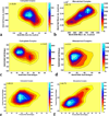
Fig. 9. Msh2 binding to the damaged DNA triggers non-specificity and destabilizing effects at the Msh2-Msh6 interface and within Msh6 subunit
2D histograms for the representative ensemble of structures are presented. (a&b) Uncorrelated (r=0.07) Msh2-DNA and Msh2-Msh6 strong hydrogen bonding in the mismatched DNA recognition complex are anti-correlated (r=−0.40) in the damaged DNA recognition complex. (c&d) Correlated (r=0.56) Msh2-DNA and Msh6-Msh6 strong hydrogen bonding in the mismatched DNA recognition complex are anti-correlated (r=−0.18) in the damaged DNA recognition complex. (e&f) Highly correlated (r=0.63) Msh2-DNA electrostatic interactions and Msh6 self-electrostatic interactions in the mismatched DNA recognition complex are weakly anti-correlated (r=−0.27) in the damaged DNA recognition complex. (g&h) Correlated (r=0.45) solvent exposed area of charged residues in Msh2 and Msh6 in the mismatched DNA recognition complex are anti-correlated (r=−0.34) in the damaged DNA recognition complex.
Fig. 10
Fig. 10. Msh6 binding to the damaged DNA triggers non-specificity and destabilizing effects within both subunits
2D histograms for the representative ensemble of structures are presented. (a&b) Synchronous Msh6-DNA electrostatic interactions and self-electrostatic interactions of the Msh2 subunit in the mismatched DNA recognition complex (r=0.72) become anti-correlated in response to damaged DNA recognition (r=−0.38). (c&d) Uncorrelated in response to mismatched DNA recognition (r=0.02), an increase in the number of specific ionic interactions at the Msh6-DNA interface triggers a decrease in the number of specific ionic interactions within the Msh6 subunit (r=−0.44). (e&f) Highly correlated in response to mismatched DNA recognition (r=0.75), protein-DNA and protein-protein specific ionic interactions (which includes salt bridges at the protein-protein interface and within subunits) are weakly correlated in response to damaged DNA recognition (r=0.21).

References

    1. Kunkel TA. DNA Replication Fidelity. J Biol Chem. 1992;267:18251–18254. - PubMed
    1. Iyer RR, Pluciennik A, Burdett V, Modrich PL. DNA Mismatch Repair: Functions and Mechanisms. Chem Rev. 2006;106:302–323. - PubMed
    1. Peltomaki P. Role of DNA Mismatch Repair Deffects in the Pathogenesis of Human Cancer. J Clin Oncol. 2003;21:1174–1179. - PubMed
    1. Wang D, Lippard SJ. Cellular Processing of Platinum Anticancer Drugs. Nature. 2005;4:307–321. - PubMed
    1. Vasilyeva A, Clodfelter JE, Reactor B, Hills T, Scarpinato KD, Salsbury FR., Jr Small molecule induction of Msh2-dependent cell death suggests a vital role of mismatche repair proteins in cell death. DNA Repair. 2009;8:103–113. - PMC - PubMed

Publication types

MeSH terms

LinkOut - more resources