. 2013 Dec;84(6):844-53.
doi: 10.1124/mol.113.088278. Epub 2013 Sep 23.

A novel noncanonical signaling pathway for the μ-opioid receptor

Affiliations

A novel noncanonical signaling pathway for the μ-opioid receptor

Lei Zhang et al. Mol Pharmacol. 2013 Dec.

Abstract

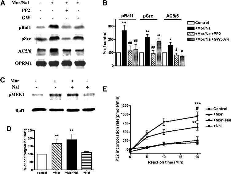
The µ-opioid receptor (OPRM1) signals as a classic G protein-coupled receptor by activating heterotrimeric Gi/Go proteins resulting in adenylyl cyclase (AC) inhibition. Such AC inhibition is desensitized after prolonged agonist treatment. However, after receptor desensitization, the intracellular cAMP level remains regulated by OPRM1, as demonstrated by the intracellular cAMP level increase or AC superactivation upon removal of an agonist or addition of an antagonist. We now demonstrate that such intracellular cAMP regulation is mediated by a novel noncanonical signaling pathway resulting from OPRM1 being converted to a receptor tyrosine kinase (RTK)-like entity. This noncanonical OPRM1 signaling is initiated by the receptor recruiting and activating Src kinase within the receptor complex, leading to phosphorylation of the OPRM1 Tyr(336) residue. Phospho-Tyr(336) serves as the docking site for growth factor receptor-bound protein/son of sevenless, leading to the recruitment and activation of the Ras/Raf-1 and subsequent phosphorylation and activation of AC5/6 by Raf-1. Such sequence of events was established by the absence of Ras/Raf1 recruitment and activation by the OPRM1-Y336F mutant, by the presence of Src kinase inhibitor 4-amino-5-(4-chlorophenyl)-7-(t-butyl)pyrazolo[3,4-d]pyrimidine (PP2) or the absence of Src activity, by the presence of specific Raf-1 inhibitor GW5074 (5-iodo-3-[(3,5-dibromo-4-hydroxyphenyl) methylene]-2-indolinone) or the absence of Raf-1, or by the dominant negative RasN17 mutant. Src together with Ras activates Raf1 which was established by the inability of the Raf1-Tyr(340/341) mutant to activate AC. Hence, the phosphorylation of OPRM1 at Tyr(336) by Src serves as the trigger for the conversion of a classic Gi/Go-coupled receptor into an RTK-like entity, resulting in a noncanonical pathway even after the original Gi/Go signals are blunted.


Figures

Fig. 1.
Fig. 1.
Grb/SOS/Ras is involved in chronic morphine-induced AC superactivation. (A) HEKMT cells were treated with 1 μM morphine for 4 hours (■ HEKMT) or were added to 10 μM Grb2 and SOS1 interaction inhibitor, SOS1 SH3 peptide, for 3 hours during the 4 hour-morphine treatment (● + SH3 peptide). The cAMP assays were performed as described in Materials and Methods. **P < 0.01; ***P < 0.001 (n = 6) when compared with the 4-hour morphine treatment (■), unpaired t test. (B) HEKMT cells were either transiently transfected with 0.5 μg mock cDNA (■ HEKMT) or dominant negative RasN17 cDNA (◇ +RasN17). After 48 hours, the cells were treated with 1 μM morphine for 4 hours, then cAMP assays were performed. The Ras inhibitor farnesylthiosalicylic acid (FTS; 50 μM) was added for 3 hours during the morphine treatment (● + FTS). ***P < 0.001 (n = 6) compared with the 4-hour morphine treatment (■), one-way analysis of variance. Forskolin-induced cAMP in cells without drug treatment is used as 100% control.
Fig. 2.
Fig. 2.
Ras is activated and colocalized with OPRM1 during chronic morphine treatment. (A) The effect of naloxone or Src and Ras inhibitors on Ras activation. HEKMT cells were treated with 1 μM morphine for 0 minutes, 10 minutes, or 4 hours, as indicated, followed by the addition of 0 or 10 μM of naloxone (Nal) for 15 minutes. Src inhibitor (PP2; 2 μM) or Ras inhibitor [farnesylthiosalicylic acid (FTS), 50 μM] was added for 1 or 3 hours during the 4-hour morphine treatment. The assays were performed as described in Materials and Methods. **P < 0.01 compared with control (the ratio of the density of active Ras to the density of input Ras from untreated cells was used as 100% control), unpaired t test. $P < 0.05 compared with 4-hour morphine treatment, unpaired t test. ##P < 0.01 compared with 4-hour morphine and naloxone treatment, unpaired t test. (B) Co-IP of Ras with OPRM1. HEKMT cells were treated with or without 1 μM morphine for 4 hours with or without 10 μM naloxone for 15 minutes, as indicated. The co-IP was performed as described in Materials and Methods. The ratio of the density of Ras to the density of OPRM1 from untreated cells was used as 100% control. **P < 0.01; ***P < 0.001 compared with control, unpaired t test. The no-IP Ab control was performed the same as earlier with the absence of IP Ab (anti-HA). (C) RasN17 inhibited Ras and OPRM1 interaction. HEKMT cells were either transiently transfected with 0.5 μg mock cDNA (Ctrl) or dominant negative RasN17 cDNA (RasN17). After 48 hours, the cells were treated with morphine (1 μM, 4 hours) and naloxone (10 μM, 15 minutes), then the co-IP was performed as described in Materials and Methods. The ratio of the density of Ras to the density of OPRM1 from mock cDNA transfected cells was used as 100% control. **P < 0.01 compared with control, unpaired t test. (D) Ras was not activated in cells expressing Y336F mutant OPRM1. Cells were treated with or without 1 μM morphine for 4 hours followed by with or without 10 μM naloxone for 15 minutes, and assays were performed as described in Materials and Methods. Each experiment was repeated 3 times. Ctrl, control; Mor, morphine.
Fig. 3.
Fig. 3.
AC superactivation is EGFR or fibroblast growth factor receptor (FGFR) independent. HEKMT cells were treated with 1 μM morphine for 4 hours (■) or pretreated with EGFR inhibitor lavendustin C (HDBA) (A) or AG-1478 (B) at a concentration of 10 nM (▼), 50 nM (♦), or 100 nM (●); or pretreated with FGF inhibitor PD-173074 (C) at a concentration of 20 nM (▼), 50 nM (♦), or 100 nM (●) for 2 hours during the 4-hour morphine treatment before cAMP assays were performed (n = 6). Forskolin-induced cAMP in cells without drug treatment is used as 100% control.
Fig. 4.
Fig. 4.
Raf-1 is activated during chronic morphine treatment. (A) Raf-1 is activated within the OPRM1 signal complex in a chronic morphine– and Src kinase–dependent manner. The cells were treated with or without morphine (Mor; 1 μM, 4 hours) and naloxone (Nal, 10 μM, 15 minutes), as indicated. Src inhibitor (PP2; 2 μM) or Raf-1 inhibitor GW5074 (GW; 10 μM) was added for 1 or 2 hours during the 4-hour morphine treatment, respectively. Co-IP was performed as described in Materials and Methods. (B) Bar graph summary of Western blots as shown in A. The ratio of the density of pRaf1, pSrc, or AC5/6 to the density of OPRM1 from untreated cells was used as 100% control. *P < 0.05; **P < 0.01; ***P < 0.001 compared with control; #P < 0.05; ##P < 0.01 compared with morphine and naloxone treatment, unpaired t test. The bars represent averages from three separate Western blots. (C) Raf-1 activation requires chronic morphine treatment. The cells were treated with or without morphine (1 μM, 4 hours) and naloxone (10 μM, 15 minutes), as indicated. Raf-1 activity assays were performed as described in Materials and Methods. The blot was probed with anti-pMEK1 and anti–Raf-1. (D) Bar graph summary of Western blots as shown in C. The ratio of the density of pMEK1 to the density of Raf-1 from untreated cells was used as 100% control. **P < 0.01 compared with control, unpaired t test. The bars represent averages from three separate Western blots. (E) Kinetics of Raf-1 kinase activation. The cells were treated with or without morphine (1 μM, 4 hours) and naloxone (10 μM, 15 minutes), as indicated. The assays were performed following the procedure in Materials and Methods. *P < 0.05; **P < 0.01; ***P < 0.001 compared with control; #P < 0.05 for morphine and naloxone treatment (▼ + Mor + Nal) compared with morphine alone treatment (▴ + Mor), unpaired t test. Each experiment was repeated 3 times.
Fig. 5.
Fig. 5.
Raf-1 activation is not observed in cells expressing Tyr mutant OPRM1. (A) Raf-1 activation. HEKMT or HEKMT-Y336F cells were treated with or without 1 μM morphine (Mor) for 4 hours followed by 0 or 10 μM naloxone (Nal) for 15 minutes, as indicated. Raf-1 activity assays were performed as described in Materials and Methods. The blots were probed with anti-pMEK1, anti–Raf-1, and anti-Gβ. (B) Bar graph summary of Western blots as shown in A. The ratio of the density of pMEK1 to the density of Raf-1 from untreated cells was used as 100% control. *P < 0.05; **P < 0.01 compared with control, unpaired t test. The bars represent averages from 3 separate Western blots. Ctrl, control.
Fig. 6.
Fig. 6.
Raf-1 is required for chronic morphine-induced AC superactivation. (A) RT-PCR analysis of Raf-1 wild type (Raf-1 WT) or Raf-1 knockout (Raf-1 KO) MEF cells transiently expressing OPRM1 and/or Raf-1. The Cells were transfected with cDNA, as indicated. After 48 hours, the cells were lysed, and the total RNA was purified. RT-PCR was performed as described in Materials and Methods. β-Actin was used as the control. (B) Both OPRM1 and Raf-1 are required for AC superactivation. MEF-Raf-1 WT cells were transfected with OPRM1 (△ Raf-1 WT + OPRM1) or Raf-1 (◇ Raf-1 WT + Raf-1) cDNA. Raf-1 KO cells were transfected with OPRM1 (■ Raf-1 KO + OPRM1), Raf-1 (● Raf-1 KO + Raf-1), or both (▼ Raf-1 KO + OPRM1 + Raf-1) cDNA. After 48 hours, the cells were treated with morphine (1 μM, 4 hours), and cAMP assays were performed in the presence of various concentrations of naloxone. Forskolin-induced cAMP in cells without drug treatment is used as 100% control. (C) Bar graph representation of the results from B when 10−4 M naloxone was added in the assay. **P < 0.01; ***P < 0.001 compared with control, unpaired t test (n = 6).
Fig. 7.
Fig. 7.
Tyr 340 and 341 on Raf-1 are critical for chronic morphine-induced AC superactivation. (A) RT-PCR analysis of MEF-Raf-1 KO cells transiently expressing OPRM1 and WT or Tyr mutant Raf-1. Cells were transfected with cDNA, as indicated. After 48 hours, cells were lysed, and total RNA was isolated. RT-PCR was performed as described in Materials and Methods. β-Actin was used as control. (B) AC activity in the presence of naloxone after 4 hours of morphine treatment. MEF-Raf-1 KO cells were transfected with OPRM1 and WT Raf-1 (■ OPRM1 + Raf-1), or Raf-1 Y340A mutant (● OPRM1 + Y340A), or Raf-1 Y341A mutant (□ OPRM1 + Y341A), or double Tyr mutant Y340/341AA (▼ OPRM1+Y340/341AA), or constitutive active mutant Y340/341DD (◇ OPRM1+Y340/341DD) cDNA. After 48 hours, the cells were treated with 1 μM morphine for 4 hours, then cAMP assays were performed in the presence of the indicated naloxone concentrations. Forskolin-induced cAMP in cells without drug treatment is used as 100% control. (C) Bar graph representation of the results from B in the presence of 10−4 M naloxone. **P < 0.01 compared with the cells expressing OPRM1 and WT Raf-1, unpaired t test (n = 6).
Fig. 8.
Fig. 8.
Schematic summary of OPRM1 differential signal pathways upon acute and chronic morphine administration. During acute morphine treatment, OPRM1 interacts with Gαi2 (Zhang et al., 2006) and causes inhibition of adenylyl cyclase activity and a decrease of the intracellular cAMP level. Under prolonged treatment, Src kinase is recruited by the OPRM1 signal complex, where it is activated. The activated Src phosphorylates OPRM1 at Tyr336. The phosphorylated Tyr336 serves as a docking site to recruit Grb/SOS/Ras and Raf-1, which converts the OPRM1 from a classic Gi/Go-coupled receptor into an RTK-like entity. The activated Raf-1 eventually phosphorylates and activates the AC isozymes, most likely AC5/6, resulting in AC superactivation. By doing so, OPRM1 can lead to an alternative signaling pathway that is initiated by the same agonist but is different depending on the duration of agonist exposure.

Similar articles

Cited by

References

    1. Avidor-Reiss T, Nevo I, Levy R, Pfeuffer T, Vogel Z. (1996) Chronic opioid treatment induces adenylyl cyclase V superactivation. Involvement of Gβγ. J Biol Chem 271:21309–21315 - PubMed
    1. Avidor-Reiss T, Nevo I, Saya D, Bayewitch M, Vogel Z. (1997) Opiate-induced adenylyl cyclase superactivation is isozyme-specific. J Biol Chem 272:5040–5047 - PubMed
    1. Baccarini M. (2005) Second nature: biological functions of the Raf-1 “kinase”. FEBS Lett 579:3271–3277 - PubMed
    1. Belcheva MM, Haas PD, Tan Y, Heaton VM, Coscia CJ. (2002) The fibroblast growth factor receptor is at the site of convergence between mu-opioid receptor and growth factor signaling pathways in rat C6 glioma cells. J Pharmacol Exp Ther 303:909–918 - PubMed
    1. Cao C, Huang X, Han Y, Wan Y, Birnbaumer L, Feng GS, Marshall J, Jiang M, Chu WM. (2009) Gα(i1) and Gα(i3) are required for epidermal growth factor-mediated activation of the Akt-mTORC1 pathway. Sci Signal 2:ra17. - PMC - PubMed

Publication types

MeSH terms