. 2014 Jan;42(1):340-8.
doi: 10.1093/nar/gkt882. Epub 2013 Sep 22.

Condensin aids sister chromatid decatenation by topoisomerase II

Affiliations

Condensin aids sister chromatid decatenation by topoisomerase II

Adrian Charbin et al. Nucleic Acids Res. 2014 Jan.

Abstract

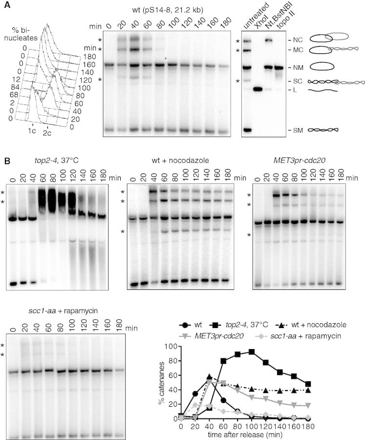
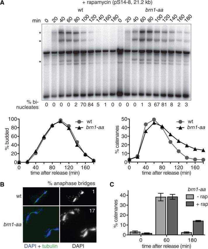
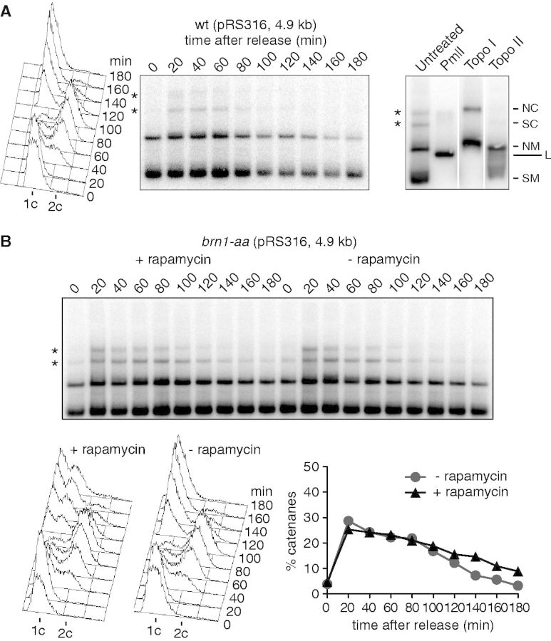
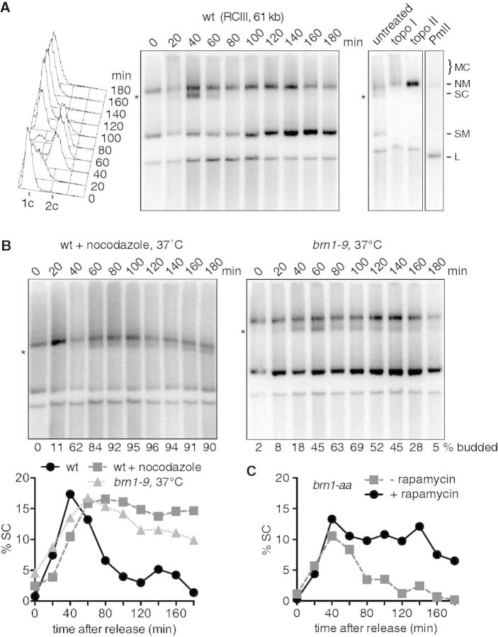
The condensin complex is a key determinant of mitotic chromosome architecture. In addition, condensin promotes resolution of sister chromatids during anaphase, a function that is conserved from prokaryotes to human. Anaphase bridges observed in cells lacking condensin are reminiscent of chromosome segregation failure after inactivation of topoisomerase II (topo II), the enzyme that removes catenanes persisting between sister chromatids following DNA replication. Circumstantial evidence has linked condensin to sister chromatid decatenation but, because of the difficulty of observing chromosome catenation, this link has remained indirect. Alternative models for how condensin facilitates chromosome resolution have been put forward. Here, we follow the catenation status of circular minichromosomes of three sizes during the Saccharomyeces cerevisiae cell cycle. Catenanes are produced during DNA replication and are for the most part swiftly resolved during and following S-phase, aided by sister chromatid separation. Complete resolution, however, requires the condensin complex, a dependency that becomes more pronounced with increasing chromosome size. Our results provide evidence that condensin prevents deleterious anaphase bridges during chromosome segregation by promoting sister chromatid decatenation.


Figures

Figure 1.
Figure 1.
Catenane formation during DNA replication and their resolution. (A) A wild-type strain carrying minichromosome pS14-8 was synchronized in G1 by α-factor treatment and samples taken at 20-min intervals after release. α-factor was added back to the culture for re-arrest in the next G1. FACS analysis of DNA content, the percentage of binucleate cells and the Southern blot to visualize pS14-8 are shown. Enzyme digests of the 40-min time point, shown on the right, allow band identification as indicated. Nt.BstNBI is a nicking endonuclease. NC, nicked catenanes; MC, mixed catenanes; NM, nicked monomers; SC, supercoiled catenanes; L, linear form; SM, supercoiled monomers. Catenated species are highlighted by asterisks (*). (B) as (A), but the indicated strains were used. FACS analysis of DNA content as a marker of cell-cycle progression for each culture is contained in Supplementary Figure S1. The top2-4 strain was released from α-factor arrest at 37°C, a wild-type control at that temperature is shown in Supplementary Figure S3. Each band species on the Southern blots was quantified and the percentage catenanes is plotted over time.
Figure 2.
Figure 2.
Condensin promotes minichromosome decatenation. (A) Condensin was depleted by rapamycin-induced anchor-away of its Brn1 subunit in the brn1-aa cells, at the time of release from an a-factor induced G1 arrest. a-factor was added back to the culture for re-arrest in the next G1. The budding index and the percentage of binucleated cells are shown as markers of cell-cycle progression, as well as the Southern blot to visualize the pS14-8 minichromosome and the quantification of the catenanes, highlighted by asterisks (*). (B) Immunofluorescence microscopy of cells stained with an α-tubulin antibody and the DNA dye 4′,6-diamidino-2-phenylindole (DAPI) reveals anaphase bridges after condensin anchor-away. The frequency of these bridges in late anaphase cells with fully elongated spindles, at 140 min, is indicated (n = 100). (C) An experiment as shown in (A) was repeated in triplicate in the brn1-aa strain in the absence or presence of rapamycin. The percentage of catenanes at the indicated time points was quantified. The means and standard error are shown.
Figure 3.
Figure 3.
A less pronounced condensin requirement for small plasmid decatenation. (A) The catenation status of the centromeric plasmid pRS316 was analyzed during synchronous cell-cycle progression. FACS analysis of DNA content and the Southern blot to visualize pRS316 are shown. Enzyme digests of the 40 min time point allow band identification as indicated. Abbreviations are as in Figure 1, catenated species are highlighted by asterisks (*). (B) The pRS316 catenation status was analyzed during synchronous cell-cycle progression in the presence or absence of rapamycin to anchor-away condensin using the brn1-aa allele. FACS analysis of DNA content is shown, as well as the Southern blot to visualize pRS316 and the quantification of catenanes.
Figure 4.
Figure 4.
A marked role for condensin in ring chromosome III (RCIII) decatenation. (A) The RCIII catenation status was analyzed in synchronized wild-type cells. FACS analysis of DNA content is shown, as well as the Southern blot to visualize RCIII together with enzyme digests that allow the indicated band assignments. Abbreviations are as in Figure 1. Note that the diagnostic band used for quantification, highlighted by an asterisk (*), represents only the supercoiled catenanes (SC) and therefore only a subset of all catenated forms. (B) Catenanes persist in a mitotic arrest or if condensin is inactivated by the brn1-9 allele. As in (A), but cells were released from G1 into nocodazole-containing medium or at 37°C to inactivate the brn1-9 allele. The fraction of supercoiled catenanes (SC) was quantified and is plotted as a function of time. (C) RCIII catenation and decatenation was assessed in a synchronous culture of brn1-aa cells in the absence or presence of rapamycin to anchor-away condensin. The FACS analysis of DNA content, as an indicator of cell-cycle progression, is shown in Supplementary Figure S1. The percentage of supercoiled catenanes is plotted as a function of time.

References

    1. Hirano T, Mitchison TJ. A heterodimeric coiled-coil protein required for mitotic chromosome condensation in vitro. Cell. 1994;79:449–458. - PubMed
    1. Saka Y, Sutani T, Yamashita Y, Saitoh S, Takeuchi M, Nakaseko Y, Yanagida M. Fission yeast cut3 and cut14, members of a ubiquitous protein family, are required for chromosome condensation and segregation in mitosis. EMBO J. 1994;13:4938–4952. - PMC - PubMed
    1. Strunnikov A, Hogan E, Koshland D. SMC2, a Saccharomyces cerevisiae gene essential for chromosome segregation and condensation, defines a subgroup within the SMC family. Genes Dev. 1995;9:587–599. - PubMed
    1. Bhat MA, Philp AV, Glover DM, Bellen HJ. Chromatid segregation at anaphase requires the barren product, a novel chromosome-associated protein that interacts with topoisomerase II. Cell. 1996;87:1103–1114. - PubMed
    1. Bhalla N, Biggins S, Murray AW. Mutation of YCS4, a budding yeast condensin subunit, affects mitotic and nonmitotic chromosome behavior. Mol. Biol. Cell. 2002;13:632–645. - PMC - PubMed

Publication types

MeSH terms