Review
. 2013 Dec;15(4):760-70.
doi: 10.1007/s12017-013-8262-x. Epub 2013 Sep 24.

Sumoylation of critical proteins in amyotrophic lateral sclerosis: emerging pathways of pathogenesis

Affiliations
Review

Sumoylation of critical proteins in amyotrophic lateral sclerosis: emerging pathways of pathogenesis

Emily Foran et al. Neuromolecular Med. 2013 Dec.

Abstract

Emerging lines of evidence suggest a relationship between amyotrophic lateral sclerosis (ALS) and protein sumoylation. Multiple studies have demonstrated that several of the proteins involved in the pathogenesis of ALS, including superoxide dismutase 1, fused in liposarcoma, and TAR DNA-binding protein 43 (TDP-43), are substrates for sumoylation. Additionally, recent studies in cellular and animal models of ALS revealed that sumoylation of these proteins impact their localization, longevity, and how they functionally perform in disease, providing novel areas for mechanistic investigations and therapeutics. In this article, we summarize the current literature examining the impact of sumoylation of critical proteins involved in ALS and discuss the potential impact for the pathogenesis of the disease. In addition, we report and discuss the implications of new evidence demonstrating that sumoylation of a fragment derived from the proteolytic cleavage of the astroglial glutamate transporter, EAAT2, plays a direct role in downregulating the expression levels of full-length EAAT2 by binding to a regulatory region of its promoter.


Conflict of interest statement

Conflict of interest

The authors declare that they have no conflict of interest.

Figures

Figure 1
Figure 1. Schematic representation of the glutamate transporter EAAT2 (a.k.a. Glt-1 in rodents) transmembrane topology
(A) Caspase-3 cleavage occurs at a conserved consensus site within the cytosolic c-terminus domain of EAAT2. (B) The site used by caspase-3 is unique within the EAAT2 sequence. Experiments with in vitro translated EAAT2 have shown that caspase-3 cleaves EAAT2 by generating only two fragments and that mutagenesis of the caspase-3 consensus site in the c-terminus abolishes this cleavage (Boston-Howes et al. 2006). The sumoylation consensus site is shown in the c-terminus domain of EAAT2 (CTE).
Figure 2
Figure 2. CTE-SUMO1 decreases mRNA and protein levels of the full-length transporter EAAT2
(A) Spinal cord astrocytes in culture were transduced with Adeno-Virus construct expressing CTE-SUMO1 fused protein (M.O.I. 3), as per our published protocol (Foran et al. 2011). AdV-empty vector and AdV-GFP (M.O.I. 3) treated astrocytes were used as controls. Homogenates were collected after 7 days of incubation post-treatment and western blot analysis was performed with an affinity-purified anti-EAAT2 polyclonal antibody recognizing an epitope in the c-terminus domain of EAAT2 downstream the caspase-3 cleavage consensus site (a.a. 518–536; anti–CTE domain), affinity purified anti-EAAT1 antibody (epitope a.a. 522–536; anti-EAAT1) and anti-GFAP antibodies. Intensity of the bands was quantified by densitometry using a Chemidoc system (Biorad). EAAT2 and EAAT1 signals were normalized against GFAP expression levels. The antibody anti-EAAT2 was produced by AffinityBioreagent (15 mg/ml) and used at 0.1. ug/ml. Anti-EAAT1 antibody was a generous gift from Dr. Danbolt (0.2 ug/ml, rabbit #8D0161). (B) Quantification of EAAT2 and EAAT1 changes as a result of CTE-SUMO1 transduction is shown (** P<0.01; Dunnett's multiple comparisons test). (C) Spinal cord astrocytes were transduced with AdV expressing CTE-SUMO1, GFP, or empty vector as control (MOI 1). Total RNA was collected after 7 days of incubation with virus and qPCR performed. First strand cDNA was synthesized by reverse transcription from 1 g of total RNA using High Capacity RNA-to-cDNA Master Mix (applied Biosystems). The thermocycler parameters were 50 °C for 2 min, 95 °C for 10 min, followed by 40 cycles of 95 °C for 15 sec, and 60 °C for 1 min. The results were analyzed by the comparative Ct method and normalized against GFAP expression levels (* P<0.05; Dunnett's multiple comparisons test).
Figure 3
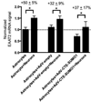
Figure 3. Astrocytic expression of CTE-SUMO1 does not block neuron-stimulated EAAT2 transcriptional activation
Astrocytes, grown in a 6 cm petri dish, were transduced with adenovirus expressing CTE-SUMO1, with AdV-empty for control (MOI=3) or left untreated. After 48 hours post-transduction, purified cortical neurons (8×10⁴ cells) were added to the astrocytes and kept for the next 7 days. Total RNA was used for qPCR analysis. EAAT2 signal was normalized against GFAP mRNA levels (* P<0.05; Dunnett's multiple comparisons test).
Figure 4
Figure 4. Occupancy of CTE-SUMO1 on EAAT2 promoter in spinal cord of SOD1-G93A mouse model of ALS increases in disease
(A) Schematic representation of a portion of the promoter region of the mouse EAAT2 gene is shown. Upstream Probe, Probe A, Probe B, and Downstream Probe indicate the locations of TaqMan probes used to amplify EAAT2 specific promoter sequences pulled down by chromatin immunoprecipitation (ChIP). (B) ChIP assay of EAAT2 promoter with antibody specific for EAAT2 c-terminus (CTE). For the preparation of chromatin samples, two spinal cords for each group were fixed in 1% formaldehyde solution for 10 min., and then a nuclear pellet was prepared following the protocol specified in the NE-PER Nuclear and Cytoplasmic Extraction Kit (Thermo Scientific, Cat#78833). The pellet was resuspended in 700 l of shearing buffer and the chromatin fragments were generated according to a protocol specified in the ChIP-IT® Express Kit and Sonication Shearing Kit Manual (ActiveMotif, Cat#53008) by sonication. Sixty l of sheared chromatin was incubated with the anti-CTE antibody (epitope 518–536; 2 g in 200 l of ChIP reaction) and Protein G magnetic beads. Chromatin elution, cross-linking reversion and DNA isolation were done with reagents and according to protocol provided by ChIP-IT® Express Kit. The DNA that was bound to CTE was amplified by PCR with the 4 different TaqMan probes. Precipitated EAAT2 promoter segments were detected using quantitative real-time PCR and relative chromatin occupancy was calculated as fold increase over non-transgenic spinal cord ChIP. Control regions in the downstream and 1,280 bp upstream EAAT2 mRNA start transcription site (Upstream and Downstream probes) were also quantitated using real-time PCR in parallel as further demonstration of assay specificity. (* P<0.05; Dunnett's multiple comparisons test).

References

    1. Arai T, Hasegawa M, Akiyama H, Ikeda K, Nonaka T, Mori H, et al. TDP-43 is a component of ubiquitin-positive tau-negative inclusions in frontotemporal lobar degeneration and amyotrophic lateral sclerosis. Biochem Biophys Res Commun. 2006;351(3):602–611. - PubMed
    1. Arredondo C, Orellana M, Vecchiola A, Pereira LA, Galdames L, Andres ME. PIASgamma enhanced SUMO-2 modification of Nurr1 activation-function-1 domain limits Nurr1 transcriptional synergy. PLoS One. 2013;8(1):e55035. - PMC - PubMed
    1. Barmada SJ, Skibinski G, Korb E, Rao EJ, Wu JY, Finkbeiner S. Cytoplasmic mislocalization of TDP-43 is toxic to neurons and enhanced by a mutation associated with familial amyotrophic lateral sclerosis. J Neurosci. 2010;30(2):639–649. - PMC - PubMed
    1. Bigio EH, Weintraub S, Rademakers R, Baker M, Ahmadian SS, Rademaker A, et al. Frontotemporal lobar degeneration with TDP-43 proteinopathy and chromosome 9p repeat expansion in C9ORF72: clinicopathologic correlation. Neuropathology. 2012 - PMC - PubMed
    1. Boston-Howes W, Gibb SL, Williams EO, Pasinelli P, Brown RH, Jr, Trotti D. Caspase-3 cleaves and inactivates the glutamate transporter EAAT2. J Biol Chem. 2006;281(20):14076–14084. - PubMed

Publication types

MeSH terms