. 2013 Nov 8;288(45):32149-32159.
doi: 10.1074/jbc.M113.493841. Epub 2013 Sep 23.

Redox control of the senescence regulator interleukin-1α and the secretory phenotype

Affiliations

Redox control of the senescence regulator interleukin-1α and the secretory phenotype

Donald A McCarthy et al. J Biol Chem. .

Abstract

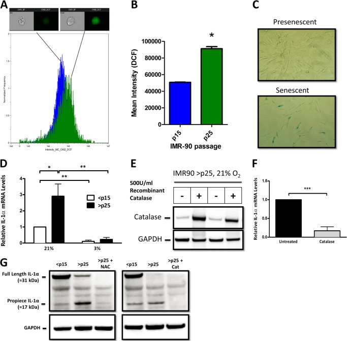
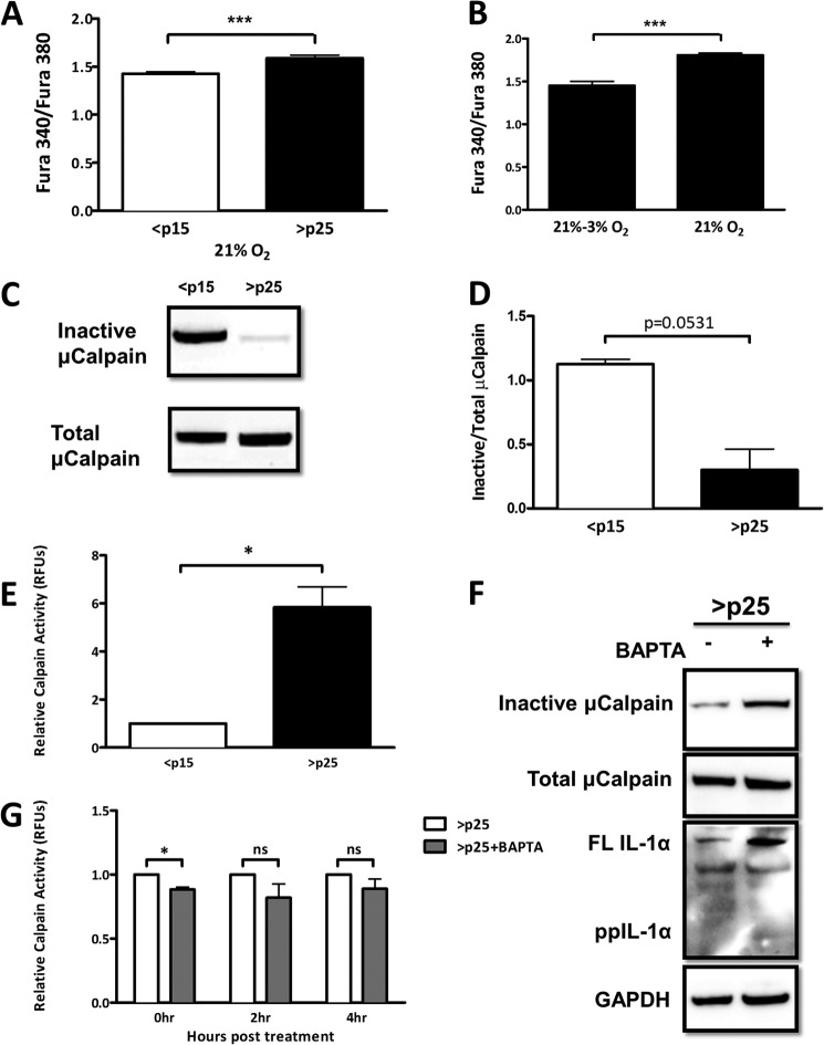
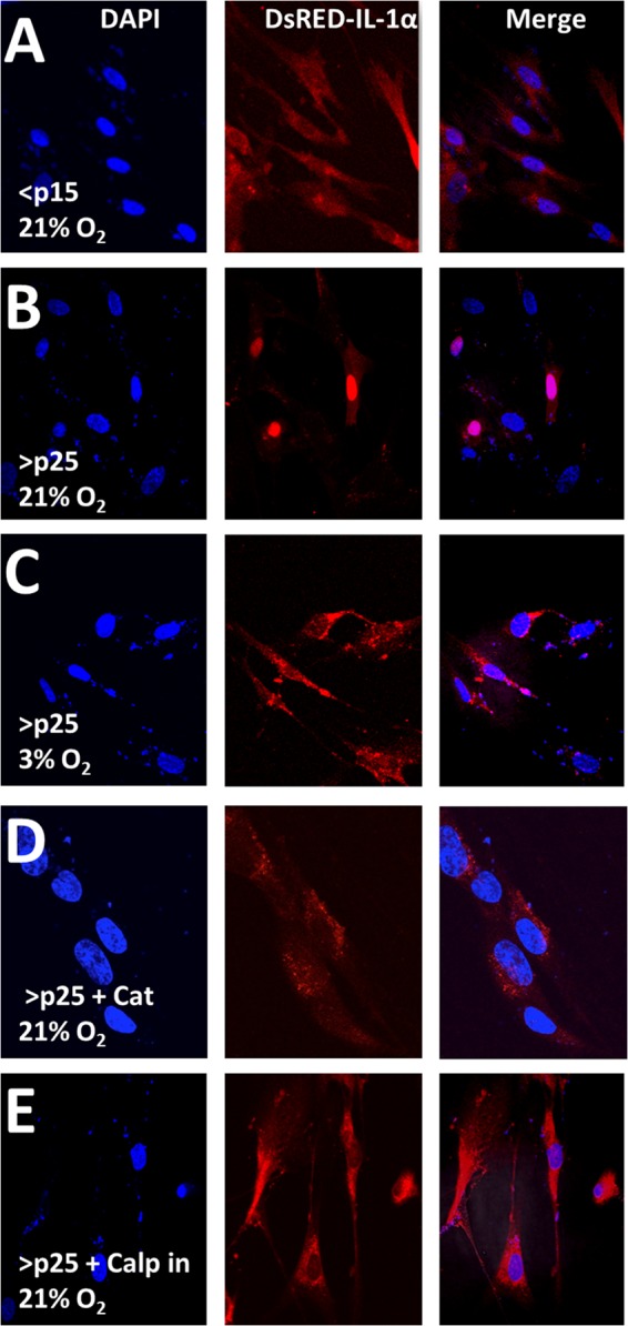
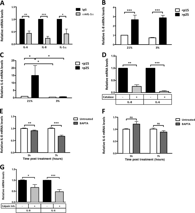
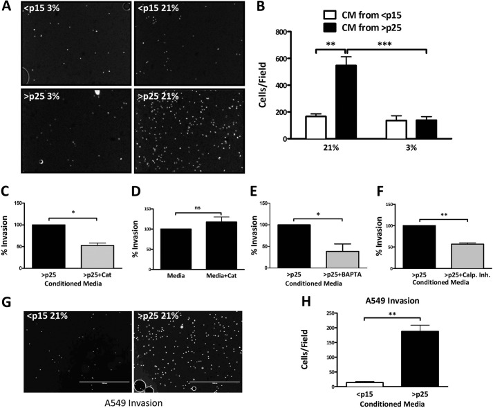
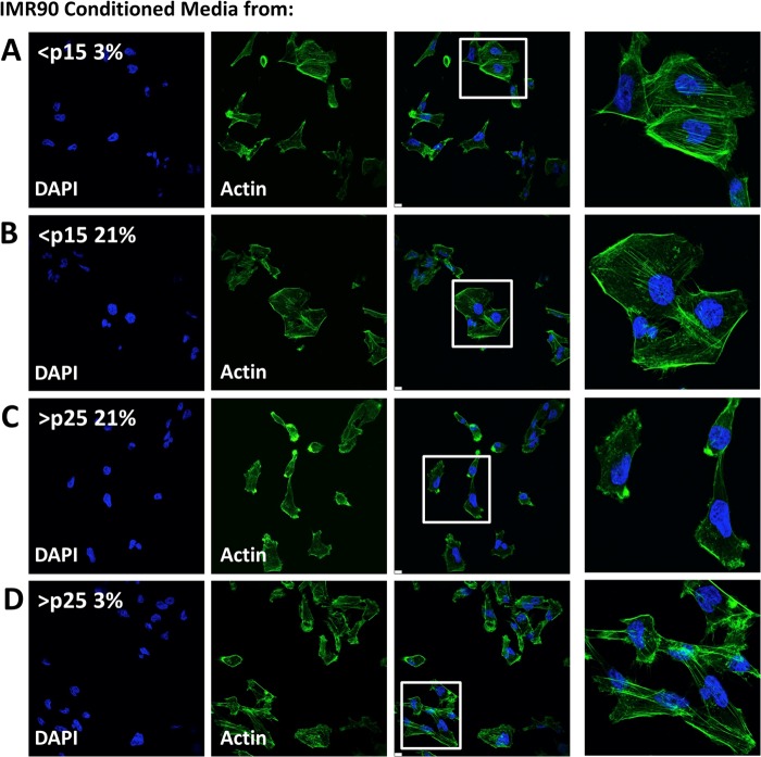
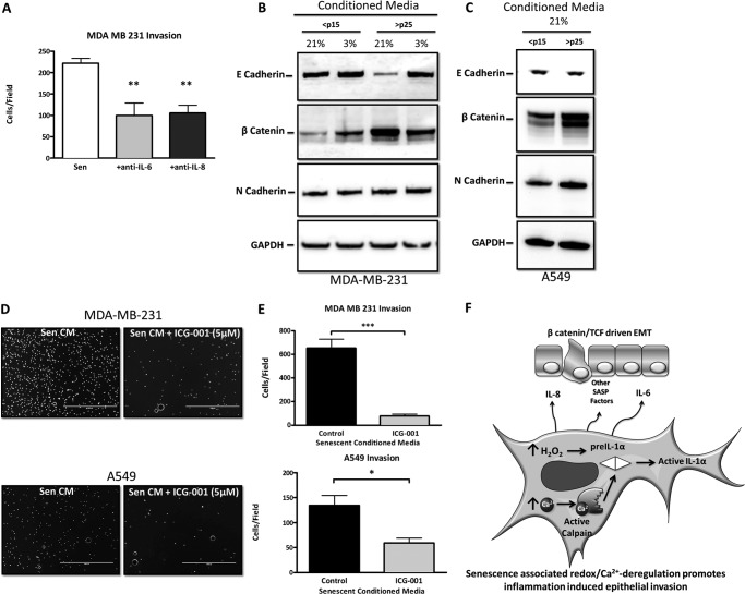
Senescent cells accumulate in aged tissue and are causally linked to age-associated tissue degeneration. These non-dividing, metabolically active cells are highly secretory and alter tissue homeostasis, creating an environment conducive to metastatic disease progression. IL-1α is a key senescence-associated (SA) proinflammatory cytokine that acts as a critical upstream regulator of the SA secretory phenotype (SASP). We established that SA shifts in steady-state H2O2 and intracellular Ca(2+) levels caused an increase in IL-1α expression and processing. The increase in intracellular Ca(2+) promoted calpain activation and increased the proteolytic cleavage of IL-1α. Antioxidants and low oxygen tension prevented SA IL-1α expression and restricted expression of SASP components IL-6 and IL-8. Ca(2+) chelation or calpain inhibition prevented SA processing of IL-1α and its ability to induce downstream cytokine expression. Conditioned medium from senescent cells treated with antioxidants or Ca(2+) chelators or cultured in low oxygen markedly reduced the invasive capacity of proximal metastatic cancer cells. In this paracrine fashion, senescent cells promoted invasion by inducing an epithelial-mesenchymal transition, actin reorganization, and cellular polarization of neighboring cancer cells. Collectively, these findings demonstrate how SA alterations in the redox state and Ca(2+) homeostasis modulate the inflammatory phenotype through the regulation of the SASP initiator IL-1α, creating a microenvironment permissive to tumor invasion.

Keywords: Antioxidants; Calcium; Calpain; Cell Invasion; Cellular Senescence; Epithelial-Mesenchymal Transition; Hydrogen Peroxide; Interleukin; Redox Regulation.


Figures

FIGURE 1.
FIGURE 1.
SA IL-1α expression is regulated by cellular H2O2. A, histogram of data obtained using an ImageStreamX flow cytometer to investigate the fluorescence intensity of the reactive oxygen species-sensitive dye H2DCFDA in IMR-90 cells. Presenescent (<p15; n = 8070) and senescent (>p25; n = 3458) cells were quantified for DCF intensity. One representative image is shown from the bin corresponding to the mean intensity of the 505–560-nm emission channel for each population. Positive DCF staining is shown in green in the cell image inlay. Mean fluorescent values for cells treated with H2DCFDA were normalized to control DCF-treated values for <p15 and >p25 cells. B, bar graph of the ImageStreamX data. The IDEAS software program was used to calculate the mean intensity of the DCF signal for each sample, and the results are plotted with error bars that represent S.D. C, SA β-galactosidase staining in presenescent (<p15) and senescent (>p25) IMR-90 fibroblasts cultured in 21% O2. D, quantitative real-time PCR (qRT-PCR) of the IL-1α transcript from presenescent (<p15) and senescent (>p25) IMR-90 fibroblasts cultured in 21 or 3% O2. E, Western blot analysis of intracellular catalase after overnight incubation with 500 units/ml recombinant catalase added to cell culture medium. GAPDH was used as a loading control. F, qRT-PCR of the IL-1α transcript from senescent cells with and without 500 units/ml recombinant catalase. G, Western blot analysis of IL-1α protein using an antibody directed against an N-terminal epitope (amino acids 2–112) in <p15 and >p25 cells. N-Acetyl-l-cysteine (NAC) was used at a concentration of 2 mm, and recombinant catalase (Cat) was used at 500 units/ml. Data represent n ≥ 3. *, p ≤ 0.05; **, p ≤ 0.01; ***, p ≤ 0.001.
FIGURE 2.
FIGURE 2.
SA shifts in intracellular Ca2+ activate calpain and promote IL-1α processing. A, ratiometric Ca2+ imaging of presenescent (<p15) and senescent (>p25) primary IMR-90 fibroblasts. B, ratiometric Ca2+ imaging of senescent (>p25) IMR-90 fibroblasts maintained at 21% oxygen tension or moved to 3% oxygen tension. Cells were assayed 1 week after changing the oxygen tension. C, Western blot analysis of inactive and total μ-calpain in presenescent and senescent cells. D, quantification of C. E, calpain activity measured using fluorescently labeled substrate in presenescent (<p15) and senescent (>p25) primary IMR-90 fibroblasts. RFU, relative fluorescence units. F, Western blot analysis of inactive and total μ-calpain and IL-1α in senescent cells 6 h post-treatment with and without 3 μm BAPTA-AM (pulsed for 1 h). FL IL-1α, full-length IL-1α; ppIL-1α, IL-1α propiece. G, calpain activity measured using fluorescently labeled substrates in senescent cells with and without 3 μm BAPTA-AM (pulsed for 1 h) at the indicated times post-treatment. Data represent n ≥ 3. *, p ≤ 0.05; ***, p ≤ 0.001; ns, not significant.
FIGURE 3.
FIGURE 3.
SA IL-1α is localized to the nucleus in a redox- and calpain-dependent fashion. A–E, confocal microscopy of a DsRed-IL-1α construct 16 h post-transient transfection into IMR-90 fibroblasts passaged as indicated at 21 or 3% O2 tension. Catalase (Cat) was added exogenously at 500 units/ml 3 h post-transfection. The calpain inhibitor (Calp) was used at a concentration of 40 μm and was added to the medium 3 h post-transfection. Data represent n ≥ 3.
FIGURE 4.
FIGURE 4.
Oxidative stress and Ca2+ regulate the SASP. A, qRT-PCR of IL-6, IL-8, and IL-1α transcripts in senescent cells with or without neutralizing antibody to secreted mouse IL-1α (αmIL-1α). B, qRT-PCR of the IL-8 transcript from presenescent (<p15) and senescent (>p25) IMR-90 fibroblasts cultured in 21 or 3% O2. C, qRT-PCR of the IL-6 transcript from presenescent (<p15) and senescent (>p25) IMR-90 fibroblasts cultured in 21 or 3% O2. D, qRT-PCR of IL-6 and IL-8 transcripts from senescent IMR-90 fibroblasts with and without 500 units/ml recombinant catalase. E, qRT-PCR of the IL-8 transcript from senescent IMR-90 fibroblasts with and without 3 μm BAPTA-AM (pulsed for 1 h) at the indicated times post-treatment. F, qRT-PCR of the IL-6 transcript from senescent IMR-90 fibroblasts with and without 3 μm BAPTA-AM (pulsed for 1 h) at the indicated times post-treatment. G, qRT-PCR of IL-6 and IL-8 transcripts from senescent IMR-90 fibroblasts with and without 4 μm calpain inhibitor (Inh.) 18 h post-treatment. Data represent n ≥ 3. *, p ≤ 0.05; **, p ≤ 0.01; ***, p ≤ 0.001; ns not significant.
FIGURE 5.
FIGURE 5.
Limiting SA oxidant production or intracellular Ca2+ restricts SASP-mediated tumor cell invasion. A, representative images of invaded MDA-MB-231 breast cancer cells submerged in CM collected over 18 h from IMR-90 fibroblasts cultured and passaged as indicated. B, quantification of Matrigel invasion assay as described for A. Data represent n ≥ 3 ± S.E. C, Matrigel invasion assay of MDA-MB-231 breast cancer cells submerged in CM from senescent IMR-90 fibroblasts with and without treatment with 500 units/ml catalase (Cat) prior to CM collection. D, Matrigel invasion assay of invaded MDA-MB-231 breast cancer cells in serum-containing medium collected over 18 h with and without the addition of 500 units/ml recombinant catalase. E, Matrigel invasion assay of MDA-MB-231 breast cancer cells submerged in CM from senescent IMR-90 fibroblasts with and without 3 μm BAPTA-AM (pulsed for 1 h). CM was collected 18 h post-treatment. F, Matrigel invasion assay of MDA-MB-231 breast cancer cells submerged in CM from senescent IMR-90 fibroblasts with and without 4 μm calpain inhibitor. CM was collected 18 h post-treatment. G, representative images of invaded A549 lung carcinoma cells submerged in CM collected over 18 h from IMR-90 fibroblasts passaged as indicated. H, quantification of Matrigel invasion assay as described for G. Data represent n ≥ 3. *, p ≤ 0.05; **, p ≤ 0.01; ***, p ≤ 0.001; ns not significant.
FIGURE 6.
FIGURE 6.
Actin remodeling in MDA-MB-231 cells is driven by exposure to CM from senescent cells. A–D, confocal microscopy of filamentous actin (stained with Alexa Fluor 488-phalloidin conjugate) in MDA-MB-231 breast cancer cells after incubation with CM from IMR-90 fibroblasts cultured to senescence at 21 or 3% O2 tension. Nuclei were stained with DAPI. Data represent n ≥ 3.
FIGURE 7.
FIGURE 7.
Redox control of SASP-mediated tumor cell invasion and transformation. A, Matrigel invasion assay of MDA-MB-231 breast cancer cells submerged in CM from senescent IMR-90 fibroblasts (Sen) with and without neutralizing antibody against IL-6 or IL-8. B, Western blot analysis of epithelial marker E-cadherin and mesenchymal markers β-catenin and N-cadherin in MDA-MB-231 cells cultured in IMR-90 CM. C, Western blot analysis of epithelial marker E-cadherin and mesenchymal markers β-catenin and N-cadherin in A549 cells cultured in IMR-90 CM. D, representative images of invaded MDA-MB-231 breast cancer cells and A549 lung carcinoma cells submerged in CM collected over 18 h from senescent IMR-90 fibroblasts with and without 5 μm ICG-001 added to CM immediately prior to use. E, quantification of D. F, model demonstrating how redox/Ca2+-stressed senescent fibroblasts synergistically promote epithelial invasion in adjacent epithelial cells through their secretory phenotype. Data represent n ≥ 3 ± S.E. *, p ≤ 0.05; **, p ≤ 0.01; ***, p ≤ 0.001. TCF, T cell factor.

Similar articles

Cited by

References

    1. Campisi J. (2005) Senescent cells, tumor suppression, and organismal aging: good citizens, bad neighbors. Cell 120, 513–522 - PubMed
    1. Baker D. J., Wijshake T., Tchkonia T., LeBrasseur N. K., Childs B. G., van de Sluis B., Kirkland J. L., van Deursen J. M. (2011) Clearance of p16Ink4a-positive senescent cells delays ageing-associated disorders. Nature 479, 232–236 - PMC - PubMed
    1. Mariotti M., Castiglioni S., Bernardini D., Maier J. A. (2006) Interleukin 1α is a marker of endothelial cellular senescent. Immun. Ageing 3, 4. - PMC - PubMed
    1. Kumar S., Millis A. J., Baglioni C. (1992) Expression of interleukin 1-inducible genes and production of interleukin 1 by aging human fibroblasts. Proc. Natl. Acad. Sci. U.S.A. 89, 4683–4687 - PMC - PubMed
    1. Garfinkel S., Brown S., Wessendorf J. H., Maciag T. (1994) Post-transcriptional regulation of interleukin 1α in various strains of young and senescent human umbilical vein endothelial cells. Proc. Natl. Acad. Sci. U.S.A. 91, 1559–1563 - PMC - PubMed

Publication types

MeSH terms

LinkOut - more resources