. 2013 Sep 30;202(7):1041-55.
doi: 10.1083/jcb.201302139. Epub 2013 Sep 23.

Cyclin B1 mRNA translation is temporally controlled through formation and disassembly of RNA granules

Affiliations

Cyclin B1 mRNA translation is temporally controlled through formation and disassembly of RNA granules

Tomoya Kotani et al. J Cell Biol. .

Abstract

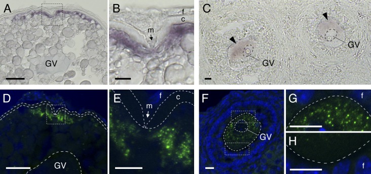
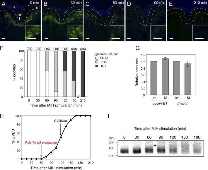
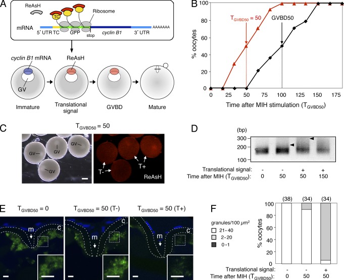
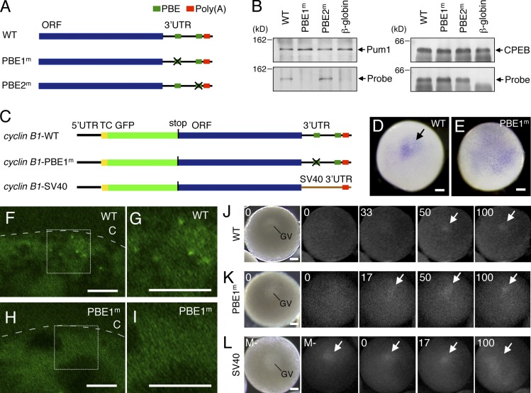
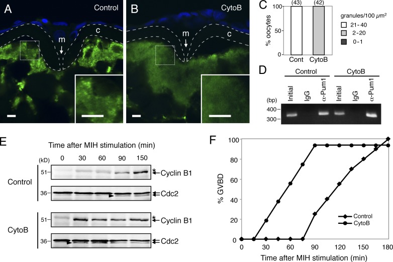
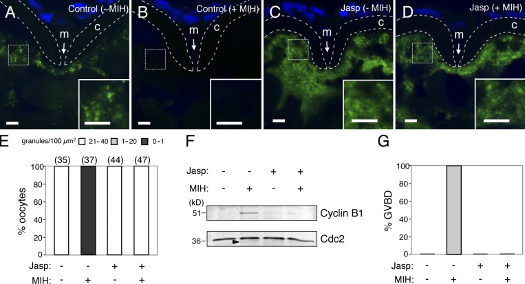
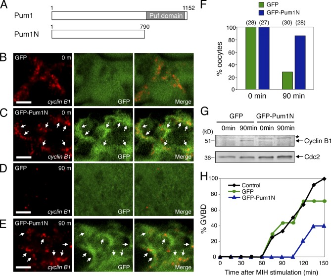
Temporal control of messenger RNA (mRNA) translation is an important mechanism for regulating cellular, neuronal, and developmental processes. However, mechanisms that coordinate timing of translational activation remain largely unresolved. Full-grown oocytes arrest meiosis at prophase I and deposit dormant mRNAs. Of these, translational control of cyclin B1 mRNA in response to maturation-inducing hormone is important for normal progression of oocyte maturation, through which oocytes acquire fertility. In this study, we found that dormant cyclin B1 mRNA forms granules in the cytoplasm of zebrafish and mouse oocytes. Real-time imaging of translation revealed that the granules disassemble at the time of translational activation during maturation. Formation of cyclin B1 RNA granules requires binding of the mRNA to Pumilio1 protein and depends on actin filaments. Disruption of cyclin B1 RNA granules accelerated the timing of their translational activation after induction of maturation, whereas stabilization hindered translational activation. Thus, our results suggest that RNA granule formation is critical for the regulation of timing of translational activation.


Figures

Figure 1.
Figure 1.
Zebrafish and mouse oocytes store dormant cyclin B1 mRNAs as asymmetrically distributed RNA granules. (A and B) Distribution of cyclin B1 mRNA in the zebrafish oocyte. B is an enlarged view of the boxed region in A. GV, germinal vesicle; m, micropile; f, follicle cells; c, chorion. (C) Distribution of cyclin B1 mRNA (arrowheads) in the mouse oocyte. (D and E) FISH analysis of cyclin B1 mRNA (green) in the zebrafish oocyte. DNA is shown in blue. E is an enlarged view of the boxed region in D. The chorion is outlined by broken lines. (F–H) FISH analysis of cyclin B1 mRNA in the mouse oocyte. G and H are enlarged views of the boxed regions in F. The oocyte is outlined by broken lines. Bars: (A and D) 50 µm; (B, C, and E–H) 10 µm.
Figure 2.
Figure 2.
Mouse cyclin B1 RNA granules disassemble during oocyte maturation. (A–C) FISH analysis of cyclin B1 mRNA (green) in mouse oocytes in GV stage (A), prometaphase I (B), and metaphase II (C). DNA is shown in blue. Oocytes are outlined by broken lines. GV, germinal vesicle; PB, polar body. Bars, 10 µm. (D) The number of RNA granules per 100 µm2 in individual oocytes in GV stage (GV), prometaphase I (PMI), and metaphase II (MII) was counted and categorized as dense (21–40), partially disassembled (2–20), and disassembled (0–1). The numbers in parentheses indicate the total number of oocytes analyzed. Similar results were obtained from two independent experiments. (E) cyclin B1 and β-actin mRNAs from equal numbers of oocytes in GV stage and metaphase II were assayed by quantitative RT-PCR (means ± standard deviations; n = 3).
Figure 3.
Figure 3.
Zebrafish cyclin B1 RNA granules disassemble during oocyte maturation. (A–E) FISH analysis of cyclin B1 mRNA (green) in zebrafish oocytes at 0 (A), 30 (B), 60 (C), 90 (D), and 210 min (E) after induction of oocyte maturation. DNA is shown in blue. Insets are enlarged views of the boxed regions. Chorions are outlined by broken lines. f, follicle cells; m, micropile; c, chorion. Bars, 5 µm. (F) The number of RNA granules per 100 µm2 in individual oocytes was counted and categorized as indicated. The numbers in parentheses indicate the total number of oocytes analyzed. Similar results were obtained from two independent experiments. (G) cyclin B1 and β-actin mRNAs from equal numbers of immature (Im) and mature (M) oocytes were assayed by quantitative RT-PCR (means ± standard deviations; n = 4). (H) Time course of GVBD after MIH stimulation. (I) Time course of PAT assay for cyclin B1 mRNA after MIH stimulation. An arrowhead indicates an initial signal of poly(A) tail elongation.
Figure 4.
Figure 4.
Timing of cyclin B1 RNA granule disassembly coincides with that of translational activation. (A) Schematic views for visualization of site and timing of translation during oocyte maturation. (top) The cyclin B1 reporter mRNA carries sequences encoding the TC tag (TC) and GFP downstream of cyclin B1 5′UTR. The biarsenical dye ReAsH emits no fluorescence in the absence of interaction with the TC tag. Translational activation of the reporter mRNA is visualized by binding of ReAsH to the nascent TC tag peptide, which immediately emits fluorescence. (bottom) The cyclin B1 reporter mRNA localized at the animal polar cytoplasm of an immature oocyte shows a translational signal at approximately half of the time point at which GVBD occurred. (B) Time course of translational signals (red triangles) and GVBD (black squares). (C) Translational signals at the time TGVBD50 = 50. Half of the oocytes show a translational signal (T+), whereas the remaining oocytes show no translational signal (T−). GV, germinal vesicle. Bar, 200 µm. (D) PAT assay of cyclin B1 mRNA at the time TGVBD50 = 0, 50, and 150 of the oocytes exhibiting no translational signal (−) or the oocytes exhibiting a translational signal (+). Arrowheads indicate poly(A) tails at the time TGVBD50 = 50. (E) FISH analysis of cyclin B1 mRNA (green) at the time TGVBD50 = 0 and 50 of the oocytes exhibiting no translational signal (T−) or the oocytes exhibiting a translational signal (T+). DNA is shown in blue. Insets are enlarged views of the boxed regions. Chorions are outlined by broken lines. m, micropile; c, chorion. Bars, 5 µm. (F) The number of RNA granules per 100 µm2 in individual oocytes was counted and categorized as indicated. The numbers in parentheses indicate the total number of oocytes analyzed. Similar results were obtained from three independent experiments.
Figure 5.
Figure 5.
Mouse and zebrafish Pum1 proteins associate with cyclin B1 mRNAs. (A) Schematic diagrams of mouse and zebrafish cyclin B1 3′UTRs. Green rectangles indicate putative PBEs, and red rectangles indicate the poly(A) signal. (B, top) Immunoblotting of mouse ovary extracts before IP (Initial) and IP with goat IgG (IgG) or anti-Pum1 goat antibody (α-Pum1). (bottom) RT-PCR amplification for cyclin B1, mos, β-actin, and α-tubulin transcripts. (C, top) Immunoblotting of zebrafish ovary extracts before IP (Initial) and IP with rabbit IgG (IgG) or anti-Pum1 rabbit antibody (α-Pum1). (bottom) RT-PCR amplification for cyclin B1, mos, β-actin, and α-tubulin transcripts. Similar results were obtained from three independent experiments.
Figure 6.
Figure 6.
Binding of cyclin B1 mRNA to Pum1 is required for granule formation and regulation of accurate timing of translational activation. (A) Schematic diagrams of RNA probes used for UV cross-linking assay. The WT probe consists of cyclin B1 ORF and 3′UTR. The PBE1m probe carries mutation in PBE at nucleotides 1,338–1,350 of cyclin B1 cDNA. The PBE2m probe carries mutation in PBE at nucleotides 1,465–1,468. (B) UV cross-linking assay of cyclin B1 probes with Pum1 (left) and CPEB (right). (C) Schematic diagrams of reporter mRNAs used for production of transgenic fish. (D and E) Whole-mount in situ hybridization of cyclin B1-WT (D) and cyclin B1-PBE1m (E) mRNAs. (F–I) FISH analysis of cyclin B1-WT (F and G) and cyclin B1-PBE1m (H and I) mRNAs. Oocytes are outlined by broken lines. c, chorion. G and I are enlarged views of the boxed regions in F and H. (J–L) Real-time images of translational activation of cyclin B1-WT (J), -PBE1m (K), and -SV40 (L) mRNAs. GV, germinal vesicle. Arrows indicate translational signals of the reporter mRNAs. Bars: (D, E, and J–L) 100 µm; (F–I) 5 µm.
Figure 7.
Figure 7.
Depolymerization of actin filaments causes disassembly of cyclin B1 RNA granules and acceleration of translational activation. (A and B) FISH analysis of cyclin B1 mRNA (green) in control (A) and cytochalasin B (CytoB)–treated (B) zebrafish oocytes. DNA is shown in blue. Insets are enlarged views of the boxed regions. Chorions are outlined by broken lines. m, micropile; c, chorion. Bars, 5 µm. (C) The number of RNA granules per 100 µm2 in individual oocytes was counted and categorized as indicated. The numbers in parentheses indicate the total number of oocytes analyzed. Similar results were obtained from three independent experiments. (D) RT-PCR amplification for cyclin B1 of extracts from control (Control) and CytoB-treated oocytes before IP (Initial) and IP with rabbit IgG (IgG) or anti-Pum1 rabbit antibody (α-Pum1). (E) Time course of Cyclin B1 synthesis after induction of oocyte maturation. Arrowheads indicate active forms of the Cdc2 kinase. Asterisks show nonspecific bands. (F) Time course of GVBD after induction of oocyte maturation in the control and CytoB-treated oocytes. Similar results were obtained from three independent experiments.
Figure 8.
Figure 8.
Stabilization of actin filaments prevents disassembly of cyclin B1 RNA granules and their translational activation. (A–D) FISH analysis of cyclin B1 mRNA (green) in control (A and B) and jasplakinolide (Jasp)-treated oocytes (C and D), stimulated with (B and D) or without (A and C) MIH. DNA is shown in blue. Insets are enlarged views of the boxed regions. Chorions are outlined by broken lines. m, micropile; c, chorion. Bars, 5 µm. (E) The number of RNA granules per 100 µm2 in individual oocytes was counted and categorized as indicated. The numbers in parentheses indicate the total number of oocytes analyzed. Similar results were obtained from two independent experiments. (F) Immunoblotting of Cyclin B1 and Cdc2 in oocytes stimulated with (+) or without (−) Jasp and MIH. An arrowhead indicates the active form of Cdc2 kinase. (G) Percentage of oocytes stimulated with (+) or without (−) Jasp and MIH that induced GVBD. Similar results were obtained from four independent experiments.
Figure 9.
Figure 9.
N terminus of Pum1 stabilizes cyclin B1 RNA granules and hinders their translational activation. (A) Schematic diagrams of zebrafish Pum1 and Pum1N. The Puf domain is responsible for binding to PBE in target mRNAs. (B–E) FISH analysis of cyclin B1 mRNA (red) and immunostaining of GFP (green) in oocytes expressing GFP (B and D) or GFP-Pum1N (C and E) before (B and C) or at 90 min after induction of oocyte maturation (D and E). Merged images are shown (Merge). Arrows indicate aggregation of GFP-Pum1N surrounding cyclin B1 RNA granules. Bars, 5 µm. (F) The number of RNA granules per 100 µm2 in individual oocytes was counted and categorized. Percentage of oocytes categorized as dense (21–40) is shown. The numbers in parentheses indicate the total number of oocytes analyzed. Similar results were obtained from three independent experiments. (G) Immunoblotting of Cyclin B1 and Cdc2 in oocytes expressing GFP and GFP-Pum1N before (0 min) and at 90 min after induction of oocyte maturation. An asterisk shows a nonspecific band. The active form of Cdc2 kinase was undetectable in the oocytes at these time points. (H) Time course of GVBD after induction of oocyte maturation in the control, GFP-expressing, and GFP-Pum1N–expressing oocytes. Similar results were obtained from three independent experiments.

Comment in

Similar articles

Cited by

References

    1. Anderson P., Kedersha N. 2009. RNA granules: post-transcriptional and epigenetic modulators of gene expression. Nat. Rev. Mol. Cell Biol. 10:430–436 10.1038/nrm2694 - DOI - PubMed
    1. Baez M.V., Luchelli L., Maschi D., Habif M., Pascual M., Thomas M.G., Boccaccio G.L. 2011. Smaug1 mRNA-silencing foci respond to NMDA and modulate synapse formation. J. Cell Biol. 195:1141–1157 10.1083/jcb.201108159 - DOI - PMC - PubMed
    1. Bally-Cuif L., Schatz W.J., Ho R.K. 1998. Characterization of the zebrafish Orb/CPEB-related RNA binding protein and localization of maternal components in the zebrafish oocyte. Mech. Dev. 77:31–47 10.1016/S0925-4773(98)00109-9 - DOI - PubMed
    1. Boag P.R., Atalay A., Robida S., Reinke V., Blackwell T.K. 2008. Protection of specific maternal messenger RNAs by the P body protein CGH-1 (Dhh1/RCK) during Caenorhabditis elegans oogenesis. J. Cell Biol. 182:543–557 10.1083/jcb.200801183 - DOI - PMC - PubMed
    1. Chapman D.L., Wolgemuth D.J. 1992. Identification of a mouse B-type cyclin which exhibits developmentally regulated expression in the germ line. Mol. Reprod. Dev. 33:259–269 10.1002/mrd.1080330305 - DOI - PubMed

Publication types

MeSH terms