. 2013 Oct 8;110(41):16486-91.
doi: 10.1073/pnas.1314819110. Epub 2013 Sep 23.

Precocious cerebellum development and improved motor functions in mice lacking the astrocyte cilium-, patched 1-associated Gpr37l1 receptor

Affiliations

Precocious cerebellum development and improved motor functions in mice lacking the astrocyte cilium-, patched 1-associated Gpr37l1 receptor

Daniela Marazziti et al. Proc Natl Acad Sci U S A. .

Abstract

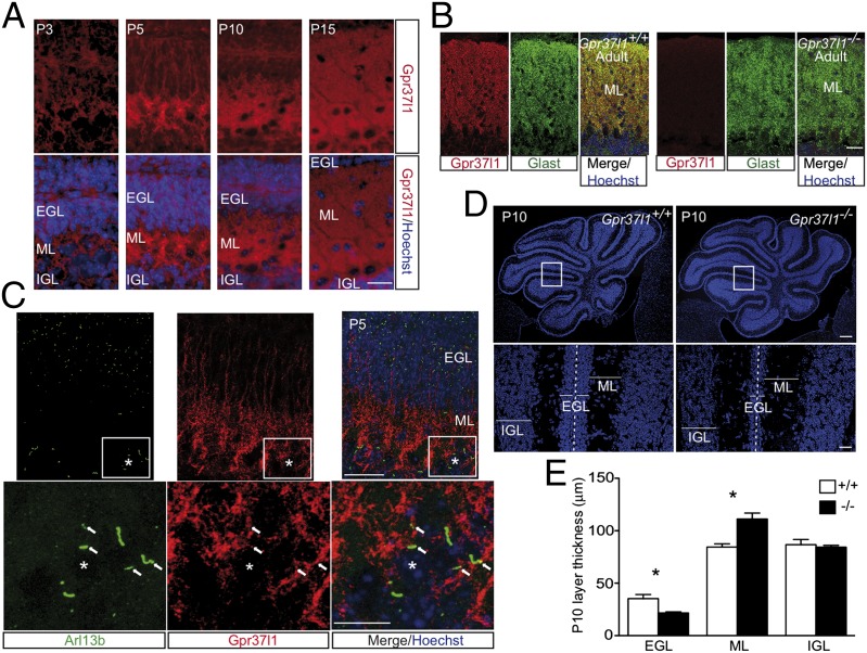
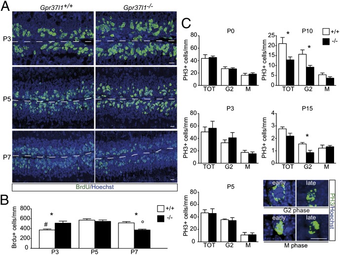
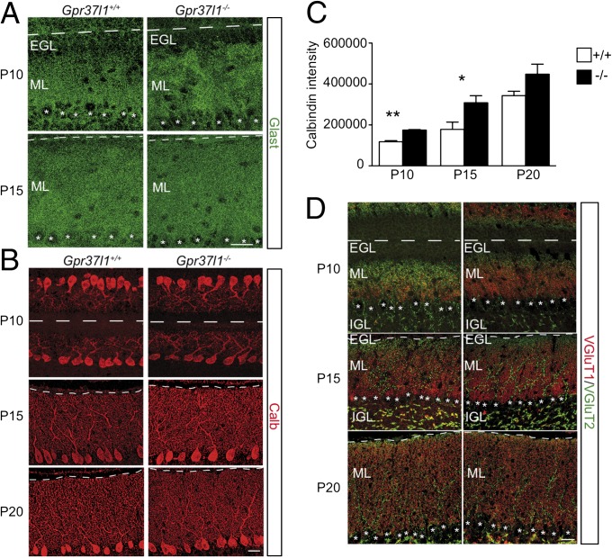
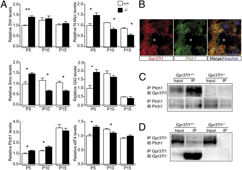
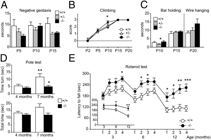
In the developing cerebellum, the proliferation and differentiation of glial and neuronal cell types depend on the modulation of the sonic hedgehog (Shh) signaling pathway. The vertebrate G-protein-coupled receptor 37-like 1 (GPR37L1) gene encodes a putative G-protein-coupled receptor that is expressed in newborn and adult cerebellar Bergmann glia astrocytes. This study shows that the ablation of the murine Gpr37l1 gene results in premature down-regulation of proliferation of granule neuron precursors and precocious maturation of Bergmann glia and Purkinje neurons. These alterations are accompanied by improved adult motor learning and coordination. Gpr37l1(-/-) mice also exhibit specific modifications of the Shh signaling cascade. Specific assays show that in Bergmann glia cells Gpr37l1 is associated with primary cilium membranes and it specifically interacts and colocalizes with the Shh primary receptor, patched 1. These findings indicate that the patched 1-associated Gpr37l1 receptor participates in the regulation of postnatal cerebellum development by modulating the Shh pathway.

Keywords: mitogenic signaling; mutant mouse model.


Conflict of interest statement

The authors declare no conflict of interest.

Figures

Fig. 1.
Fig. 1.
Gpr37l1 expression and cerebellar morphology of adult and developing Gpr37l1+/+ and Gpr37l1−/− male mice. (A) Cerebellar immunofluorescence labeling of Gpr37l1 and nuclear Hoechst staining in P3, P5, P10, and P15 pups and (B) immunofluorescence labeling of Gpr37l1 (red) and Glast (green) in Gpr37l1+/+ and Gpr37l1−/− adult mice. (C) Double immunofluorescence staining of Gpr37l1 and Arl13b in sagittal cerebellar sections of P5 wild-type pups. (D) Nuclear Hoechst staining in P10 cerebella of Gpr37l1+/+ and Gpr37l1−/− pups. (E) Quantification of the average thickness of the EGL, ML, and IGL in the two genotype groups at P10 (mean ± SEM, n = 3 per group). *P < 0.05 +/+ vs. −/−, unpaired t test. (Scale bars in AC, Upper, and D, Lower, 25 μm and in C, Lower, 10 μm and D, Upper, 250 μm.) EGL, external granular layer; IGL, internal granular layer; ML, molecular layer. Asterisks indicate Purkinje neuron somata; arrows point to Gpr37l1 and Arl13b doubly positive primary cilium structures.
Fig. 2.
Fig. 2.
Postnatal proliferation of cerebellar granule neuron precursors in Gpr37l1+/+ and Gpr37l1−/− male mice. (A) Immunofluorescence labeling of BrdU and Hoechst staining of EGL cells at P3, P5, and P7 in Gpr37l1+/+ and Gpr37l1−/− littermates. (B) Quantification of the average number of BrdU-positive cells normalized by the EGL length (mean ± SEM), *P < 0.05 +/+ vs. −/−; #P < 0.05 P3 vs. P5, P7 in Gpr37l1+/+ mice; °P < 0.05 P7 vs. P3, P5 in Gpr37l1−/− mice, unpaired t test. (C) Quantification of the average number of phosphohistone H3 (PH3)-positive and G2- or M-phase cells at P0, P3, P5, P10, and P15 in the EGL of Gpr37l1+/+ and Gpr37l1−/− littermates (mean ± SEM n = 3 per group), *P ≤ 0.05 +/+ vs. −/−, unpaired t test. (Lower) Representative images of anti-PH3 immunofluorescence labeling and nuclear Hoechst counterstaining of EGL cells in P10 Gpr37l1+/+ pups, showing distinct PH3-positive cells in either early or late G2 phase and either early or late (mitotic division) M phase. (Scale bars in A and C, 10 μm.)
Fig. 3.
Fig. 3.
Postnatal maturation of Bergmann glia and Purkinje neurons in Gpr37l1+/+ and Gpr37l1−/− male mice. (A) Immunofluorescence labeling of Glast in P10 and P15 cerebella of Gpr37l1+/+ and Gpr37l1−/− littermates. (B) Immunofluorescence labeling of Calb in P10, P15, and P20 cerebella of Gpr37l1+/+ and Gpr37l1−/− littermates. (C) Quantification of Calb immunostaining intensity. Data are expressed in arbitrary units (mean ± SEM, n = 3 per group), *P = 0.057, **P < 0.001 +/+ vs. −/−, unpaired t test. (D) Double immunofluorescence staining of VGluT1 and VGluT2 in P10, P15, and P20 cerebella of Gpr37l1+/+ and Gpr37l1−/− littermates. (Scale bars in A, B, and D, 25 μm.) EGL, external granular layer; IGL, internal granular layer; ML, molecular layer. Asterisks indicate Purkinje neuron somata.
Fig. 4.
Fig. 4.
Postnatal cerebellar Shh signaling and Gpr37l1–Ptch1 interaction in Gpr37l1+/+ and Gpr37l1−/− male mice. (A) Immunoblotting and densitometric quantification of Shh pathway proteins in whole cerebellar extracts prepared at P5, P10, and P15 from Gpr37l1+/+ and Gpr37l1−/− littermate pups (mean ± SEM, n = 3 mice per group), *P < 0.05, **P < 0.005 +/+ vs. −/−, unpaired t test. (B) Double immunofluorescence staining of Gpr37l1 and Ptch1 in sagittal cerebellar sections of P3 wild-type pups. (C and D) Coimmunoprecipitation analysis of Ptch1 and Gpr37l1 in cerebellar extracts from P10 Gpr37l1+/+ and Gpr37l1−/− littermates. Immunoprecipitations (IP) were performed with the Ptch1 or Gpr37l1 antibody followed by immunoblotting for the indicated proteins. A representative result from three distinct experiments is shown. (Scale bars, 10 μm.) Asterisks indicate Purkinje neuron somata; arrows point to doubly positive Bergmann glia cells.
Fig. 5.
Fig. 5.
Behavioral analysis of newborn and adult Gpr37l1+/+ and Gpr37l1−/− littermates. (AC) Negative geotaxis latency, climbing score, bar holding, and wire hanging time measured in male mice at P2, P5, P10, P15, and P20. *P < 0.05 +/+ vs. −/− or +/− vs. −/−, unpaired t test. +/+ n = 7–8; +/− n = 13–15; −/− n = 5–6. (D) Pole test performed in adult male mice at 4 and 7 mo. Bars represent the time to turn downward (Upper graph) and the total time to climb down (Lower graph). *P < 0.05, +/+ vs. −/−, **P < 0.005, 4 vs. 7 mo in +/+ group, unpaired t test. +/+ n = 16; −/− n = 13. (E) Rotarod test performed in adult male mice at 3, 6, and 12 mo; open (+/+) and solid (−/−) circles represent the latency to fall off the rod over 4 d (mean of three trials per day); in the Inset the latency to fall at 3, 6, and 12 mo, averaged across 4 d per age group (mean ± SEM). *P < 0.05; **P < 0.005; ***P < 0.0001, +/+ vs. −/− unpaired t test. +/+ n = 17; −/− n = 12.

References

    1. Sotelo C. Cellular and genetic regulation of the development of the cerebellar system. Prog Neurobiol. 2004;72(5):295–339. - PubMed
    1. Gao WO, Heintz N, Hatten ME. Cerebellar granule cell neurogenesis is regulated by cell-cell interactions in vitro. Neuron. 1991;6(5):705–715. - PubMed
    1. Rakic P. Neuron-glia relationship during granule cell migration in developing cerebellar cortex. A Golgi and electronmicroscopic study in Macacus Rhesus. J Comp Neurol. 1971;141(3):283–312. - PubMed
    1. Yue Q, et al. PTEN deletion in Bergmann glia leads to premature differentiation and affects laminar organization. Development. 2005;132(14):3281–3291. - PubMed
    1. Tanaka S, Shaikh IM, Chiocca EA, Saeki Y. The Gs-linked receptor GPR3 inhibits the proliferation of cerebellar granule cells during postnatal development. PLoS ONE. 2009;4(6):e5922. - PMC - PubMed

Publication types

MeSH terms