. 2013 Oct 8;110(41):16420-5.
doi: 10.1073/pnas.1310097110. Epub 2013 Sep 23.

Structural insights into proteoglycan-shaped Hedgehog signaling

Affiliations

Structural insights into proteoglycan-shaped Hedgehog signaling

Daniel M Whalen et al. Proc Natl Acad Sci U S A. .

Abstract

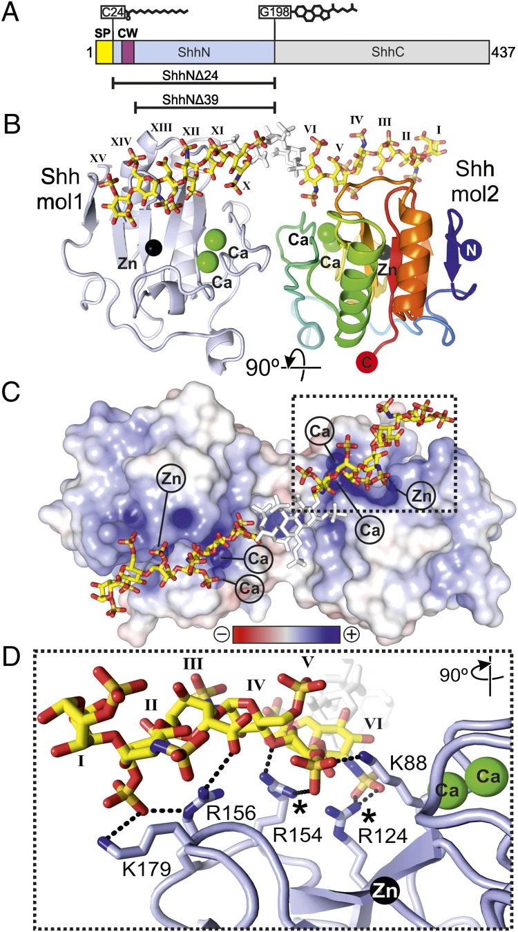
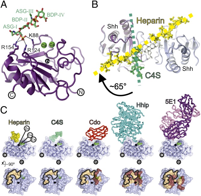
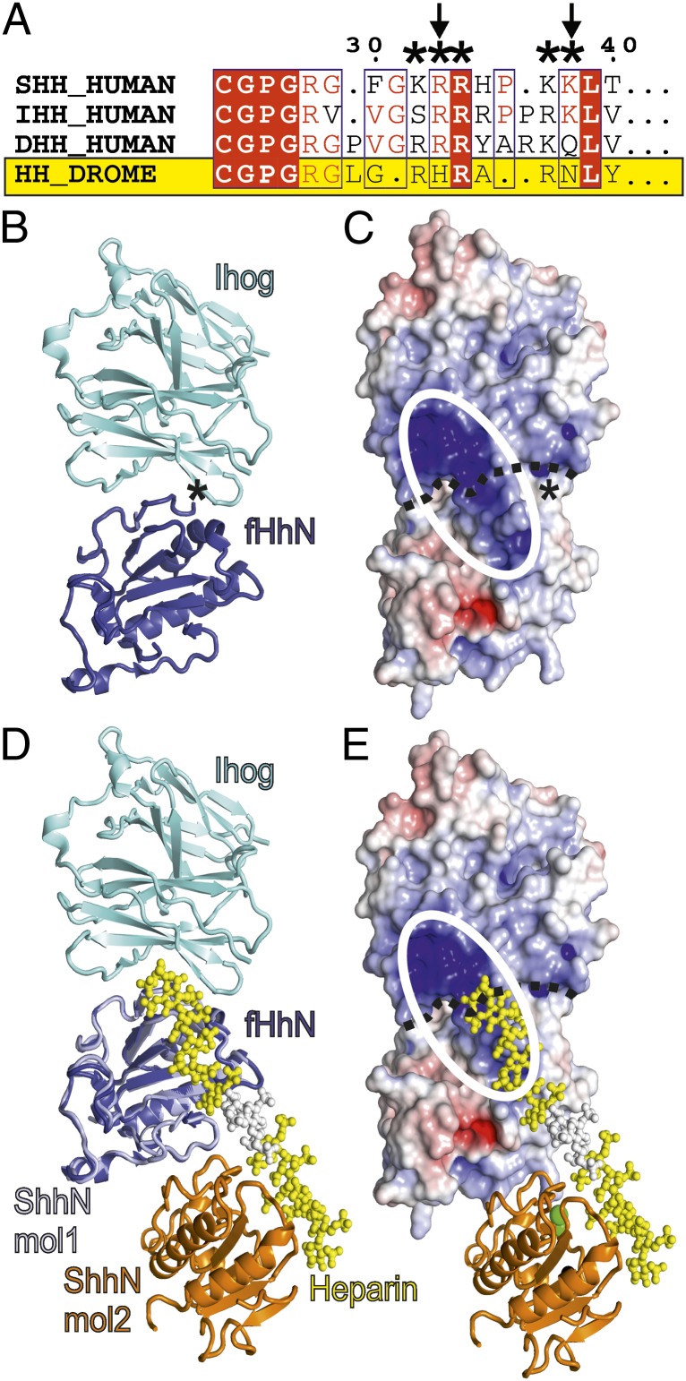
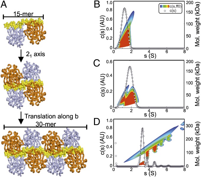
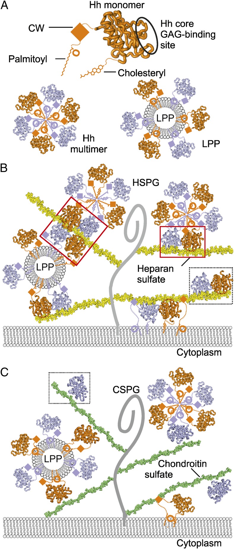
Hedgehog (Hh) morphogens play fundamental roles during embryogenesis and adulthood, in health and disease. Multiple cell surface receptors regulate the Hh signaling pathway. Among these, the glycosaminoglycan (GAG) chains of proteoglycans shape Hh gradients and signal transduction. We have determined crystal structures of Sonic Hh complexes with two GAGs, heparin and chondroitin sulfate. The interaction determinants, confirmed by site-directed mutagenesis and binding studies, reveal a previously not identified Hh site for GAG binding, common to all Hh proteins. The majority of Hh residues forming this GAG-binding site have been previously implicated in developmental diseases. Crystal packing analysis, combined with analytical ultracentrifugation of Sonic Hh-GAG complexes, suggests a potential mechanism for GAG-dependent Hh multimerization. Taken together, these results provide a direct mechanistic explanation of the observed correlation between disease and impaired Hh gradient formation. Moreover, GAG binding partially overlaps with the site of Hh interactions with an array of protein partners including Patched, hedgehog interacting protein, and the interference hedgehog protein family, suggesting a unique mechanism of Hh signaling modulation.


Conflict of interest statement

The authors declare no conflict of interest.

Figures

Fig. 1.
Fig. 1.
Structure of the Shh–heparin complex. (A) Schematic of Shh. CW, Cardin–Weintraub sequence; SP, signal peptide. Lipid modifications are shown. Constructs used are indicated. (B) Ribbon representation of the ShhNΔ39–heparin complex. One molecule is shown in blue, the other in rainbow coloring. Calciums (green), zinc (black), and heparin (yellow, carbon; red, oxygen; blue, nitrogen; and orange, sulfur) are depicted. The three disordered sugar residues (VII, VIII, and IX) are shown in white. Sugar residues are marked in roman numerals (even, N,O6-disulfoglucosamine; odd, O2-sulfoiduronic acid). (C) Electrostatic potential from red (−8 kbT/ec) to blue (+8 kbT/ec). (D) Close-up view of the Shh–heparin-binding site showing the marked region in C. Color coding is as in B. Shh molecule 2 has a similar interaction pattern and architecture due to the pseudosymmetric heparin-binding site (Fig. S3). Equivalent Shh residues of Ihh disease mutations (27, 28) are marked.
Fig. 2.
Fig. 2.
Structure of the Shh–C4S complex and analysis of the ShhN core GAG-binding site. (A) Ribbon representation of the ShhNΔ39–C4S complex [Shh, violet; calciums, green; zinc, black; C4S, atomic coloring (green, carbon; red, oxygen; blue, nitrogen; orange, sulfur)]. (B) Comparison of the Shh–C4S and Shh–heparin structures. View is as in Fig. 1C. The Shh–C4S complex is superimposed onto the Shh–heparin complex (Shh, light blue; heparin, yellow). Directions of the GAG chains are shown. (C, Upper) Ribbon representation of Shh (light blue) complexes with (from Left to Right) heparin, C4S, Cdo [Protein Data Bank (PDB) ID 3D1M], Hhip (PDB ID 2WFX), and the antigen-binding fragment of the antibody 5E1 (PDB ID 3MXW). (Lower) Binding footprints mapped on Shh corresponding to Upper. Hydrophilic interactions, beige; hydrophobic interactions, brown. The heparin binding footprint on Shh is marked.
Fig. 3.
Fig. 3.
Implications of the Shh–GAG-binding site for fly Hh signaling. (A) Sequence alignment of the Shh CW. The CW is marked (*) and is only partially conserved in fly (residue exchanges are indicated with arrows). DROME, Drosophila melanogaster. (BE) Comparison of the core GAG-binding site in mouse and fly. (B and C) The fHh–Ihog complex (PDB ID 2IBG) (fHh, blue; Ihog, cyan). In C, the surface is color coded according to electrostatic potential from red (−8 kbT/ec) to blue (+8 kbT/ec). The continuous basic surface stretch formed by both fHh and Ihog is marked as a white ellipse. The asterisk marks the fHh N terminus. (D and E) Superposition of the ShhNΔ39–heparin complex onto fHh (Shh, lightblue/orange; heparin, yellow/white) in the same orientation as in B and C. Extension of the heparin chain would result in covering the positively charged patch on Ihog.
Fig. 4.
Fig. 4.
Implications for Shh multimerization. (A) Crystal packing analysis of the ShhNΔ39–heparin structure. Heparin lies parallel to the crystallographic b axis, which has 21 symmetry (Top). This operation results in sandwich arrangement of two Shh dimers packed around the heparin chain (Middle). Extension along the b axis reveals a structure comprising Shh molecules attached to heparin like beads on a string (Bottom). (BD) AUC sedimentation velocity experiments of ShhNΔ39 with heparin. (B) ShhNΔ39 shows a monodisperse peak at 1.2 S corresponding to monomeric ShhNΔ39 (rmsd 0.0322). (C) ShhNΔ39 mixed with a 6-mer heparin shows reduction in the monomeric peak and the appearance of a major peak at 1.8 S indicating a 1:1 Shh:heparin complex (rmsd 0.0183). The minor species observed was residual Shh (at ∼1.2 S). (D) ShhNΔ39 mixed with a 30-mer heparin reveals multiple peaks corresponding to discrete Shh–heparin complexes (rmsd 0.0112). AU, arbitrary unit.
Fig. 5.
Fig. 5.
Model of Hh–GAG interactions at the cell surface. (A) The Hh monomer (orange) is covalently linked to palmitoyl and cholesteryl moieties at the N- and C-termini, respectively. CW is located between the palmitoyl and Hh core domain. Our identified Hh core GAG-binding site is circled. Hh monomers assemble into homomultimers by burying lipid moieties within the hydrophobic core (Lower Left) or as part of lipoprotein particles (LPP) (Lower Right). (B) Different monomeric and oligomeric forms of Hh bind to HS chains (yellow) linked to the stalk regions of HSPGs (gray curve). Both CW and the Hh core GAG-binding site mediate Hh binding to HS. High degree of HS sulfation mediates HS-dependent Hh oligomerization. HS-induced tetramer and dimer are shown in red boxes, the Shh–heparin complex observed in the crystallographic asymmetric unit, in a dashed box. (C) Similarly to HS, CS chains (green) bind to multiple forms of Hh (monomers or lipid-induced oligomers). However, in contrast to HS, the less sulfated CS provides a platform for a lower degree of Hh oligomerization on the cell surface. A Shh–C4S complex as observed in the crystal structure is framed within a dashed box.

References

    1. Ingham PW, McMahon AP. Hedgehog signaling in animal development: Paradigms and principles. Genes Dev. 2001;15(23):3059–3087. - PubMed
    1. Belloni E, et al. Identification of Sonic hedgehog as a candidate gene responsible for holoprosencephaly. Nat Genet. 1996;14(3):353–356. - PubMed
    1. Rubin LL, de Sauvage FJ. Targeting the Hedgehog pathway in cancer. Nat Rev Drug Discov. 2006;5(12):1026–1033. - PubMed
    1. Ingham PW, Nakano Y, Seger C. Mechanisms and functions of Hedgehog signalling across the metazoa. Nat Rev Genet. 2011;12(6):393–406. - PubMed
    1. Yan D, Lin X. Shaping morphogen gradients by proteoglycans. Cold Spring Harb Perspect Biol. 2009;1(3):a002493. - PMC - PubMed

Publication types