. 2013 Nov;19(11):1483-96.
doi: 10.1261/rna.037945.112. Epub 2013 Sep 23.

FMRP regulates actin filament organization via the armadillo protein p0071

FMRP regulates actin filament organization via the armadillo protein p0071

Alexander Nolze et al. RNA. 2013 Nov.

Abstract

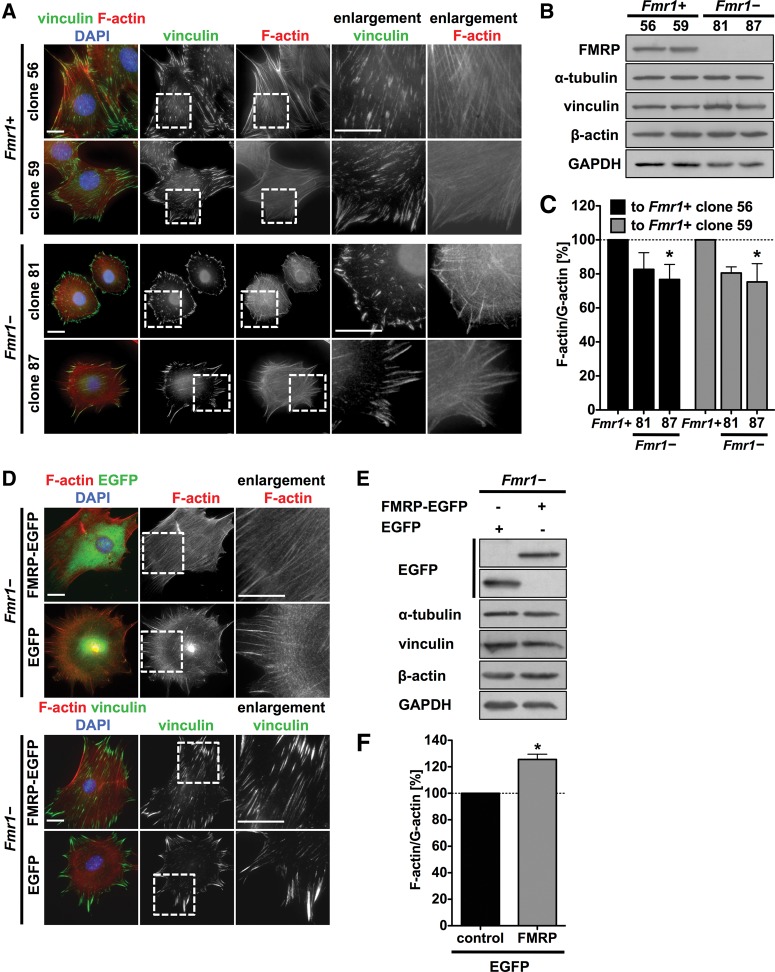
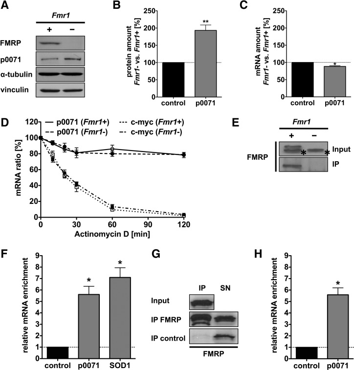
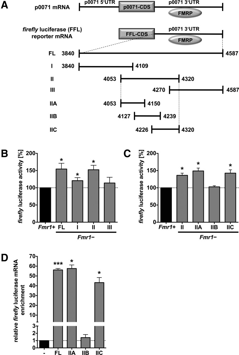
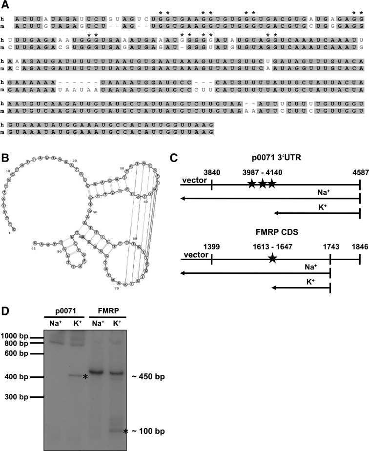
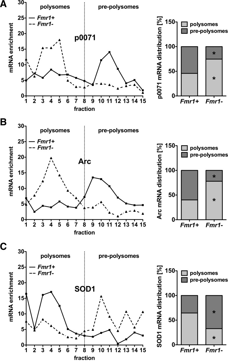
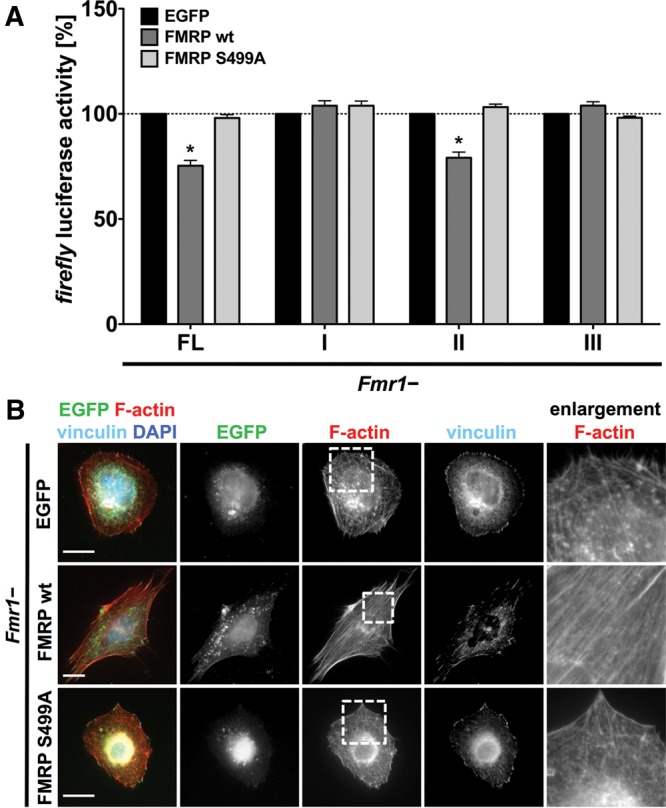
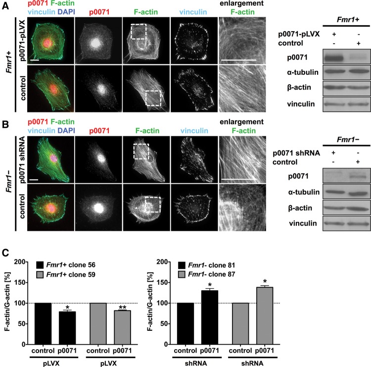
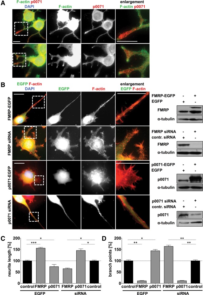
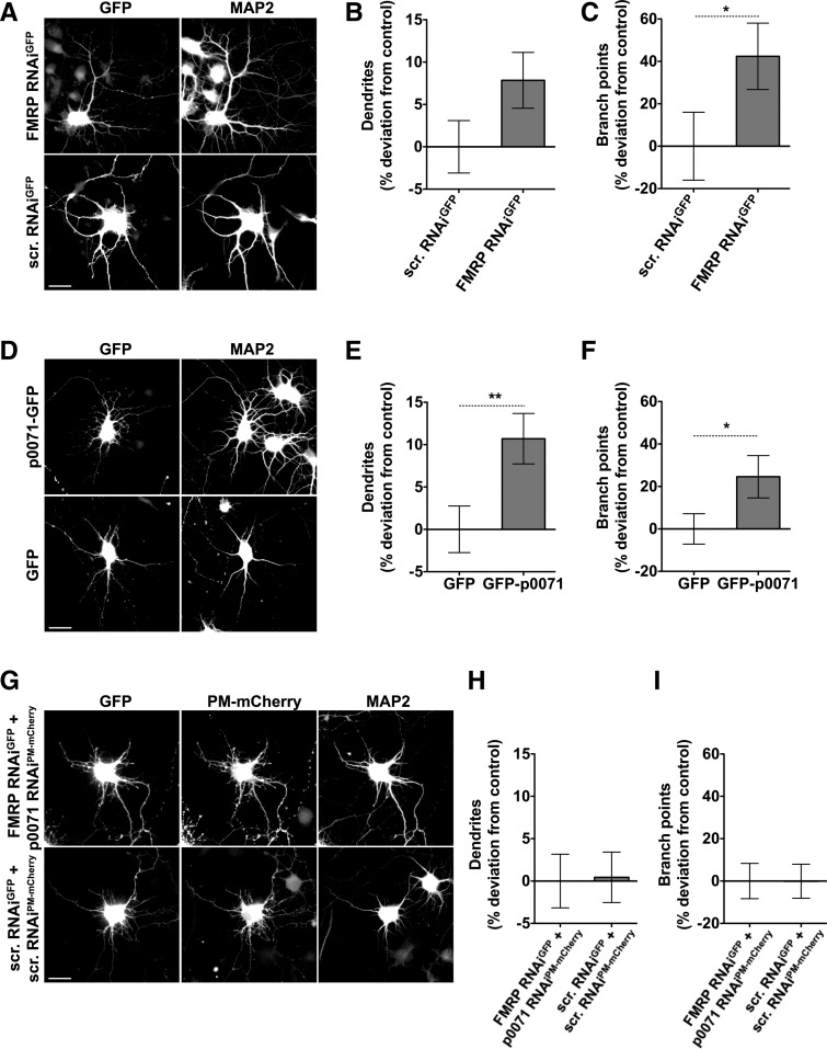
Loss of fragile X mental retardation protein (FMRP) causes synaptic dysfunction and intellectual disability. FMRP is an RNA-binding protein that controls the translation or turnover of a subset of mRNAs. Identifying these target transcripts is an important step toward understanding the pathology of the disease. Here, we show that FMRP regulates actin organization and neurite outgrowth via the armadillo protein p0071. In mouse embryonic fibroblasts (MEFs) lacking FMRP (Fmr1-), the actin cytoskeleton was markedly reorganized with reduced stress fibers and F-actin/G-actin ratios compared to fibroblasts re-expressing the protein. FMRP interfered with the translation of the p0071 mRNA in a 3'-UTR-dependent manner. Accordingly, FMRP-depleted cells revealed elevated levels of p0071 protein. The knockdown of p0071 in Fmr1- fibroblasts restored stress fibers and an elongated cell shape, thus rescuing the Fmr1- phenotype, whereas overexpression of p0071 in Fmr1+ cells mimicked the Fmr1- phenotype. Moreover, p0071 and FMRP regulated neurite outgrowth and branching in a diametrically opposed way in agreement with the negative regulation of p0071 by FMRP. These results identify p0071 as an important and novel FMRP target and strongly suggest that impaired actin cytoskeletal functions mediated by an excess of p0071 are key aspects underlying the fragile X syndrome.

Keywords: FMRP; actin organization; dendrite formation; neuromorphogenesis; p0071.


Figures

FIGURE 1.
FIGURE 1.
The loss of FMRP alters the morphology and F-actin/G-actin ratios of MEFs. (A) Immunofluorescence studies of FMRP re-expressing MEF clones (Fmr1+, clones 56, 59, top panels) with parallel stress fibers and Fmr1− cell clones (clones 81, 87, bottom panels) with reduced stress fiber formation. Enlargements of boxed regions are shown in the right panels; bar, 20 µm. (B) Representative Western blotting of Fmr1+ and Fmr1− cell lysates confirming FMRP loss in the Fmr1− cell clones and FMRP re-expression in clones 56 and 59. (C) F-actin/G-actin assay of Fmr1+ and Fmr1− cell clones showing a reduced F-actin/G-actin ratio in Fmr1− cell clones. The F-actin organization of both Fmr1− cell clones (81 and 87) was compared to both WT cell clones re-expressing FMRP (56 and 59). n = 3, (*) P ≤ 0.05. (D) Fluorescence microscopy studies of Fmr1− cells after lentiviral transduction with the indicated constructs showing F-actin- (top panels) and vinculin-staining (bottom panels). Re-expression of FMRP (FMRP-EGFP) reverted the Fmr1− phenotype compared to control transduction (EGFP); bar, 20 µm. (E) Representative Western blotting of Fmr1− cell lysates after lentiviral transduction with FMRP-EGFP or EGFP alone confirming FMRP re-expression in Fmr1− cells. (F) F-actin/G-actin assay of lentivirally transduced Fmr1− MEFs showing an increased F-actin/G-actin ratio in FMRP-EGFP-expressing cells. n = 3, (*) P ≤ 0.05.
FIGURE 2.
FIGURE 2.
FMRP associates with the p0071 mRNA and regulates p0071 expression. (A,B) Representative Western blotting (A) and quantification (B) of p0071 protein levels in Fmr1+ and Fmr1− cell lysates indicating an increased expression of p0071 in Fmr1− cells. n = 4, (**) P ≤ 0.005. (C) Quantification of p0071 mRNA levels in Fmr1+ and Fmr1− cells indicating a slightly reduced p0071 mRNA level in Fmr1− cells. mRNA levels were calculated via the ΔΔCt-method and normalized to the control mRNA encoding RPLP0. n = 4, (*) P ≤ 0.05. (D) For p0071 mRNA decay analyses, cells were treated with 2.5 µM Actinomycin D, and RNA levels were determined by qRT-PCR. p0071 mRNA decayed with the same rate in both, Fmr1+ (black line) and Fmr1− cells (black dashed line). C-myc mRNA stability was not affected by FMRP and served as a control. n = 3. (E) Western blotting of FMRP coimmunoprecipitation experiments. FMRP was specifically precipitated from Fmr1+ cells but not from Fmr1− cells (top panel). The asterisk denotes an unspecific side reaction detected in the input fraction only. (F) qRT-PCR analysis of mRNAs that coprecipitated with FMRP in Fmr1+ vs. Fmr1− cells, showing a p0071- and SOD1 mRNA enrichment. Enrichment was calculated via the ΔΔCt-method and normalized to the control mRNA encoding CyclophilinA. n = 3, (*) P ≤ 0.05. (G) Western blotting of FMRP immunoprecipitated from HEK293 cells with immobilized FMRP antibody. A control fraction was incubated with beads in the absence of FMRP antibody. (IP) immunoprecipitation, (SN) supernatant. (H) qRT-PCR analysis revealed an enrichment of the p0071 mRNA after FMRP precipitation but not in the bead control. Enrichment was calculated via the ΔΔCt-method using CyclophilinA mRNA abundance for normalization. n = 3, (*) P ≤ 0.05.
FIGURE 3.
FIGURE 3.
FMRP represses p0071 mRNA translation in a 3′-UTR-dependent manner. (A) Schematic of luciferase reporters used to determine FMRP-dependent regulation of p0071 mRNA translation. Constructs used in a reporter assay to analyze the effect of FMRP binding to the p0071 3′ UTR. (5′ UTR) 5′ untranslated region, (3′ UTR) 3′ untranslated region, (CDS) coding sequence, (FFL) firefly luciferase, (FL) full length. Nucleotide numbers refer to the p0071 mRNA accession number of NM_003628. (B) Luciferase reporter assays performed in Fmr1+ vs. Fmr1− cells after transfection with the indicated constructs revealed increased FFL activity in Fmr1− cells expressing the full-length p0071 3′ UTR or fragment II. FFL activity was normalized to Renilla luciferase activity. n = 3, (*) P ≤ 0.05. (C) Luciferase reporter assays performed in Fmr1+ vs. Fmr1− cells after transfection with the indicated constructs. Elevated FFL activity was observed with fragments IIA and IIC. FFL activity was normalized to Renilla luciferase activity. n = 3, (*) P ≤ 0.05. (D) qRT-PCR analysis of mRNAs that coprecipitated with FMRP in HEK293 cells transfected with the indicated reporter constructs harboring fragments of the p0071 3′ UTR. Enrichment was calculated via the ΔΔCt-method and normalized to the control mRNA encoding CyclophilinA. n = 3, (***) P ≤ 0.0005, (*) P ≤ 0.05.
FIGURE 4.
FIGURE 4.
The FMRP p0071 mRNA association depends on two cis-elements in the p0071 3′ UTR. (A) Sequence homology of the human (h) and mouse (m) fragment II nucleotide sequences of the p0071 3′ UTR. Fragments IIA and IIC of the 3′ UTR encompass a G- and U-rich region, respectively, which could function as potential FMRP-binding sites. Asterisks indicate putative G-quadruplex-forming nucleotides as identified by the QGRS-mapper program (Kikin et al. 2006) (http://bioinformatics.ramapo.edu/QGRS/analyze.php). (B) Prediction of RNA secondary structures for the fragment IIC sequence using the in silico prediction program CyloFold (http://cylofold.abcc.ncifcrf.gov; Bindewald et al. 2010) suggests formation of two stem–loops in the p0071 3′ UTR that serve as putative recognition sites for the KH-domains of FMRP, the “kissing complex” motif. (C) Schematic of the p0071 3′ UTR and the FMRP CDS with the position of putative G-quadruplexes (asterisks, p0071) and a known G-quadruplex motif (asterisk, FMRP) and the expected products after reverse transcription in the presence of K+ or Na+ ions. (D) Reverse transcription in the presence of K+ or Na+ shows pausing of the reverse transcriptase in the presence of K+, indicative of a G-quadruplex structure in the p0071 3′ UTR. The FMRP fragment, which is known to contain a G-quadruplex, served as a positive control. Asterisks denote incomplete products originating from reverse transcriptase pausing at G-quadruplex structures in the presence of K+.
FIGURE 5.
FIGURE 5.
p0071 mRNA distribution is shifted to polysomal fractions in Fmr1− cells. (AC) Cytoplasmic Fmr1+ and Fmr1− cell extracts were fractionated by centrifugation on a linear 10%–45% sucrose gradient. Diagrams on the right show mean values of three independent experiments with pre-polysomal vs. polysomal distribution of the investigated mRNAs. n = 3, (*) P ≤ 0.05. (A) The p0071 mRNA distribution revealed a shift to polysomal fractions in Fmr1− cells (black dashed line) in comparison to Fmr1+ cells (black line), indicating a translational repression of the p0071 mRNA by FMRP. (B) The Arc mRNA is known to be translationally repressed by FMRP and reveals a similar shift to polysomal fractions in Fmr1− cells (black dashed line). (C) The SOD1 mRNA translation is positively regulated by FMRP and served as a control for translational repression as indicated by an increased pre-polysomal mRNA abundance in Fmr1− cells (black dashed line).
FIGURE 6.
FIGURE 6.
FMRP controls p0071 protein synthesis and actin organization in a S499-dependent manner. (A) Luciferase reporter assay with the indicated constructs (as described in Fig. 3A) showing that translational repression of the p0071 3′ UTR depends on S499 of FMRP. Transfection of the FMRP S499A mutant in Fmr1− MEFs had no influence on luciferase activity. n = 3, (*) P ≤ 0.05. (B) Immunofluorescence studies of Fmr1− MEFs overexpressing EGFP, EGFP-FMRP WT, or EGFP-FMRP S499A. Whereas the expression of FMRP WT promotes parallel organization of stress fibers and elongated cell morphology, expression of FMRP S499A fails to modulate actin organization or cell shape. Bars, 20 µm.
FIGURE 7.
FIGURE 7.
p0071 overexpression and knockdown mimic the Fmr1− and Fmr1+ phenotype. (A,B) Immunofluorescence studies and Western blotting of Fmr1+ cells and Fmr1− cells after lentiviral transduction with the indicated vectors. Overexpression of EGFP-p0071 in Fmr1+ cells mimicked the Fmr1− phenotype, whereas the expression of EGFP alone had no effect (A). The shRNA-mediated knockdown of p0071 in Fmr1− cells mimicked the Fmr1+ phenotype (B). Bars, 20 µm. (C) F-actin/G-actin assay after lentiviral p0071 overexpression in Fmr1+ cells (left panel) and after p0071 knockdown in Fmr1− cells (right panel). Overexpression of p0071 reduced the F-actin/G-actin ratio in both Fmr1+ MEF clones (56 and 59), whereas the ratio was increased upon p0071 depletion in both Fmr1− MEF clones (81 and 87). n = 3, (**) P ≤ 0.005, (*) P ≤ 0.05.
FIGURE 8.
FIGURE 8.
Role of p0071 in Neuro-2a neuroblastoma derived cells. (A) Immunofluorescence studies of differentiated Neuro-2a cells showing p0071 localization. F-actin was labeled with Alexa Fluor 594-conjugated Phalloidin. Bar, 20 µm. (B) Immunofluorescence studies and Western blotting of differentiated Neuro-2a cells after transfection with the indicated constructs. Bars, 20 µm. (C,D) Quantitation of neurite length (C) and branch point number (D) of 100 transfected cells as shown in B, compared to pEGFP-C2 or control siRNA transfection (control). n = 3, (***) P ≤ 0.0005, (**) P ≤ 0.005, (*) P ≤ 0.05.
FIGURE 9.
FIGURE 9.
Promoted dendritogenesis observed upon FMRP RNAi is phenocopied by p0071 overexpression and suppressed by p0071 RNAi. (A,D,G) Dissociated primary hippocampal neurons were transfected at DIV4 and processed for immunofluorescence microscopy after 48 h. MAP2 staining was used for morphological evaluation of transfected neurons. Bars, 20 µm. (B,C,E,F,H,I) Quantitative analyses evaluating number of dendrites (B,E,H) and dendritic branch points (C,F,I), respectively. Data represent mean ± SEM. FMRP RNAiGFP, n = 76; scrambled (scr.) RNAiGFP, n = 73; GFP-p0071, n = 80; GFP, n = 80; FMRP RNAiGFP + p0071 RNAiPM-mCherry, n = 70; scrambled (scr.) RNAiGFP + scrambled (scr.) RNAiPM-mCherry, n = 66. (*) P < 0.05, (**) P < 0.01. FMRP-deficient neurons showed a trend toward an increased number of dendrites and a significant increase in dendritic branch points compared to control cells (AC). Neurons transfected with GFP-p0071 mimicked this morphological phenotype. The amount of dendritic branch points as well as of dendrites was significantly higher in comparison to GFP-transfected cells (DF). If both FMRP and p0071 are knocked down, the phenotype is rescued, showing dendrite number and dendritic branch points at control level (GI).

Similar articles

Cited by

References

    1. Anastasiadis PZ, Reynolds AB 2001. Regulation of Rho GTPases by p120-catenin. Curr Opin Cell Biol 13: 604–610 - PubMed
    1. Arikkath J, Peng I-F, Ng YG, Israely I, Liu X, Ullian EM, Reichardt LF 2009. δ-catenin regulates spine and synapse morphogenesis and function in hippocampal neurons during development. J Neurosci 29: 5435–5442 - PMC - PubMed
    1. Ascano M, Mukherjee N, Bandaru P, Miller JB, Nusbaum JD, Corcoran DL, Langlois C, Munschauer M, Dewell S, Hafner M, et al. 2012. FMRP targets distinct mRNA sequence elements to regulate protein expression. Nature 492: 382–386 - PMC - PubMed
    1. Bechara EG, Didiot M-C, Melko M, Davidovic L, Bensaid M, Martin P, Castets M, Pognonec P, Khandjian EW, Moine H, et al. 2009. A novel function for Fragile X Mental Retardation Protein in translational activation. PLoS Biol 7: e16. - PMC - PubMed
    1. Bindewald E, Kluth T, Shapiro BA 2010. CyloFold: Secondary structure prediction including pseudoknots. Nucleic Acids Res 38: W368–W372 - PMC - PubMed

Publication types

MeSH terms

LinkOut - more resources