

Lipids in Regulated Exocytosis: What are They Doing?
- PMID: 24062727
- PMCID: PMC3775428
- DOI: 10.3389/fendo.2013.00125
Lipids in Regulated Exocytosis: What are They Doing?
Abstract
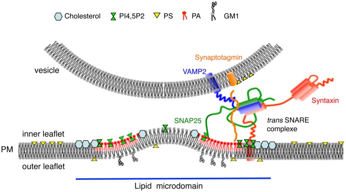
The regulated secretory pathway in neuroendocrine cells ends with the release of hormones and neurotransmitters following a rise in cytosolic calcium. This process known as regulated exocytosis involves the assembly of soluble N-ethylmaleimide-sensitive factor attachment protein receptor (SNARE) proteins, the synaptic vesicle VAMP (synaptobrevin), and the plasma membrane proteins syntaxin and SNAP-25. Although there is much evidence suggesting that SNARE proteins play a key role in the fusion machinery, other cellular elements regulating the kinetics, the extent of fusion, and the preparation of vesicle for release have received less attention. Among those factors, lipids have also been proposed to play important functions both at the level of secretory vesicle recruitment and late membrane fusion steps. Here, we will review the latest evidence supporting the concept of the fusogenic activity of lipids, and also discuss how this may be achieved. These possibilities include the recruitment and sequestration of the components of the exocytotic machinery, regulation of protein function, and direct effects on membrane topology.
Keywords: cholesterol; chromaffin cell; exocytosis; membrane fusion; phosphatidic acids; phosphoinositides.
Figures
References
Publication types
LinkOut - more resources
Full Text Sources
Other Literature Sources