

Pituitary macroadenoma presenting with pituitary apoplexy, acromegaly and secondary diabetes mellitus - a case report
- PMID: 24062868
- PMCID: PMC3779461
- DOI: 10.11604/pamj.2013.15.39.2054
Pituitary macroadenoma presenting with pituitary apoplexy, acromegaly and secondary diabetes mellitus - a case report
Abstract
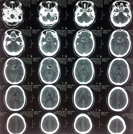
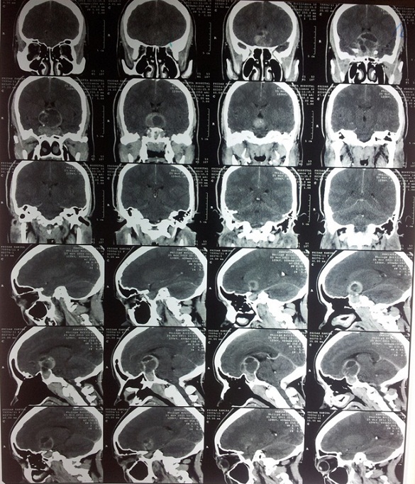
Pituitary adenomas are associated with significant morbidity. The usual symptoms on presentation are of endocrine dysfunction and mass effects. A 31-year-old African female presented with headache, irregular menses, blurring of vision in the right eye and complete loss of vision in the left eye for 1 year. She had coarse facial features, enlarged hands and feet. Her right eye had temporal hemianopia with decreased visual acuity and her left eye had no perception of light. Investigations revealed an elevated fasting blood sugar and an elevated prolactin and growth hormone level. A CT scan and MRI done showed a hemorrhagic pituitary macroadenoma. She was put on bromocriptine, ocreotide, analgesics and insulin. Thereafter, she underwent transphenoidal surgery, where near total resection of the tumor was achieved. Patient is doing well post-operatively. This case highlights the importance of the use of a high clinical index of suspicion and radiological findings in diagnosis.
Keywords: CT scan; MRI; Pitutary macroadenoma; acromegaly; diabetes mellitus.
Figures
Similar articles
-
Spontaneous remission of acromegaly: apoplexy mimicking meningitis or meningitis as a cause of apoplexy?Arq Bras Endocrinol Metabol. 2014 Feb;58(1):76-80. doi: 10.1590/0004-2730000002701. Arq Bras Endocrinol Metabol. 2014. PMID: 24728169
-
Pituitary macroadenoma presenting with brow mass and acromegaly.Ophthalmic Plast Reconstr Surg. 2009 Jan-Feb;25(1):56-7. doi: 10.1097/IOP.0b013e3181936850. Ophthalmic Plast Reconstr Surg. 2009. PMID: 19273930
-
Sudden visual loss and headache: important symptoms of pituitary disease.Hosp Med. 2000 Jan;61(1):60-1. doi: 10.12968/hosp.2000.61.1.1867. Hosp Med. 2000. PMID: 10735158 No abstract available.
-
[Pituitary apoplexy following coronary bypass surgery: A case report and literature review].Rev Med Interne. 2020 Dec;41(12):852-857. doi: 10.1016/j.revmed.2020.07.001. Epub 2020 Aug 13. Rev Med Interne. 2020. PMID: 32800377 Review. French.
-
Gestational pituitary apoplexy: Case series and review of the literature.J Gynecol Obstet Hum Reprod. 2019 Dec;48(10):873-881. doi: 10.1016/j.jogoh.2019.05.005. Epub 2019 May 3. J Gynecol Obstet Hum Reprod. 2019. PMID: 31059861 Review.
Cited by
-
Pituitary Macroadenoma Presenting as Acromegaly and Subacute Pituitary Apoplexy: Case Report and Literature Review.Cureus. 2020 Aug 8;12(8):e9612. doi: 10.7759/cureus.9612. Cureus. 2020. PMID: 32923214 Free PMC article.
-
Stubborn hiccups as a sign of massive apoplexy in a naive acromegaly patient with pituitary macroadenoma.Endocrinol Diabetes Metab Case Rep. 2017 May 18;2017:17-0044. doi: 10.1530/EDM-17-0044. eCollection 2017. Endocrinol Diabetes Metab Case Rep. 2017. PMID: 28567295 Free PMC article.
-
Chorio-retinal thickness measurements in patients with acromegaly.Eye (Lond). 2014 Nov;28(11):1350-4. doi: 10.1038/eye.2014.216. Epub 2014 Sep 19. Eye (Lond). 2014. PMID: 25233822 Free PMC article.
References
-
- James RM. Pituitary Macroadenomas. 201 http://emedicine.medscape.com/article/123223. Accessed 1 April 2012.
-
- Chahal HS, Stals K, Unterlander M, et al. AIP mutation in pituitary adenomas in the 18th century and today. N Engl J Med. 2011;364(1):43–50. - PubMed
-
- Hagiwara A, Inoue Y, Wakasa K, et al. Comparison of growth hormone-producing and non-growth hormone-producing pituitary adenomas: imaging characteristics and pathologic correlation. Radiology. 2003;228(2):533–8. - PubMed
-
- Kricheff II. The radiologic diagnosis of pituitary adenoma: an overview. Radiology. 1979;131(1):263–5. - PubMed
-
- Loeffler JS, Shih HA. Radiation therapy in the management of pituitary adenomas. J Clin Endocrinol Metab. 2011;96(7):1992–2003. - PubMed
Publication types
MeSH terms
LinkOut - more resources
Full Text Sources
Other Literature Sources
Medical
