. 2013:2013:474931.
doi: 10.1155/2013/474931. Epub 2013 Aug 25.

Sublethal oxidative stress induces the premature senescence of human mesenchymal stem cells derived from endometrium

Affiliations

Sublethal oxidative stress induces the premature senescence of human mesenchymal stem cells derived from endometrium

Elena Burova et al. Oxid Med Cell Longev. 2013.

Abstract

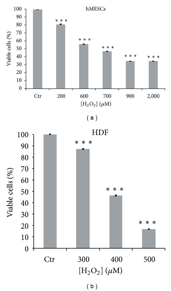
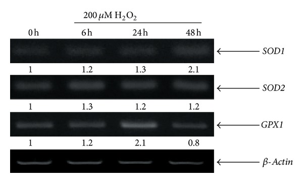
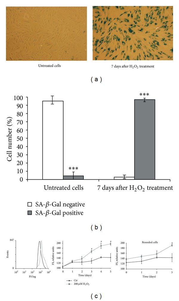
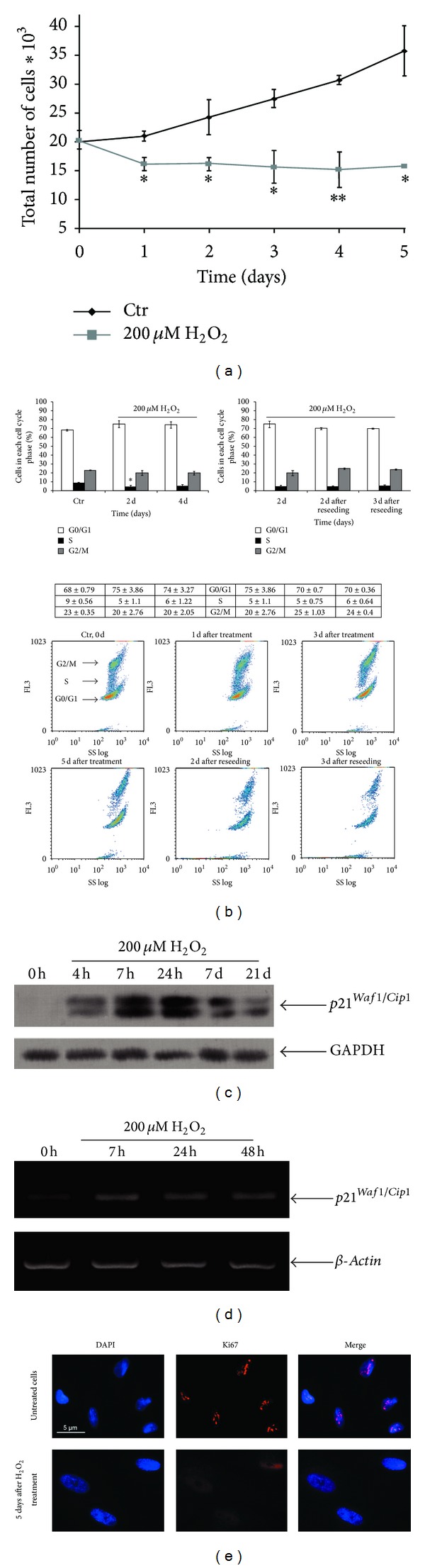
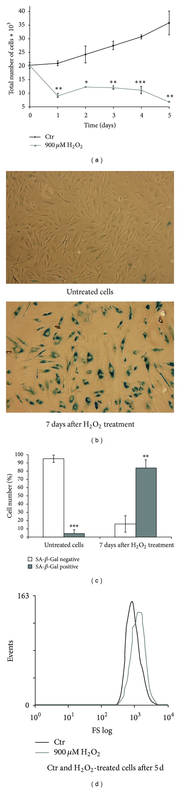
The specific responses of mesenchymal stem cells to oxidative stress may play a crucial role in regulation of tissue homeostasis as well as regeneration of organs after oxidative injury. The responses of human endometrium-derived mesenchymal stem cells (hMESCs) to oxidative stress remain still unknown. Herein, we examined the impact of H2O2 on cell viability, induction of premature senescence, and apoptosis. hMESCs were highly resistant to H2O2 compared with human diploid fibroblasts. To test a hypothesis whether hMESCs may undergo oxidative stress-induced premature senescence, cells were briefly exposed to the sublethal H2O2 doses. H2O2-treated cells were permanently arrested, lost Ki67 proliferation marker, and exhibited a senescent phenotype including cell hypertrophy and increased SA- β -Gal activity. Additionally, in stressed cells the expression levels of p21Cip1, SOD1, SOD2, and GPX1 were elevated. hMESCs survived under stress were not able to resume proliferation, indicating the irreversible loss of proliferative potential. While the low H2O2 doses promoted senescence in hMESCs, the higher H2O2 doses induced also apoptosis in a part of the cell population. Of note, senescent hMESCs exhibited high resistance to apoptosis. Thus, we have demonstrated for the first time that hMESCs may enter a state of premature senescence in response to sublethal oxidative stress.


Figures

Figure 1
Figure 1
The viability of hMESCs and HDF under oxidative stress. Cells were either treated or not with H2O2 at indicated concentrations for 1 h. The percentage of viable cells was evaluated in 24 h after treatment using MTT assay as described in Section 2. Results are shown as a percent of control. Data represent mean ± SEM of at least three independent experiments. ***P < 0.001 significantly different from the untreated control cells. LD values were 600–700 and 370–400 μM for hMESCs and HDF, respectively. Control (Ctr): untreated cells.
Figure 2
Figure 2
Gene expression of SOD1, SOD2, and GPX1 is enhanced in hMESCs in response to H2O2 treatment. Exponentially growing cells were treated with the sublethal dose (200 μM) of H2O2 for 1 h with following H2O2 replacement and then cultured under normal conditions for the indicated time. Total RNA isolated from hMESCs was amplified with specific primers for referred genes. β-Actin was used as a loading control.
Figure 3
Figure 3
The sublethal dose of H2O2 induces senescent phenotype in hMESCs. Cells were treated as indicated in the legend of Figure 2. (a) SA-β-Gal staining. (b) Quantitative assay of SA-β-Gal-positive cells. (c) H2O2-induced cell size increase. Typical presentation of forward scatter (FS), reflecting the average cell size (left). Cell size was determined daily: H2O2-treated cells were either cultured for 5 days under standard conditions (middle) or were reseeded in 2 days and additionally cultivated for 3 days (right). Data were obtained by light-scattering cytometry with using Win MDI program version 2.8. Data represent mean ± SEM of at least three independent experiments. Significant difference was based on the Student's t-test (*P < 0.05, ***P < 0.001). Control (Ctr): untreated cells.
Figure 4
Figure 4
Induction of the premature senescence in hMESCs under oxidative stress leads to the permanent arrest of cell cycle and irreversible loss of proliferative potential. Cell treatment was done as described in Figure 2. (a) Growth curve of both H2O2-treated and untreated cells. Cell number was determined daily after cell exposure to H2O2 by FACS analysis (M ± SEM, n = 3, *P < 0.05, **P < 0.01). (b) H2O2-treated cells were either cultured for 5 days or were reseeded in 2 days and additionally cultured for 3 days. Flow cytometry analysis of cell cycle phase distribution: the percentage of cells in the G0/G1, S, and G2/M phases (upper panel) (*P < 0.05); visualization of phase distribution based on light-scattering analysis (lower panel); SS: side scattering, FL3: PI fluorescence. (c) The expression levels of p21 protein. Representative results of the three experiments are shown in the figure. (d) The levels of p21 mRNA expression. GAPDH and β-actin were used as loading controls. (e) The nuclear localization of Ki67 was tested in control or H2O2-treated cells by immunofluorescence and DAPI staining. Representative photomicrographs of the staining are shown. Images were taken at magnification 100x. Control (Ctr): untreated cells.
Figure 5
Figure 5
Dot plots of FITC-annexin V/PI flow cytometry. hMESCs were subjected to 900 μM or 3,000 μM H2O2 for 1 h with following H2O2 replacement and cell cultivation under normal conditions. Apoptosis was detected at indicated time points. Control (Ctr): untreated cells.
Figure 6
Figure 6
The senescent phenotype in hMESCs treated with 900 μM H2O2. (a) H2O2-induced inhibition of cell proliferation (M ± SEM, n = 3). (b) SA-β-Gal staining. (c) Quantitative assay of SA-β-Gal-positive cells. (d) Cell hypertrophy detected by light-scattering cytometry. Significant difference was based on the Student's t-test (*P < 0.05, **P < 0.01, and ***P < 0.001). Control (Ctr): untreated cells.

References

    1. Otto WR, Wright NA. Mesenchymal stem cells: from experiment to clinic. Fibrogenesis and Tissue Repair. 2011;4(1, article 20) - PMC - PubMed
    1. Pittenger MF, Mackay AM, Beck SC, et al. Multilineage potential of adult human mesenchymal stem cells. Science. 1999;284:143–144. - PubMed
    1. Toma C, Pittenger MF, Cahill KS, Byrne BJ, Kessler PD. Human mesenchymal stem cells differentiate to a cardiomyocyte phenotype in the adult murine heart. Circulation. 2002;105(1):93–98. - PubMed
    1. Majumdar MK, Thiede MA, Mosca JD, Moorman M, Gerson SL. Phenotypic and functional comparison of cultures of marrow-derived mesenchymal stem cells (MSCs) and stromal cells. Journal of Cellular Physiology. 1998;176:57–66. - PubMed
    1. Sharpless NE, DePinho RA. How stem cells age and why this makes us grow old. Nature Reviews Molecular Cell Biology. 2007;8(9):703–713. - PubMed

Publication types

Substances

LinkOut - more resources