

Nosology of juvenile muscular atrophy of distal upper extremity: from monomelic amyotrophy to Hirayama disease--Indian perspective
- PMID: 24063005
- PMCID: PMC3770029
- DOI: 10.1155/2013/478516
Nosology of juvenile muscular atrophy of distal upper extremity: from monomelic amyotrophy to Hirayama disease--Indian perspective
Abstract
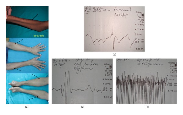
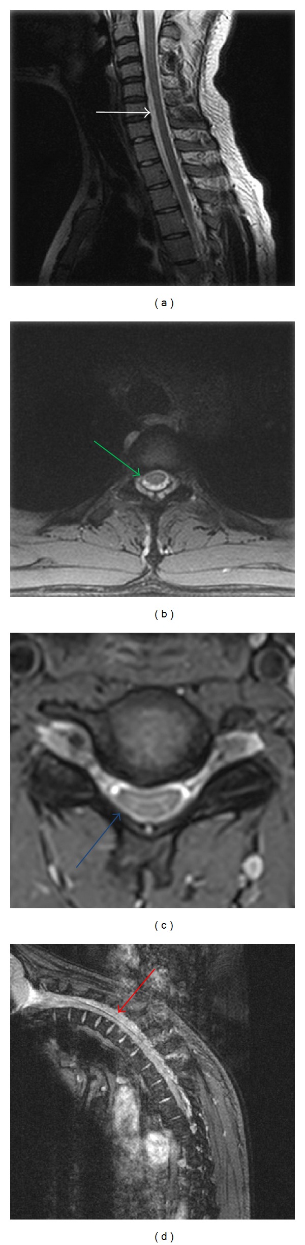
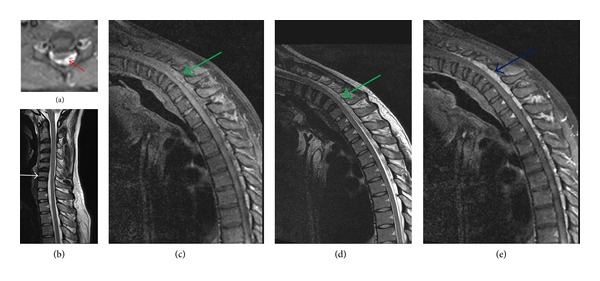
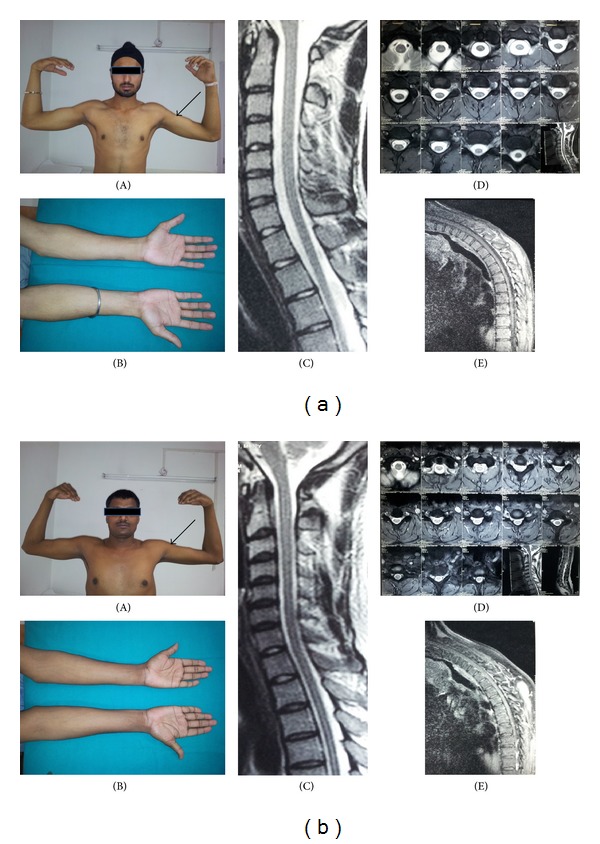
Since its original description by Keizo Hirayama in 1959, "juvenile muscular atrophy of the unilateral upper extremity" has been described under many nomenclatures from the east. Hirayama disease (HD), also interchangeably referred to as monomelic amyotrophy, has been more frequently recognised in the west only in the last two decades. HD presents in adolescence and young adulthood with insidious onset unilateral or bilateral asymmetric atrophy of hand and forearm with sparing of brachioradialis giving the characteristic appearance of oblique amyotrophy. Symmetrically bilateral disease has also been recognized. Believed to be a cervical flexion myelopathy, HD differs from motor neuron diseases because of its nonprogressive course and pathologic findings of chronic microcirculatory changes in the lower cervical cord. Electromyography shows features of acute and/or chronic denervation in C7, C8, and T1 myotomes in clinically affected limb and sometimes also in clinically unaffected contralateral limb. Dynamic forward displacement of dura in flexion causes asymmetric flattening of lower cervical cord. While dynamic contrast magnetic resonance imaging is diagnostic, routine study has high predictive value. There is a need to lump all the nomenclatures under the rubric of HD as prognosis in this condition is benign and prompt diagnosis is important to institute early collar therapy.
Figures
References
-
- Hirayama K. Juvenile muscular atrophy of unilateral upper extremity (Hirayama disease)—half-century progress and establishment since its discovery. Brain and Nerve. 2008;60(1):17–29. - PubMed
-
- Kikuchi S, Tashiro K, Kitagawa M, Iwasaki Y, Abe H. A mechanism of juvenile muscular atrophy localized in the hand and forearm (Hirayama’s disease)—flexion myelopathy with tight dural canal in flexion. Clinical Neurology. 1987;27(4):412–419. - PubMed
-
- Gourie-Devi M, Suresh TG, Shankar SK. Monomelic amyotrophy. Archives of Neurology. 1984;41(4):388–394. - PubMed
Publication types
MeSH terms
Supplementary concepts
LinkOut - more resources
Full Text Sources
Other Literature Sources
Miscellaneous
