. 2013 Sep 24;12(1):109.
doi: 10.1186/1476-4598-12-109.

Astrocyte elevated gene-1 is associated with metastasis in head and neck squamous cell carcinoma through p65 phosphorylation and upregulation of MMP1

Affiliations

Astrocyte elevated gene-1 is associated with metastasis in head and neck squamous cell carcinoma through p65 phosphorylation and upregulation of MMP1

Yi-Ping Wang et al. Mol Cancer. .

Abstract

Background: The survival rate of head and neck squamous cell carcinoma (HNSCC) at advanced stage is poor, despite contemporary advances in treatment modalities. Recent studies have indicated that astrocyte elevated gene-1 (AEG-1), a single transmembrane protein without any known functional domains, is overexpressed in various malignancies and is implicated in both distant metastasis and poor survival.

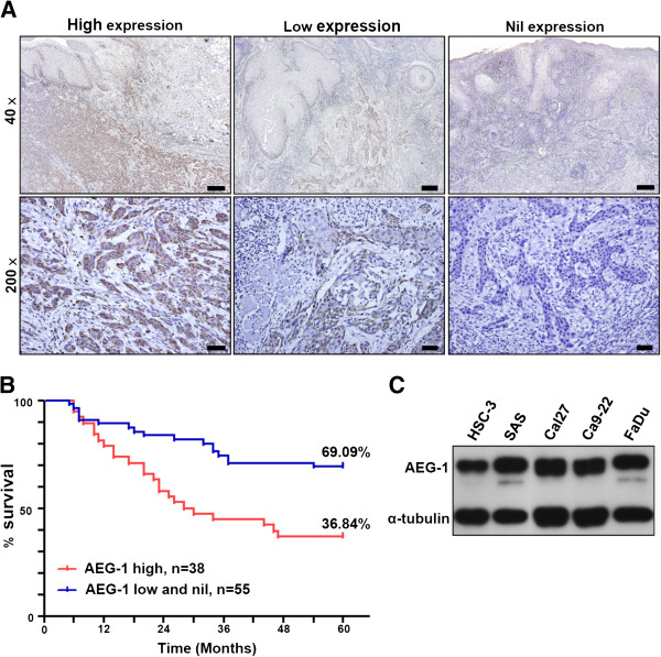
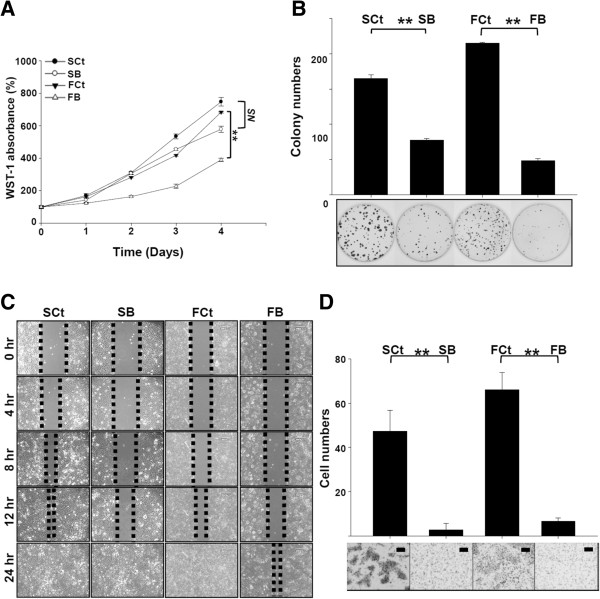
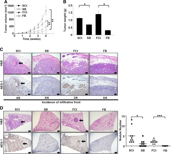
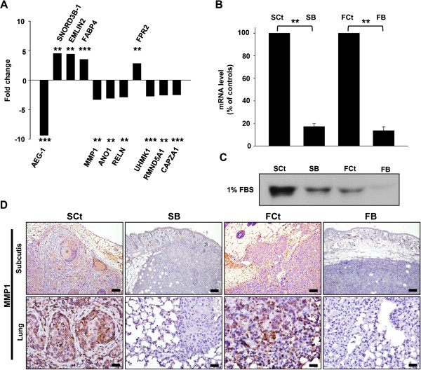
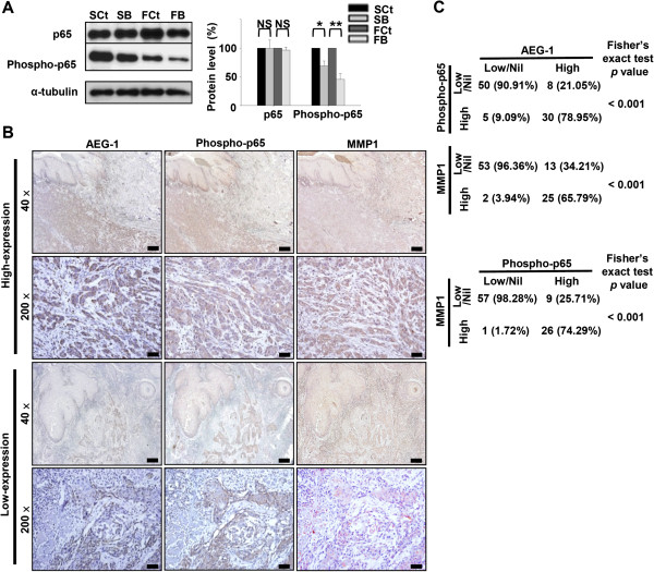
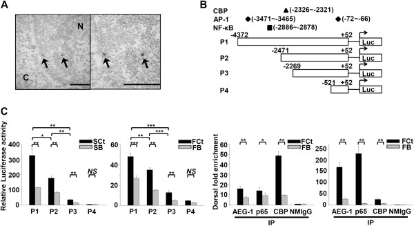
Results: High expression of AEG-1 in HNSCC was positively correlated with regional lymph node metastasis and a poor 5-year survival rate. Knockdown of AEG-1 in HNSCC cell lines reduced their capacity for colony formation, migration and invasion. Furthermore, decreased tumor volume and metastatic foci were observed after knockdown of AEG-1 in subcutaneous xenografts and pulmonary metastasis assays in vivo, respectively. We also demonstrated that AEG-1 increased phosphorylation of the p65 subunit of NF-κB, and regulated the expression of MMP1 in HNSCC cells. Moreover, compromised phosphorylation of the p65 (RelA) subunit of NF-κB at serine 536 was observed upon silencing of AEG-1 in both HNSCC cell lines and clinical specimens.

Conclusion: High expression of AEG-1 is associated with lymph node metastasis and its potentially associated mechanism is investigated.


Figures

Figure 1
Figure 1
AEG-1 expression in clinical specimens of OSCC and cell lines of HNSCC. (A) immunohistochemical staining of formalin-fixed, paraffin embedded OSCC specimens. Scale bar: 40 ×, 300 μm; 200 ×, 45 μm. (B) Kaplan-Meier 5-year survival analysis of 93 cases of OSCC segregated by expression status of AEG-1 protein. (C) AEG-1 protein expression in HNSCC cell lines.
Figure 2
Figure 2
Impact of AEG-1 knockdown on the function of HNSCC cell lines. (A) WST-1 cell proliferation assay. (B) colony formation assay. (C) wound-healing migration assay. Initial gap: 500 μm. (D) transwell Matrigel invasion assay. All values are the average of three independent experiments. SB, AEG-1 knock-down SAS cells. FB, AEG-1 knock-down FaDu cells. SCt, SAS cells transfected with scrambled control shRNA. FCt, FaDu cells transfected with scrambled control shRNA. All data were expressed as mean ± SEM; n = 3. NS, not significant (p > 0.05); **, p < 0.01. Scale Bar: 130 μm.
Figure 3
Figure 3
AEG-1 knockdown compromises tumorigenecity (n = 6) and pulmonary metastasis (n = 10) of SAS and FaDu cells. (A) time-course plot of tumor volumes. (B) tumor weights at the end-point. All data were expressed as mean ± SEM. *, p < 0.05; **, p < 0.01. (C) microscopy images of xenograft tumors at the end-point. Note the infiltrated invasion fronts in the SCt and FCt groups and the expansile tumor borders in the SB and FB groups. Arrow, focus of perineural invasion. Upper row, H&E stain; lower row, immunohistochemical stain with Lyric 4–7. Scale bar, 100 μm. (D)In vivo lung metastasis assay. Arrow, metastatic focus. Upper row, H&E stain; lower row, immunohistochemical stain with anti- AEG-1 antibody. Scale bar, 500 μm.
Figure 4
Figure 4
AEG-1 knockdown down-regulated MMP1 expression in HNSCC cells in vitro and in vivo. (A) microarray analysis of gene expression after AEG-1 knockdown in SAS cells. Genes with an absolute fold change greater than 2.5 are shown. SNORD3B-1, Homo sapiens small nucleolar RNA, C/D box 3B-1; EMILIN2, elastin microfibril interfacer 2; FABP4, fatty acid binding protein 4; MMP1, matrix metallopeptidase 1; ANO1, anoctamin 1; RELN, reelin; FPR2, formyl peptide receptor 2; UHMK1, U2AF homology motif kinase 1; RMND5A, required for meiotic nuclear division 5 homolog A; CAPZA1; capping protein (actin filament) muscle Z-line, alpha 1. *, p < 0.05; **, p < 0.01; ***, p < 0.001. (B) RT-QPCR confirmation of gene expression profiles after knockdown of AEG-1 in SAS and FaDu cells. All experiments were performed in triplicate (n = 3) and data were normalized to GAPDH. (C) secreted MMP1 protein in cell-conditioned culture media. Each cell lines were seeded in equal numbers (1 × 10⁶ cells) and were cultured in starving condition (1% fetal bovine serum, FBS) for 24 hr and the media were harvested. (D) immunohistochemical staining of MMP1 in murine subcutaneous xenograft tumors (upper row) and metastatic lesions from in vivo lung metastasis assays (lower row). Scale bar: upper row, 100 μm; lower row, 25 μm.
Figure 5
Figure 5
AEG-1 knockdown suppressed phosphorylation of serine 536 of the p65 subunit (RelA) of NF-κB. (A) Western blot of total p65 and phosphorylated p65 (serine 536). Data were normalized to respective controls and are were expressed as mean ± SEM; n = 3. NS, not significant (p > 0.05); *, p < 0.05; **, p < 0.01. (B and C) immunohistochemical staining of phosphorylated p65 (Ser536) and MMP1 in formalin-fixed, paraffin embedded OSCC specimens. Expression of phosphorylated p65 and MMP1 was positively cor related with that of AEG-1 both intratumorally (B) and intertumorally (C). Scale bar: 40 ×, 300 μm; 200 ×, 45 μm.
Figure 6
Figure 6
AEG-1 regulates MMP1 expression at the transcriptional level through enhancing the binding of p65 to MMP1 promoter. (A) nuclear translocation of AEG-1 protein was revealed by transmission electron microscopy. Arrow, 18 nm gold particles conjugated to anti-AEG-1 antibody. N, nucleus. C, cytosol. Right, magnified view. Scale bar, 500 nm. (B) constructs of the MMP1 promoter region used for the lucife-rase reporter assay. (C) transactivating activity of AEG-1 on MMP1 promoter constructs in the SAS and FaDu cell lines. All data were expressed as mean ± SEM; n = 3. (D) ChIP assay showing AEG-1, p65 and CBP binding to the NF-κB binding site of the MMP1 promoter in SAS and FaDu cell lines. NMIgG served as a negative control. All data were expressed as mean ± SEM; n = 3. NS, not significant (p > 0.05); *, p < 0.05; **, p < 0.01; ***, p < 0,001.

References

    1. Jemal A, Bray F, Center MM, Ferlay J, Ward E, Forman D. Global cancer statistics. CA Cancer J Clin. 2011;61:69–90. doi: 10.3322/caac.20107. - DOI - PubMed
    1. Leemans CR, Tiwari R, Nauta JJ, van der Waal I, Snow GB. Regional lymph node involvement and its significance in the development of distant metastases in head and neck carcinoma. Cancer. 1993;71:452–456. doi: 10.1002/1097-0142(19930115)71:2<452::AID-CNCR2820710228>3.0.CO;2-B. - DOI - PubMed
    1. Karamouzis MV, Grandis JR, Argiris A. Therapies directed against epidermal growth factor receptor in aerodigestive carcinomas. JAMA. 2007;298:70–82. doi: 10.1001/jama.298.1.70. - DOI - PubMed
    1. Burtness B, Goldwasser MA, Flood W, Mattar B, Forastiere AA. Phase III randomized trial of cisplatin plus placebo compared with cisplatin plus cetuximab in metastatic/recurrent head and neck cancer: an Eastern Cooperative Oncology Group study. J Clin Oncol. 2005;23:8646–8654. doi: 10.1200/JCO.2005.02.4646. - DOI - PubMed
    1. Vermorken JB, Trigo J, Hitt R, Koralewski P, Diaz-Rubio E, Rolland F, Knecht R, Amellal N, Schueler A, Baselga J. Open-label, uncontrolled, multicenter phase II study to evaluate the efficacy and toxicity of cetuximab as a single agent in patients with recurrent and/or metastatic squamous cell carcinoma of the head and neck who failed to respond to platinum-based therapy. J Clin Oncol. 2007;25:2171–2177. doi: 10.1200/JCO.2006.06.7447. - DOI - PubMed

Publication types

MeSH terms