. 2014 Jun 1;20(16):2514-27.
doi: 10.1089/ars.2013.5223. Epub 2014 Feb 14.

STAT3 regulation by S-nitrosylation: implication for inflammatory disease

Affiliations

STAT3 regulation by S-nitrosylation: implication for inflammatory disease

Jinsu Kim et al. Antioxid Redox Signal. .

Abstract

Aims: S-nitrosylation and S-glutathionylation, redox-based modifications of protein thiols, are recently emerging as important signaling mechanisms. In this study, we assessed S-nitrosylation-based regulation of Janus-activated kinase 2/signal transducer and activator of transcription 3 (JAK2/STAT3) pathway that plays critical roles in immune/inflammatory responses and tumorigenesis.

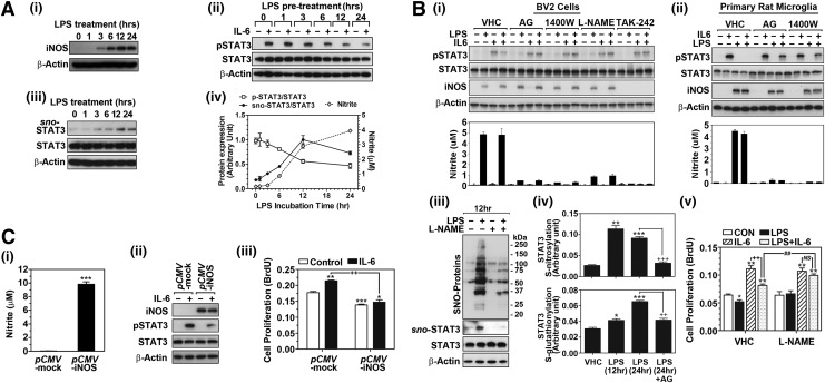
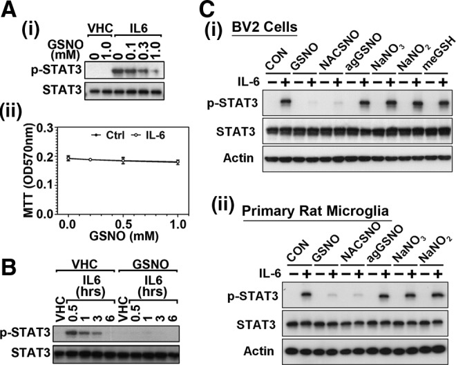
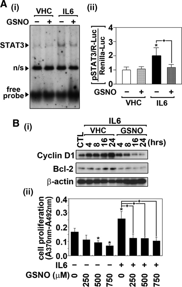
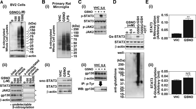
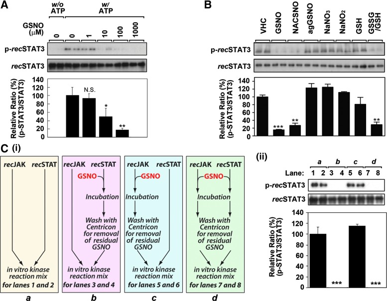
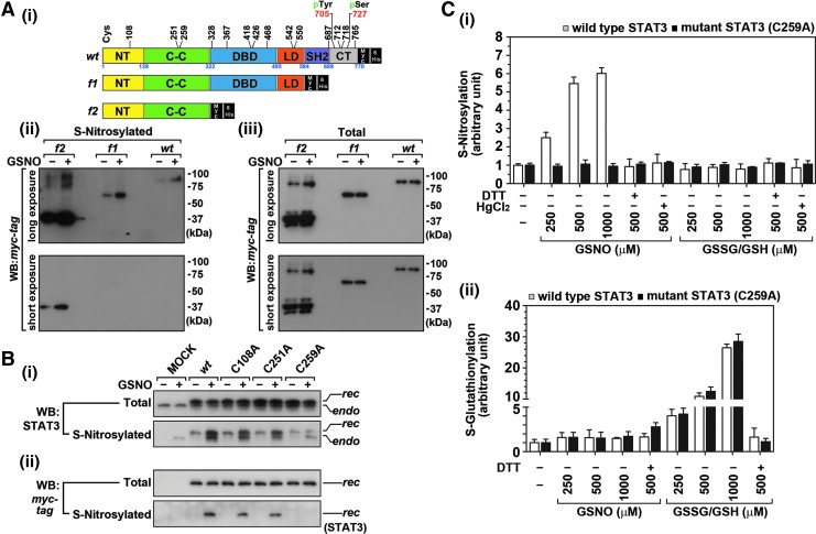
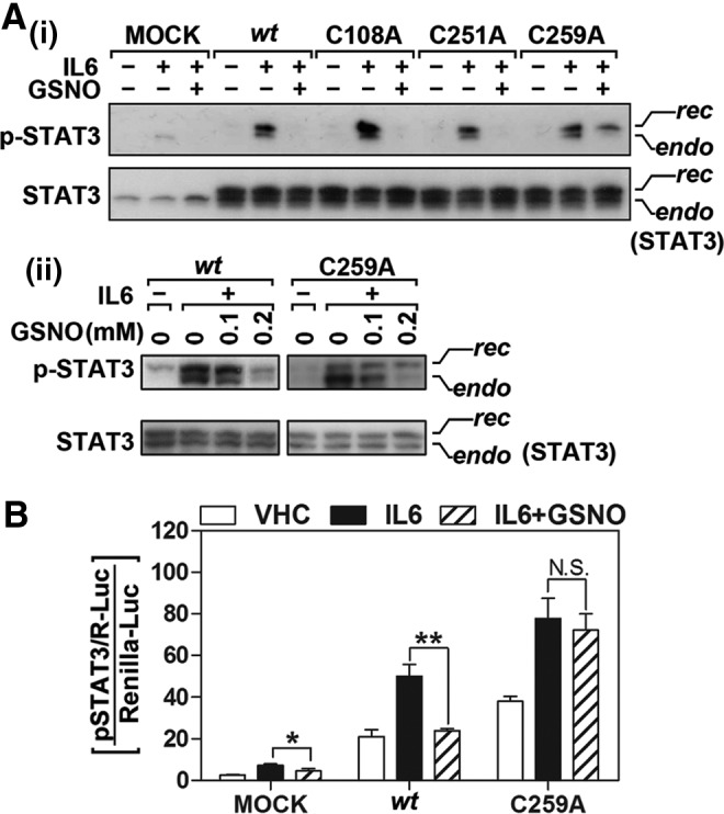
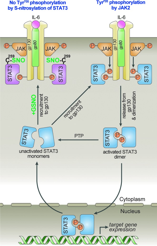
Results: Our studies show that STAT3 in stimulated microglia underwent two distinct redox-dependent modifications, S-nitrosylation and S-glutathionylation. STAT3 S-nitrosylation was associated with inducible nitric oxide synthase (iNOS)-produced nitric oxide (NO) and S-nitrosoglutathione (GSNO), whereas S-glutathionylation of STAT3 was associated with cellular oxidative stress. NO produced by iNOS or treatment of microglia with exogenous GSNO inhibited STAT3 activation via inhibiting STAT3 phosphorylation (Tyr(705)). Consequently, the interleukin-6 (IL-6)-induced microglial proliferation and associated gene expressions were also reduced. In cell-free kinase assay using purified JAK2 and STAT3, STAT3 phosphorylation was inhibited by its selective preincubation with GSNO, but not by preincubation of JAK2 with GSNO, indicating that GSNO-mediated mechanisms inhibit STAT3 phosphorylation through S-nitrosylation of STAT3 rather than JAK2. In this study, we identified that Cys(259) was the target Cys residue of GSNO-mediated S-nitrosylation of STAT3. The replacement of Cys(259) residue with Ala abolished the inhibitory role of GSNO in IL-6-induced STAT3 phosphorylation and transactivation, suggesting the role of Cys(259) S-nitrosylation in STAT3 phosphorylation.

Innovation: Microglial proliferation is regulated by NO via S-nitrosylation of STAT3 (Cys(259)) and inhibition of STAT3 (Tyr(705)) phosphorylation.

Conclusion: Our results indicate the regulation of STAT3 by NO-based post-translational modification (S-nitrosylation). These findings have important implications for the development of new therapeutics targeting STAT3 for treating diseases associated with inflammatory/immune responses and abnormal cell proliferation, including cancer.


Figures

<b>FIG. 1.</b>
FIG. 1.
NO produced by iNOS inhibits cell proliferation by inhibiting STAT3 activity. (A) Cultured BV2 murine microglial cells were treated with LPS (0.1 μg/ml) for different time periods, and the production of NO and expression of iNOS protein were analyzed (A-i, iv). To assess the role of endogenous NO produced by LPS treatment on phosphorylation of STAT3 (Tyr705), the cells were pretreated with LPS for different time periods (0–24 h), then treated with IL-6 (30 ng/ml) for 0.5 h, and cellular phosphorylated STAT3 (pSTAT3) levels were analyzed (A-ii, iv). Under the same experimental conditions, the effect of LPS on STAT3 S-nitrosylation (sno-STAT3 levels) was analyzed by biotin switch assay as described under the Materials and Methods section (A-iii, iv). The NO production by LPS treatment and its effect on IL-6-induced STAT3 phosphorylation and STAT3 S-nitrosylation were quantified by densitometry (A-iv). (B). The cells were treated with LPS (0.1 μg/ml) in the presence or absence of iNOS inhibitor, such as aminoguanidine (AG; 1 mM) or N-(3-(Aminomethyl)benzyl) acetamidine) (1400W; 50 μM), general NOS inhibitor L-Nω-nitroarginine methyl ester (L-NAME; 300 μM), or TLR4 inhibitor TAK-242 (1 μM) for 18 h and the iNOS expression, the production of NO (nitrite levels) and the effect on IL-6-induced (30 ng/ml 0.5 h) phosphorylation of STAT3 were analyzed in BV2 cells (B-i) or primary cultured rat microglia (B-ii). In addition, the levels of S-nitrosylated proteins (sno-Proteins) and S-nitrosylated and S-glutathionylated STAT3 levels (B-iii, iv) and cell proliferation (B-v) were analyzed in BV2 cells. (C) Following the transfection of CHO cells with empty pCMV vector (pCMV-mock) or pCMV vector expressing human iNOS (pCMV-iNOS), the production of NO (nitrite levels) (C-i) and the effect of IL-6 (30 ng/ml 0.5 h) on the phosphorylation of STAT3 (C-ii) and cell proliferation (C-iii) were analyzed. The vertical lines in A-iv, B-i, -ii, -iv, and -v, C-i and -iii indicate the standard error mean; *p<0.05; **p<0.01; ***p<0.001 compared with control group, ++p<0.01; +++p<0.001; ##p<0.01; not significant (N.S.) >0.05 compared with indicated group. IL-6, interleukin-6; iNOS, inducible nitric oxide synthase; LPS, lipopolysaccharide; NO, nitric oxide; STAT3, signal transducer and activator of transcription 3.
<b>FIG. 2.</b>
FIG. 2.
GSNO inhibits phosphorylation of STAT3 (Tyr705). (A) Cultured BV2 murine microglial cells were pretreated with the increasing concentrations of GSNO for 2 h in the dark and stimulated with IL-6 (30 ng/mL) for 30 min. Phosphorylated (Tyr705) and pan STAT3 protein levels in nuclear extract were determined by Western analysis as described under experimental procedures (A-i). Under the same experimental conditions, cell viability was also examined by MTT assay (A-ii). (B) Effect of GSNO pretreatment (500 μM for 2 h) on the time course changes in STAT3 phosphorylation was also examined following the IL-6 (30 ng/ml) treatment. (C) Effect of GSNO-related compounds, such as S-nitroso-N-acetyl cysteine (NACSNO; cell permeable S-nitroso donor; 500 μM) or GSNO-related metabolites, such as aged (decomposed) GSNO (agGSNO; 500 μM), cell permeable GSH (GSH monoethyl ester; meGSH; 500 μM), sodium nitrite (NaNO2; 500 μM), and sodium nitrate (NaNO3; 500 μM), on IL-6 (30 ng/ml 0.5 h)-induced STAT3 phosphorylation was examined in BV2 cells (C-i) or in primary cultured rat microglia (C-ii). GSH, glutathione; GSNO, S-nitrosoglutathione; MTT, 3-(4,5-dimethylthiazol-2-yl)-2,5-diphenyltetrazolium bromide.
<b>FIG. 3.</b>
FIG. 3.
GSNO inhibits target DNA interaction of STAT3 and its transactivation by IL-6. (A) The effects of GSNO on IL-6-induced STAT3 target DNA binding activity and its transactivity were examined by gel-shift and reporter gene assays. For the gel-shift assay, BV2 microglia were pretreated with GSNO (500 μM) for 2 h and then treated with IL-6 (30 ng/mL) for 30 min. Nonspecific reaction is indicated by n/s (A-i). For the reporter gene assay, BV2 microglia transfected with STAT3-responsive luciferase construct (pSTAT3/R-Luc) and renilla luciferase contruct (phRL-CMV) as transfection control were pretreated with GSNO (500 μM) for 2 h and then treated with IL-6 (30 ng/mL) for 24 h. STAT3 transactivities were analyzed by luciferase activity assay as described under experimental procedure (A-ii). (B) The effects of GSNO on the expression of STAT3 downstream gene expression and cell proliferation were examined in BV2 microglia. The cells were treated with GSNO (500 μM) for 2 h and then treated with IL-6 (30 ng/mL) for indicated time points. The cellular levels of cyclin D1 and Bcl-2 were then analyzed by Western analysis (B-i). The effect of pretreatment of BV2 cells with increasing concentrations of GSNO for 2 h on IL-6-induced (30 ng/mL for 14 h) cell proliferation was analyzed by BrdU incorporation assay (B-ii). The vertical lines in A-ii and B-iii indicate the standard error of mean; *p<0.05 compared with vehicle-treated (VHC; dimethylsulfoxide or phosphate-buffered saline) control groups; +p<0.01 compared with IL-6-treated groups. BrdU, 5-bromo-2′-deoxyuridine.
<b>FIG. 4.</b>
FIG. 4.
GSNO increases S-nitrosylation of STAT3. (A). The effects of GSNO (2 h treatment) on the S-nitrosylation of STAT3 or its upstream kinase JAK2 and receptor gp130 in BV2 cells were examined by biotin switch method. For analysis of total S-nitrosylated proteins, Western analysis for biotin-labeled proteins was performed (A-i). For analysis of S-nitrosylation of STAT3, JAK2, and receptor gp130, the biotinylated proteins were pulled down with avidin–agarose conjugate and the levels of STAT3, JAK2, and gp130 were determined by Western analysis (A-ii). (B) The effect of GSNO (0.5 mM for 2 h) and LPS (0.1 μg/ml for 12 h) treatment on S-nitrosylation of cellular proteins (B-i) and STAT3 (B-ii) was also examined in primary cultured rat microglia. (C) The effects of GSNO (500 μM for 2 h) on IL-6-induced (30 ng/mL for 0.5 h) phosphorylation of STAT3 (Tyr705) and JAK2 (Tyr1007/1008) were examined by the Western analysis (C-i). The phosphorylation of gp130 was examined by immunoprecipitation of phospho-Tyr containing proteins and following Western analysis of gp130 (C-ii). The levels of β-actin were used as internal loading control. (D) The effect of GSNO (2 h) on S-glutathionylation of STAT3 and other cellular proteins in BV2 cells were analyzed by immunoprecipitation and Western analysis. For positive control, the control cell lysates were incubated with oxidized and reduced glutathione mixture (GSSG/GSH; 5 mM each; pH 7.0) for 1 h at room temperature in the presence or absence of 20 mM dithiothreitol (DTT). (E) STAT3 S-nitrosylation (E-i) and S-glutathionylation (E-ii) in BV2 cells treated with GSNO (0.5 mM for 2 h) were further analyzed by sandwich ELISA using antibodies specific to S-nitrosocysteine or GSNO and STAT3. The vertical lines in E indicate the standard error mean; **p>0.01; not significant (N.S.)>0.05 compared with vehicle (VHC) treated group. ELISA, enzyme-linked immunosorbent assay; JAK2, Janus-activated kinase 2.
<b>FIG. 5.</b>
FIG. 5.
GSNO inhibits JAK2-mediated STAT3 phosphorylation in cell-free in vitro kinase assay system. (A) To examine whether GSNO is able to inhibit JAK2-mediated STAT3 phosphorylation without any involvement of other regulatory factors, in vitro kinase assay was performed using purified recombinant STAT3 (recSTAT3) and JAK2 (recJAK2). The purified recSTAT3 and recJAK2 mixture was incubated with increasing concentration of GSNO for 30 min, and kinase assay was initiated by the addition of reaction buffer without (w/o) or with (w/) ATP. The resulted phosphorylated recSTAT3 (Tyr705) levels were analyzed by Western blot. (B) To examine the effect of GSNO-related compounds on the phosphorylation of recSTAT3 in cell-free system, GSNO (100 μM), NACSNO (100 μM), aged GSNO (100 μM agGSNO; decomposed GSNO), NaNO3 (100 μM), NaNO2, (100 μM). glutathione (GSH/100 μM), or GSSG/GSH (1 mM) were added to reaction mixture before initiation of kinase reaction for 30 min. (C) To specify the target molecule of GSNO in inhibition of recSTAT3 phosphorylation, recSTAT3 or recJAK2 was preincubated with GSNO (100 μM for 30 min) individually and the residual GSNO was removed with Centricon (YM-10) before initiate the kinase reaction to minimize the unwanted effects of residual GSNO on untreated component as shown in the flow diagram (C-i). Following the in vitro kinase reaction, phosphorylated recSTAT3 was analyzed by Western blot (C-ii). For lane 1 and 2 (a), untreated recJAK2 and recSTAT3 were used. For lane 3 and 4 (b), untreated recJAK2 and GSNO-treated recSTAT3 were used. For lane 5 and 6 (c), GSNO-treated recJAK2 and untreated recSTAT3 were used. For lane 7 and 8 (d), GSNO-treated recJAK2 and recSTAT3 were used. The vertical lines in A, B, and C-ii indicate the standard error of mean; *p<0.05; **p<0.01; ***p<0.001; not significant (N.S.)>0.05 compared with vehicle-treated (VHC; dimethylsulfoxide) control groups.
<b>FIG. 6.</b>
FIG. 6.
GSNO selectively S-nitrosylates STAT3 at Cys259. (A) Wild-type (wt) STAT3 contains 14 Cys residues, which are distributed throughout the 6 domains, namely NH2-terminal (NT) domain, coiled-coil (CC) domain, DNA binding domain (DBD), in linker domain (LD), Src homology 2 (SH2) domain, and COOH terminal (CT) domain. To identify the target Cys residue(s) for S-nitrosylation, wt STAT3 conjugated with Myc and 6xHis tags was sequentially deleted; the deletion mutants were designated as f1 and f2 (A-i). Chinese hamster ovary (CHO) cells were transfected and overexpressed with wt STAT3 or its deletion mutant (f1 or f2) and treated with GSNO (300 μM) for 2 h. The S-nitrosylation of wt or mutant STAT3 was analyzed by Western analysis of Myc-tagged STAT3 followed by the biotin switch procedure (A-ii). The protein levels of myc-tagged STAT3 in whole cell lysates were used to check for equal transfection and equal expression of recombinant STAT3s between GSNO-treated group and untreated group (A-iii). (B) Because GSNO was able to increase S-nitrosylation of f2 fragment, Cys residues localized in this fragment were individually mutated to Ala (C108A, C251A, and C259) and transfected into CHO cells as described under the Materials and Methods section. Empty vector (MOCK) was used for control of transfection. Following treatment of cells with GSNO (300 μM) for 2 h, total STAT3 protein levels as well as S-nitrosylated STAT3 levels were analyzed by biotin switch procedure and Western analysis using antibody specific to STAT3 (B-i) or myc-tag (B-ii). In each panel, upper bands correspond to recombinant STAT3 (rec), which is conjugated with myc and 6xHis tags; lower bands correspond to their endogenous counterparts (endo). (C). The effect of GSNO or GSSG/GSH mix (0–500 μM) treatment for 0.5 h on S-nitrosylation (C-i) and S-glutathionylation (C-ii) of recombinant wt STAT3 or recombinant C259A mutant STAT3 was analyzed by sandwich ELISA using respective antibodies specific to S-nitrosocysteine or glutathione. The purified recombinant wt STAT3 and its C259A mutant proteins in cell-free system were used for in vitro S-nitrosylation and S-glutathionylation. For negative control for S-nitrosylation and S-glutathionylation, 10 mM of dithiothreitol or HgCl2 with GSNO was used. rec, recombinant.
<b>FIG. 7.</b>
FIG. 7.
Point mutation of Cys259 to Ala abolishes the inhibitory action of GSNO on IL-6-induced STAT3 Tyr705 phosphorylation and its transactivity. (A) CHO cells were transfected and overexpressed with empty vector (MOCK), wt STAT3 or its point mutant (C108A, C251A, or C259). The cells were then pretreated with GSNO (300 μM for 2 h) followed by IL-6 (30 ng/ml) treatment for 30 min. The total and phospho-Tyr705 STAT3 levels were analyzed by Western analysis. The upper bands corresponds to recombinant STAT3 (rec), which is conjugated with myc and 6xHis tags; lower bands correspond to their endogenous counterparts (endo) (A-i). STAT3 phosphorylation in the cells expressing wtSTAT3 and mutant STAT3 (C259A) was also analyzed following the treatment of cells with lower concentration of GSNO (A-ii). (B) Effect of point mutation of Cys259 to Ala on the inhibitory role of GSNO in IL-6-induced STAT3 transactivation was examined by STAT3 responsive element luciferase assay. CHO cells transfected with STAT3-responsive luciferase construct (pSTAT3/R-Luc) and wt or C259A mutant STAT3 constructs were pretreated with GSNO (500 μM) for 2 h and then treated with IL-6 (30 ng/mL) for 24 h. STAT3 transactivities were analyzed by luciferase activity assay as described under experimental procedure. The renilla luciferase contruct (phRL-CMV) was used as a transfection control. The vertical lines in B indicate the standard error of mean; *p<0.05; **p<0.01; N.S.>0.05 compared with IL-6-treated groups.
<b>FIG. 8.</b>
FIG. 8.
Regulation of STAT3 activation (Tyr705 phosphorylation) by S-nitrosylation-dependent mechanism. IL-6 induces STAT3 Tyr705 phosphorylation by following sequential steps: (i) homodimerization of gp130 and auto-phosphorylation of JAK2 (Tyr1007/1008), (ii) JAK2-mediated phosphorylation of Tyr residues in cytoplasmic part of gp130, (iii) recruitment of STAT3 to phospho-Tyr of gp130, (iv) STAT3 Tyr705 phosphorylation by JAK2, and (v) release of phospho-Tyr705-STAT3 from gp130 followed by STAT3 homodimerization through reciprocal interaction of the phospho-Tyr705 of one monomer and the SH2 domain of the corresponding monomer. Small molecular mass S-nitroso compounds including GSNO, which are endogenously formed by NO and thiol compounds, transfer their S-nitrosyl group to Cys259 residue of STAT3. The S-nitrosylation of Cys259 residue may inhibit JAK2-mediated phosphorylation of STAT3, which is recruited by gp130 receptor. Consequently, S-nitrosylation of STAT3 inhibits formation of STAT3 dimer and suppresses STAT3-dependent gene expression for cell cycle and cell survival.

Similar articles

Cited by

References

    1. Anand P. and Stamler JS. Enzymatic mechanisms regulating protein S-nitrosylation: implications in health and disease. J Mol Med (Berl) 90: 233–244, 2012 - PMC - PubMed
    1. Baker TL, Booden MA, and Buss JE. S-Nitrosocysteine increases palmitate turnover on Ha-Ras in NIH 3T3 cells. J Biol Chem 275: 22037–22047, 2000 - PubMed
    1. Block ML. and Hong JS. Microglia and inflammation-mediated neurodegeneration: multiple triggers with a common mechanism. Prog Neurobiol 76: 77–98, 2005 - PubMed
    1. Bromberg JF, Wrzeszczynska MH, Devgan G, Zhao Y, Pestell RG, Albanese C, and Darnell JE., Jr.Stat3 as an oncogene. Cell 98: 295–303, 1999 - PubMed
    1. Campbell IL. Cytokine-mediated inflammation, tumorigenesis, and disease-associated JAK/STAT/SOCS signaling circuits in the CNS. Brain Res Brain Res Rev 48: 166–177, 2005 - PubMed

Publication types

LinkOut - more resources