

Planarian MBD2/3 is required for adult stem cell pluripotency independently of DNA methylation
- PMID: 24063805
- PMCID: PMC3824064
- DOI: 10.1016/j.ydbio.2013.09.020
Planarian MBD2/3 is required for adult stem cell pluripotency independently of DNA methylation
Abstract
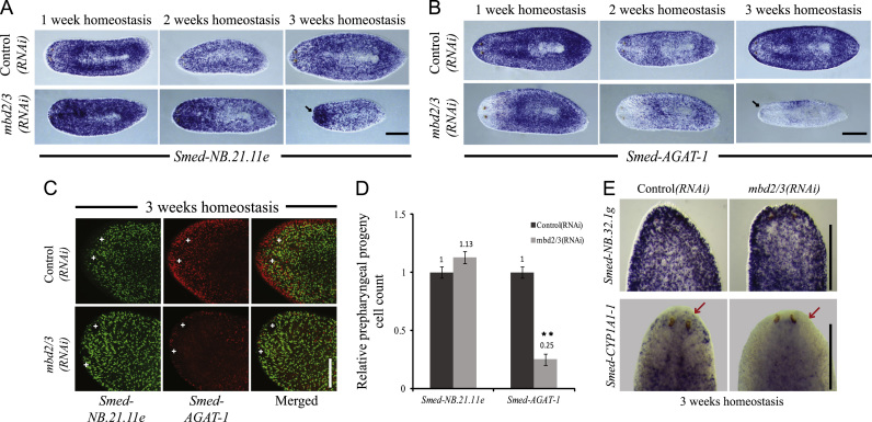
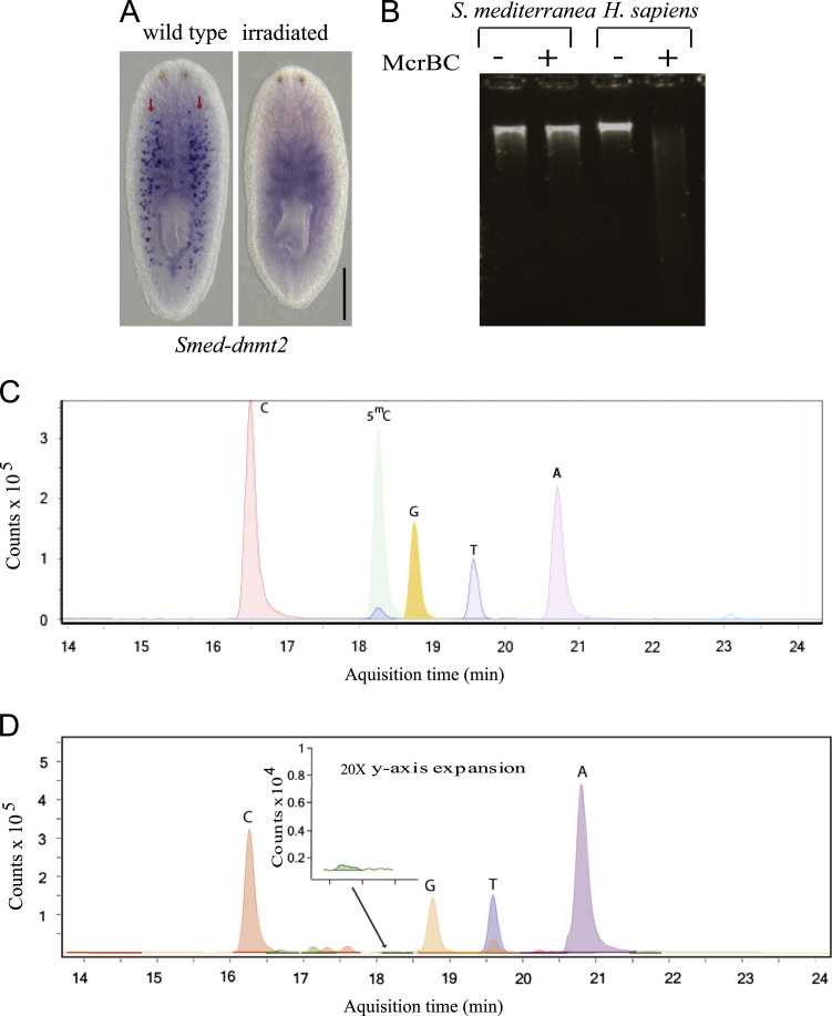
Planarian adult stem cells (pASCs) or neoblasts represent an ideal system to study the evolution of stem cells and pluripotency as they underpin an unrivaled capacity for regeneration. We wish to understand the control of differentiation and pluripotency in pASCs and to understand how conserved, convergent or divergent these mechanisms are across the Bilateria. Here we show the planarian methyl-CpG Binding Domain 2/3 (mbd2/3) gene is required for pASC differentiation during regeneration and tissue homeostasis. The genome does not have detectable levels of 5-methylcytosine (5(m)C) and we find no role for a potential DNA methylase. We conclude that MBD proteins may have had an ancient role in broadly controlling animal stem cell pluripotency, but that DNA methylation is not involved in planarian stem cell differentiation.
Keywords: Differentiation; Methyl binding domain; Pluripotency; Regeneration.
© 2013 The Authors. Published by Elsevier Inc. All rights reserved.
Figures

Similar articles
-
Genetic regulators of a pluripotent adult stem cell system in planarians identified by RNAi and clonal analysis.Cell Stem Cell. 2012 Mar 2;10(3):299-311. doi: 10.1016/j.stem.2012.01.016. Cell Stem Cell. 2012. PMID: 22385657 Free PMC article.
-
CBP/p300 homologs CBP2 and CBP3 play distinct roles in planarian stem cell function.Dev Biol. 2021 May;473:130-143. doi: 10.1016/j.ydbio.2021.02.004. Epub 2021 Feb 16. Dev Biol. 2021. PMID: 33607113
-
Different requirements for conserved post-transcriptional regulators in planarian regeneration and stem cell maintenance.Dev Biol. 2010 May 15;341(2):429-43. doi: 10.1016/j.ydbio.2010.02.037. Epub 2010 Mar 15. Dev Biol. 2010. PMID: 20230812
-
Cellular and molecular dissection of pluripotent adult somatic stem cells in planarians.Dev Growth Differ. 2010 Jan;52(1):27-41. doi: 10.1111/j.1440-169X.2009.01155.x. Dev Growth Differ. 2010. PMID: 20078652 Review.
-
What is the role of PIWI family proteins in adult pluripotent stem cells? Insights from asexually reproducing animals, planarians.Dev Growth Differ. 2020 Aug;62(6):407-422. doi: 10.1111/dgd.12688. Epub 2020 Aug 9. Dev Growth Differ. 2020. PMID: 32621324 Review.
Cited by
-
Methyl-CpG-binding (SmMBD2/3) and chromobox (SmCBX) proteins are required for neoblast proliferation and oviposition in the parasitic blood fluke Schistosoma mansoni.PLoS Pathog. 2018 Jun 28;14(6):e1007107. doi: 10.1371/journal.ppat.1007107. eCollection 2018 Jun. PLoS Pathog. 2018. PMID: 29953544 Free PMC article.
-
Epigenetics and Shared Molecular Processes in the Regeneration of Complex Structures.Stem Cells Int. 2016;2016:6947395. doi: 10.1155/2016/6947395. Epub 2015 Nov 22. Stem Cells Int. 2016. PMID: 26681954 Free PMC article. Review.
-
The genome of the crustacean Parhyale hawaiensis, a model for animal development, regeneration, immunity and lignocellulose digestion.Elife. 2016 Nov 16;5:e20062. doi: 10.7554/eLife.20062. Elife. 2016. PMID: 27849518 Free PMC article.
-
The expanding epigenetic landscape of non-model organisms.J Exp Biol. 2015 Jan 1;218(Pt 1):114-22. doi: 10.1242/jeb.110809. J Exp Biol. 2015. PMID: 25568458 Free PMC article. Review.
-
Mutational profile of the regenerative process and de novo genome assembly of the planarian Schmidtea polychroa.Nucleic Acids Res. 2024 Feb 28;52(4):1779-1792. doi: 10.1093/nar/gkad1250. Nucleic Acids Res. 2024. PMID: 38180823 Free PMC article.
References
-
- Aboobaker A.A. Planarian stem cells: a simple paradigm for regeneration. Trends in Cell Biology. 2011;21:304–311. - PubMed
-
- Aguilera C., Nakagawa K., Sancho R., Chakraborty A., Hendrich B., Behrens A. c-Jun N-terminal phosphorylation antagonises recruitment of the Mbd3/NuRD repressor complex. Nature. 2011;469:231–235. - PubMed
-
- Ahringer J. NuRD and SIN3 histone deacetylase complexes in development. Trends in Genetics. 2000;16:351–356. - PubMed
-
- Albalat R. Evolution of DNA-methylation machinery: DNA methyltransferases and methyl-DNA binding proteins in the amphioxus Branchiostoma floridae. Development Genes and Evolution. 2008;218:691–701. - PubMed
Publication types
MeSH terms
Substances
Grants and funding
LinkOut - more resources
Full Text Sources
Other Literature Sources
Miscellaneous
