. 2013 Dec;57(2):455-67.
doi: 10.1016/j.bone.2013.09.007. Epub 2013 Sep 21.

The plastic nature of the human bone-periodontal ligament-tooth fibrous joint

Affiliations

The plastic nature of the human bone-periodontal ligament-tooth fibrous joint

Sunita P Ho et al. Bone. 2013 Dec.

Abstract

This study investigates bony protrusions within a narrowed periodontal ligament space (PDL-space) of a human bone-PDL-tooth fibrous joint by mapping structural, biochemical, and mechanical heterogeneity. Higher resolution structural characterization was achieved via complementary atomic force microscopy (AFM), nano-transmission X-ray microscopy (nano-TXM), and microtomography (MicroXCT™). Structural heterogeneity was correlated to biochemical and elemental composition, illustrated via histochemistry and microprobe X-ray fluorescence analysis (μ-XRF), and mechanical heterogeneity evaluated by AFM-based nanoindentation. Results demonstrated that the narrowed PDL-space was due to invasion of bundle bone (BB) into PDL-space. Protruded BB had a wider range with higher elastic modulus values (2-8GPa) compared to lamellar bone (0.8-6GPa), and increased quantities of Ca, P and Zn as revealed by μ-XRF. Interestingly, the hygroscopic 10-30μm interface between protruded BB and lamellar bone exhibited higher X-ray attenuation similar to cement lines and lamellae within bone. Localization of the small leucine rich proteoglycan biglycan (BGN) responsible for mineralization was observed at the PDL-bone interface and around the osteocyte lacunae. Based on these results, it can be argued that the LB-BB interface was the original site of PDL attachment, and that the genesis of protruded BB identified as protrusions occurred as a result of shift in strain. We emphasize the importance of bony protrusions within the context of organ function and that additional study is warranted.

Keywords: Alveolar bone; Bone functional adaptation; Bone–PDL–tooth fibrous joint; Bundle bone; Discontinuities; Periodontal ligament.


Figures

Figure 1
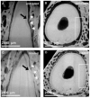
Figure 1. Qualitative assessment of conforming and nonconforming bone-tooth root surfaces at no load (TOP row) and loaded (BOTTOM row) conditions
Seemingly conforming surfaces are illustrated in an unloaded bone-PDL-tooth complex (A, B) stained with phosphotungstic acid (PTA) [3]. However, under loaded conditions the non-conforming tooth root and alveolar bone surface sites (white stars and box) are highlighted (C, D).
Figure 2
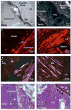
Figure 2. Fiber orientation varied across tissues of the bone-PDL-cementum complex
Higher magnification light micrograph of an ultrasectioned surface block illustrated structural heterogeneity in alveolar bone, PDL, and tooth. The tissue-level characteristics of constricted PDL-space include invasion of BB from LB and adapted cementum (black stars) (A). Picrosirius red stain coupled with polarized light microscopy of the narrowed region demonstrated differential fiber orientation within secondary cementum and alveolar bone (B), and the differential collagen fiber orientation between the cementum dentin junction (CDJ), and lamellae and interlamellae regions (characterized by birefringence) (C, D). Lower and higher magnification hematoxylin and eosin staining (H&E) of alveolar bone coupled with polarized light microscopy demonstrated the heterogeneity of fiber orientation between BB, which was dominated by radial fibers, versus LB consisting of primarily circumferential fibers (circle: cementum, white star: dentin) (E,F). Hematoxylin and eosin staining (H&E) demonstrated localization of basophilic lines in both cementum and bundle bone (white arrows). Additionally, directional bone growth illustrated by bony protrusions (black arrows) was expressed at the PDL-bone interface (G,H). LB: Lamellar bone, BB: Bundle bone, PDL: Periodontal ligament, CDJ: Cementum-dentin junction. *Note: the orientation of the bone-PDL-tooth complex varies in images.
Figure 3
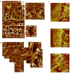
Figure 3. Tissue architecture under dry and wet conditions illustrated LB-BB interface as the original site of PDL attachment
Higher resolution AFM micrograph of an alveolar bone-PDL-cementum complex imaged under dry (A: a1) and wet (A: a2, a3) conditions. Radial fiber insertions (stars, cross sections revealed) into both BB and cementum interfacing the PDL are visible. Swelling of insertion fibers (stars) under wet conditions are indicative of polyanionic molecules and predominance of organic matter. The LB-BB interface is shown in greater detail under dry (B: b1) and wet (B: b2) conditions. Under dry conditions, lamellae regions demonstrated greater depth relative to inter-lamellae regions of LB. However, under wet conditions lamellae regions demonstrated increased hygroscopicity compared to inter-lamellae regions of LB, suggestive of differential degrees of mineral and/or organic content. A corresponding lamellar region is denoted by arrows in both dry and wet conditions. It should be a noted that angle of sectioning can illustrate a structure significantly different from the LB-BB interface, as observed in B: b2. Additionally, continuous fibers from BB through the distinct interface and attached to LB are visible (C: c1, c2, c3). All images exhibited hygroscopicity under wet conditions (C: c3) and insertion of Sharpey’s fibers across BB and into the LB region, alongside multiple osteocytes, indicates the LB–BB interface as the point of genesis for BB growth. LB: Lamellar bone, BB: Bundle bone, PDL: Periodontal ligament, Cem: Cementum, CDJ: Cementum-Dentin Junction.
Figure 4
Figure 4. Presence of fibromodulin and biglycan small leucine rich proteoglycans at the PDL-bone and PDL-cementum interfaces
Immunohistochemistry identified localized fibromodulin (FMOD) in Sharpey’s fibers of cementum and BB (arrows), and within interlamellae and lamellae regions of LB (stars) (A, B). Expression of biglycan (BGN) was localized at the PDL-cementum and PDL-alveolar bone interfaces (arrows) (C, D).
Figure 5
Figure 5. A higher resolution map of heterogeneous X-ray attenuations within alveolar bone
A composite made out of micrographs taken using a high resolution Nano-TXM illustrated varying X-ray attenuations within bundle bone (BB). Bone adjacent to the PDL-space was identified as BB by the presence of less attenuating organic Sharpey’s fibers inserts (white arrows). Interestingly, the LB-BB interface is highlighted as highly X-ray attenuating suggesting higher mineral content. Overlay is an AFM micrograph with radial PDL fibers in BB and an orientation change to circumferential in LB within a junction of 10-30 μm. It is possible that the density of fibers could have contributed to the higher X-ray attenuation (dashed white line). Highly attenuated (radiopaque) regions (star) in lamellar bone (circle). Lamellar bone: LB, Bundle bone: BB, Periodontal ligament: PDL
Figure 6
Figure 6. Complementary TEM data illustrated electron dense lamellar regions with visible collagen and possibly intrafibrillar mineral
Conventional TEM micrographs illustrate interlamellae and lamellae regions of lamellar bone (A) with visible collagen banding (B) and high resolution lattice fringes confirm crystallinity of the apatite mineral phase (C). High angle annular dark field STEM images (D) give rise to a slight compositional contrast of lamella and interlamellae regions. Selected area diffraction patterns (E) also highlight the varying and preferred texture of crystals within these regions. The preferred orientation of the c-axis of the crystal, corresponding to the (002) direction, is indicated by red lines on the diffraction pattern.
Figure 7
Figure 7. Presence of higher amounts of Zn and Ca in bony protrusions
Light micrograph of bony protrusion identified as bundle bone, protruding into the PDL-space towards cementum of the tooth (A). High resolution micro-XRF spatial maps of elemental Calcium (Ca) (B), Phosphorus (P) (C), and Zinc (Zn) (D) demonstrated higher X-ray fluorescence counts of Ca and P in BB relative to LB. Corresponding line maps (overlays) illustrated steep gradients in Ca and P at the LB-BB interface. Additionally, several lacunae in alveolar bone were identified (white arrows). Interestingly, a distinct localization of Zn, an element theorized as a potential marker for function- and disease-induced biomineralization was observed in BB and cementum. Lamellar bone: LB, Bundle bone: BB
Figure 8
Figure 8. Heterogeneity in elastic modulus within and across bundle bone and lamellar bone
Three-dimensional plot of reduced elastic modulus (Er) overlaid on an AFM image illustrating the location of site-specific nanoindentation measurements. As illustrated via frequency plots, bundle bone demonstrated higher reduced elastic modulus values ranging from 2-8 GPa compared to 0.8-6 GPa for lamellar bone. All values are from hydrated tissues. LB: Lamellar bone, BB: Bundle bone
Figure 9
Figure 9. A biomechanical model postulating events that led to observed results
Results presented within the manuscript provided us insights to develop the following model. The model represents local fluctuations in mechanical strain that could explain adaptation of bone resulting in heterogeneous properties of individual tissues that form the compromised tooth-PDL-bone complex. Zones Z1, Z2, and Z3 represent strains at the PDL-bone, PDL per se, and PDL-cementum sites. It was documented that Z1 and Z3 are strain amplification sites owing to the attachment of a dissimilar softer tissue with a harder tissue (Qian et al.). The proposed model is an extrapolation of fundamentals from orthopedics that states that the anatomical axis and loading axis should coincide for physiological function of a joint (A). However, if eccentric loading perpetuates, joint impairment is inevitable (B, C), because it promotes uneven strain fields and increased pullout forces at the PDL-bone interface (Z1) prompting movement of the mineralized front into the PDL-space. Perpetuating loading can shift an original tension based zone to a compression zone specifically at Z3 (B). The increased compression field in Z3 can cause cementum resorption in an attempt to maintain necessary PDL-space. However, it should be noted that the unequal turnover rates of bone and cementum tissues to a similar vector (magnitude and direction) is a fundamental that should be highlighted in explaining this model. V: blood vessel; T: tooth; B: bone; F: Force; Z1: zone 1 corresponding to PDL-bone interfacial region; Z2: zone 2 corresponding predominantly to PDL; Z3: zone 3 corresponding to PDL-cementum interfacial region. Note: Zones Z1 and Z3 are proposed as strain amplified sites owing to PDL-soft tissue interaction with both hard tissues, i.e. bone and cementum respectively.

Similar articles

Cited by

References

    1. Nanci A. Ten Cate’s Oral Histology. Mosby Elsevier; St. Louis: 2008.
    1. van den Bos T, Beertsen W. Alkaline phosphatase activity in human periodontal ligament: age effect and relation to cementum growth rate. J Periodontal Res. 1999;34:1–6. - PubMed
    1. Ho SP, Kurylo MP, Fong TK, Lee SSJ, Wagner HD, Ryder MI, et al. The biomechanical characteristics of the bone-periodontal ligament-cementum complex. Biomaterials. 2010;31:6635–6646. - PMC - PubMed
    1. Thompson DW. On Growth and Form. Cambridge University Press; 1961. On form and mechanical efficiency; p. 221.
    1. Wolff J. The law of bone remodelling. Springer Verlag; Berlin: 1986.

Publication types

MeSH terms